Biology Chapter 14, DNA: Genetic Material
1/119
Earn XP
Description and Tags
Mc G hill
Name | Mastery | Learn | Test | Matching | Spaced |
|---|
No study sessions yet.
120 Terms
Genes
Scientists knew chromosomes were primarily made of protein and DNA but did not know which actually made up genes
DNA composed of 4 nucleotides, proteins contained 20 distinct amino acids suggesting proteins had greater capacity for storing information
Genes
A series of experiments in
1920-1950s determines DNA is the genetic material
Fredrick Griffith- 1928
Studied
Streptococcus Pneumoniae, a pathogenic bacterium causing pneumonia
Two strains: “S” strain is Virulent, “R” strain is nonvirulent
Griffith infected mice with both strains hoping to understand the difference between the strains
Griffith’s Results
Injection of, live virulents (S) and live nonvirulent (R)
S-Strain cells killed the mice
R- Strain cells did not kill the mice
Griffith’s Result
injections
Heat killed virulent (S) strain cells did not kill the mice
Heat killed virulent (S) strain + lives nonvirulent (R) strain cells killed the mice
Transformation
Griffith called the transfer of virulence form the dead S strain cells into the live R strain cells transformation
Did not know the mechanism for movement of genetic information
Transformation
Modern interpretation is that genetic material was
Physically transferred between the cells
Avery MacLeod & MacCarty 1944
Repeted Griffith experiments experiment using purified cell extracts
Removel of all protein form the transforming material did not destroy its ability to transform R strain cells
DNA digesting enzymes destory all transformign ability
Supported DNA as the genetic material at least in bacteria
Hershey & Chase 1952
Investigated genetic material using bacteriophages ( also called phages) Virus that infect bacteria
Wanted to determine which of molecules is the genetic material is the genetic material that is injected into the bacteria
Bacteriophages are composed of
Only DNA and protein
Hershey and chase experiment
Bacteriophage DNA was labeled with
Radioactive phosphorus
Hershey & Chase Experiment
Bacteriophage proteins was labeled with
Radioactive Sulfur
Hershey & Chase Experiment
Radioactive molecules
Were tracked
Hershey & Chase Experiment
Only the bacteriophage DNA entered the
Bacteria and was used to prodice more bacteriophage
Hershey & Chase Experiment
Conclusion
DNA is the genetic material
DNA Structure
DNA is a
Nucleic acid composed of Nucleotides:
Deoxyribose (5 carbon sugar) with a Phosephate group attached
Nitrogenous base: Adnine, Thymine, Cytosine, Guanine
Free Hydroxyl Group ( attached to 3’ carbon of sugar)
Nucleotide subunits of DNA and RNA, Structure
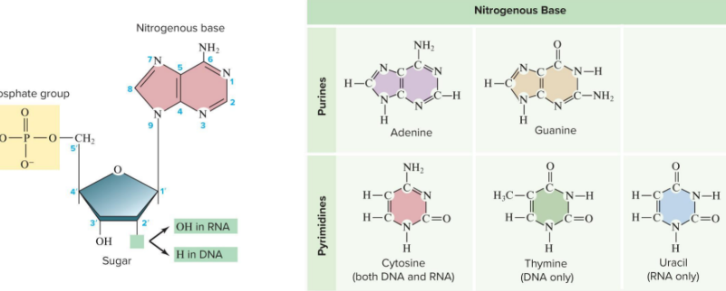
Phosphodiester Bond
Bound between adjacent nucleotides
Formed between the phosphate group of one nucleotide and the 3’ -OH of the next neucleotide
The Chain of nucleotides has 5’-to-3’ orientation
Phosphodiester Bond

Chargaff’s Rule
Erwin Chargadd determined that
Always an equal proportion of two ringed purines (A & G ) and a single ringed pyrimidines (C & T)
Chargaff’s Rule
Amount of Adenine =
Amount of Thymine
Chargaff’s Rule
Amount of Cytosine =
Amount of Guanine
Chargaff’s Rule
Ratio of A-T and G-C
Varies by species
Rosalind Franklin
Preformed X- Ray diffraction studies to identify the 3-D structure
Discovered that DNA is helical
Using Maurice Walkins DNA fibers, discovered that the molecule has a diameter of 2nm and makes a complete turn of the helix every 3.4 nm
James Watson and Francis Crick 1953
Deduced the structure of DNA using evidence form Charguff, Franklin and others
They did not preform a single experiment themselves related to DNA
Key insight of their model was each DNA molecule was made of two intertwined chains of nucleotides that is a double helix structure
Strucutre of a single DNA strand
Phosphodiester back bone repeating
Sugar and phosphate units joined by phosphodiester bonds
A single strand extends in a 5’ to 3’ direction
The double helix
Two strands arrange as a double helix
Forms two groves the larger major groove and the smaller minor groove
The double helix
Strands connect via hydrogen bonds between bases on opposite strands
Result is specific base pairs : A-T and G-C
Helix has a consistent diameter is stable because of addictive property of thousands of low energy hydrogen bonds
Base pairing
Pattern of base pairing is complementary
A forms two H bonds with T
G forms 3 H bonds with C
Base pairing
Two strands of single DNA molecule are not identical
Each strand specifies the other rby base pair complementarity
Antiparallel configuration
each phosphodiesterase strand has inherent polarity base on orientation of sugar phosphate backbone
One end terminates in 3’ OH
One end terminates in 5’ PO
Antiparallel Configuration
Strands are referred as having
5’-to-3’ or 3’-to-5’ polarity
Antiparallel Configuration
The two strands of a single DNA molecule have
Opposite polarity to one another
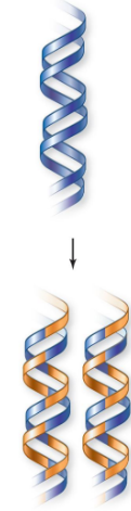
Three possible models of DNA Replication
Conservative
Semiconservative
Dispersive
DNA replication models
Conservative Model
Both strands of parental DNA remain intact, new DNA copies consist of all new molecules
DNA replication model
SemiConvservative model
Daughter stands each consist of one parental strand and one new strand
DNA replication model
Dispersive Model
New DNA is dispersed throughout each strand both daughter molecules after replication
DNA replication models
Conservative Model, Image

DNA replication models
SemiConservative Model, Image

DNA replication model
Dispersive Model, Image
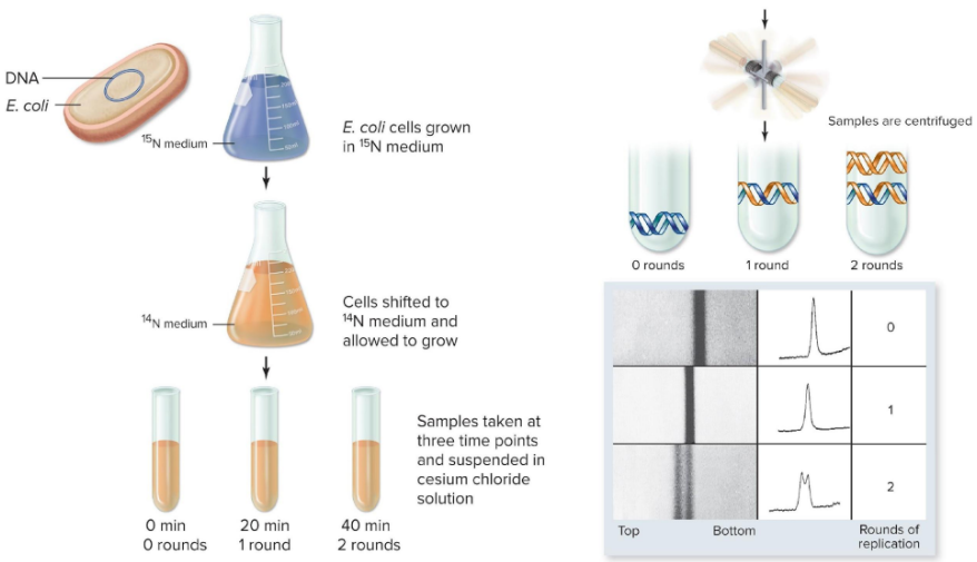
Meselson and Stahl 1958
Bacterial cells were grown in a
Heavy isotope of Nitrogen, 15^N
Meselson and Stahl 1958
After Several generation the DNA of
These Bacteria was denser than normal DNA
Meselson and Stahl 1958
Cells were switched to
Media Containing lighter 14N
Meselson and Stahl 1958
DNA was Extracted form the cells at
Various time intervals and centrifuged to separate out by weight
Meselson and Stahl’s Results
Conservative Model
Is rejected
Two density bands were not observed after round 1
Meselson and Stahl Results
Semiconservative model
is Supported
Consistent with all observations
One band after round 1
Two bands after round 2
Meselson and Stahl Results
Dispersive Model
Is rejected
1st Round Results consistent
2nd Round, did not observed one band
The Meselson Stahl experiment,
Diagram
DNA Replication
Requirements
Something to copy, parental DNA molecule
2. Something to do the copying, Enzymes
3. Building Blocks to make copy, Nucleotide Triphosphates
DNA Replication
Something to copy
Parental DNA Molecule
DNA Replication
Something to do the copying
Enzymes
DNA Replication
Building Blocks to make copy
Nucleotide Triphosphates
Stages of DNA replication
Initiation
Replication Begins
Stages of DNA Replication
Elongation
New strand of DNA are synthesized by DNA Polymerase
Stages of DNA replication
Termination
Replication is Terminated
Action of DNA polymerase
Diagram

DNA Polymerase
Match existing DNA bases with
Complementary nucleotides and links them, That is build new DNA strands
DNA Polymerase
All have several common features
Add new base to 3’ end of existing strands
Synthesize in 5’- to-3’ direction
Require a primer of RNA
RNA Polyermerase makes primer to DNA polymerase extends primer, image
Prokaryotic Replication
E. coli used as model systems for Understanding universal attributes of replication
Single circular molecule of DNA
Replication in both directions around the chromosome
Replicon
Replicon
DNA controlled by an origin
replication is bidirectional form a unique origin , Diagram

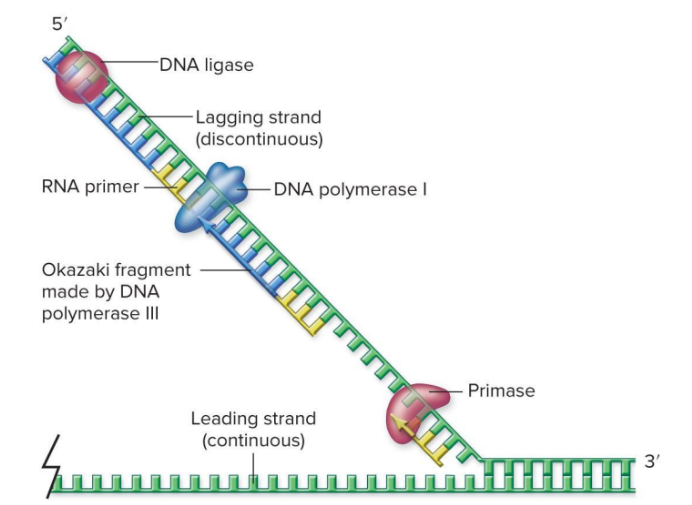
E. coli has three DNA polymerase
DNA polymerase |
Acts on lagging strand to remove primers and replace them with DNA
E. coli has three DNA polymerase
DNA polymerase ||
Involved in DNA repair processes
E. coli has three DNA polymerase
DNA polymerase |||
Main replication enzyme
E. coli has three DNA polymerase
All 3 have 3’-to- 5’ exonuclease activity: proofreading
DNA Pol | has 5’-to- 30 exonuclease activity remivug RNA primers: removing RNA primers
DNA Polyemerase activity
In addition to adding nucleotides to a growing DNA strand
Some polymerase molecules can remove nucleotides acting as nucleases
DNA Polyemerase activity
Can be
Endonucleases : Cut DNA internally
Exonuclease: Remove neucleotides form end of DNA
DNA Polymerase activity
All three E. Coli DNA polymerase have
3’-to-5’ Exonuclease activity- Proofreading
DNA polymerase activity
DNA Pol | has
5’-to-3’ Exonuclease activity : removing RNA primers
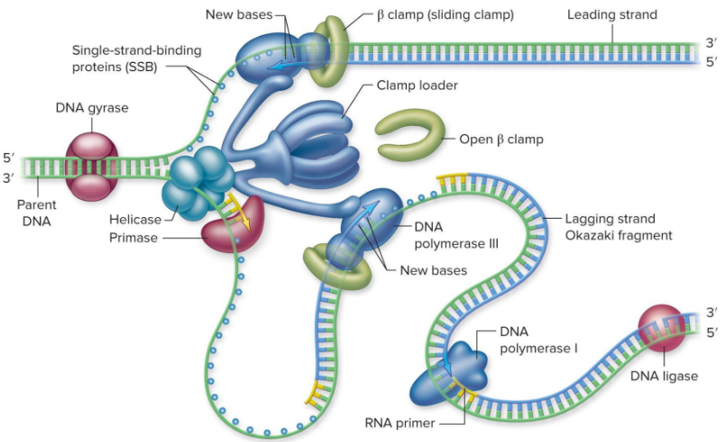
Enzymes unwind DNA
Helicases
Use energy form ATP to unwind DNA
Enzymes unwind DNA
single strand binding proteins (SSBs)
Coats strands to keep them apart
Enzymes unwind DNA
Unwinding of DNA introduces Torsional strand in the molecules that can lead to
Additional twisting of the helix called supercoiling
Enzymes unwind DNA
Topoisomerases
Are enzymes that prevent supercoiling
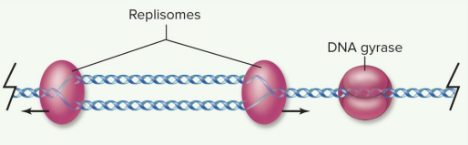
DNA gyrase is the Topoisomerase involved in DNA replication that relives the torsional strand
Unwinding the helix causes torsional strand, Supercoiling diagram

Unwinding the helix causes torsional strand, No Supercoiling diagram
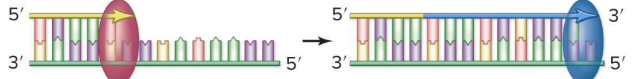
Replication is semi discontinuous
DNA polymerase can only synthesize in the
5’-to- 3’ direction
Replucation is semi discontinuous
Antiparallel nature of DNA means
New DNA strand must be synthesized in oppsite directions
Replication is semi discontinuous
Leading strand synthesized
Continuously form an initial primer
Replication is semi discontinuous
Lagging strand synthesized
Discontinuously form an initial primer
DNA fragments on the lagging strand are called OKazaki fragments must be connected together
Lagging is synthesized in pieces, Diagram
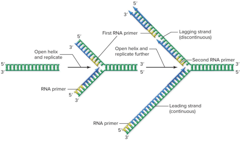
Synthesis occurs at the replication fork
Replication Fork
Is partial opening of helix formed where doubles stranded DNA is being unwound
Synthesis occurs at the replication fork
DNA Primase
RNA Polymerase that makes RNA primer
RNA will be removed and replaced with DNA later
Leading strand synthesis
Single priming event
Leading strand synthesis
strand extended by DNA Pol |||
Processivity the ability of polymerase to stay attached
β subunit forms “sliding clamp” to keep DNA Pol ||| attached to DNA ( high processivity)
Lagging strand synthesis requires additional enzymes
Discontinuous synthesis, requiers multiple Enzymes
DNA Pol ||| - like leading strand
Primase - Makes RNA primer for each Okazaki fragment
DNA Pol |- Removes all RNA primer and replaced with DNA
DNA ligase- joins Okazaki fragments to form complete strands
Lagging strand synthesis requires additional enzymes
Termination occurs at specific site
DNA Gyrase unlinks two copies
Lagging strand synthesis, Diagram latter
Replisome
Is a macromolecular assembly of enzymes involved in DNA replication
Replisome
Two main components
Primosome - Primase, helicase, accessory proteins
Complex of two DNA Pol ||| - one for each strand
Model of the structure of the replication fork diagram
Eukaryotic Replication
More complex than in prokaryotes due primarily to
Large amount of DNA in multiple chromosomes
Linear structure ( versus circular chromosomes)
Eukaryotic Replication uses multiple origins
Basic Enzymology is similar
Requiers new enzymatic activity for dealing with ends only
Eukaryotic Replication uses multiple origins
Multiple replicons, multiple origins of replications for each chromsome
Not sequence specific can be adjusted
Example: early in development when cells divide rapidly more origins can be used
Eukaryotic replication fork is more complex
Before S phase
Helicase are loaded onto possible replication origins but not activated
Eukaryotic replication fork is more complex
During S phase
Subset of these are activated and the rest of the replisome assembled
priming uses a complex of both DNA polymerase α and Primase
DNA polymerase epsilon (Pol ε) synthesizes leading strand
DNA polymerase Delta (Pol δ) synthessizes lagging strand
Archaeal and eukaryotic replication proteines are evolutionarily related
Enzymes that are similar between Eukaryotes and Archaea but different form those in Prokaryotes
DNA polymerases
Replicative helicase
Primases
Linear chromosomes have specialized ends
Telomeres
Specialized structures found on the ends of eukaryotic chromosomes
Composed of specific repeat sequences
Linear chromosomes have specialized ends
Protect ends chromosomes form nucleases
Maintain the integrity of linear chromosomes
Not made by replication complex
Replication of the end of linear DNA presents a problem, diagram
