Organisms respond to changes in their internal and external environments
1/139
There's no tags or description
Looks like no tags are added yet.
Name | Mastery | Learn | Test | Matching | Spaced | Call with Kai |
|---|
No study sessions yet.
140 Terms
What is a stimulus?
A detectable change in the environment. These changes can be detected by cells, which are called receptors.
How do organisms increase their chance of survival?
By responding to stimuli via different response mechanisms.
What are taxes and kinesis?
Simple responses which keep organisms within the favourable conditions of their environment (light, moisture, chemicals).
Taxes
Simple response in which an organism will move it’s entire body away from a favourable stimulus or away from an unfavourable stimulus.
Move towards- positive stimulus
Move away- negative stimulus
What do Earthworms show?
Negative phototaxis (light away)
Move away from light to dark environments such as soil to help avoid, dehydration, predators and to locate food.
What do bacteria show?
Positive chemotaxis, as they move towards certain chemicals to aid survival.
Kinesis
When an organism changes the speed of movement and the rate it changes directions.
If an organism moves from area where there are positive and beneficial stimuli → place with harmful stimuli, it’s kinesis response will be to increase the rate it changes direction → return to favourable conditions quickly.
If the opposite way then the rate of turning decreases to keep it moving in a relatively straight line to avoid harmful stimuli and increase chance of finding a new location with favourable conditions.
What does tropism mean?
When plants respond via growth, to stimuli.
Can be positive or negative, growing towards or away from a stimulus.
Examples: phototropism, geotropism, chemotropism, hydrotropism, aerotropism.
What are tropisms controlled by?
Specific growth factors e.g. IAA (indoleacetic acid)
What are growth factors?
Chemical substances that are released in response to a stimulus.
Act in similar way to hormones
Effect of growth factors not as quick as that of an electrical nervous system.
Why do plants have specific growth factors?
Because they do not have a nervous system, so need another major way to respond to changes in the environment.
Growth factors aren’t hormones as hormones travel in blood.
Examples: IAA
What does the effect of a plant growth factor depend on?
Concentration of growth factor
The tissue being acted on (e.g. root vs shoots)
Developmental stage of plant (affects signalling pathways e.g. IAA promotes elongation more effectively in young shoots).
Species of plant
Other growth factors present
Makes it difficult to investigate. Also the fact that they’re present in small amounts.
What is IAA?
A type of auxin.
Auxins are continually made in shoot apex and young leaves→ can diffuse or translocate (to roots).
IAA however, is synthesised in root and shoot tips.
It can control cell elongation in shoots
It inhibits growth of cells in the roots.
(Does this by affecting the growing area of plant)
(Positive) Phototropism in unilateral light (one direction of light) SHOOTS
Shoots need light for LDR in photosynthesis, which is why plants grow and then bend towards light (to receive more).
Phototropism is controlled by the IAA.
Shoot tip cells produced IAA, and this IAA diffuses to other cells (on side of plant further from the sun).
This causes that side to elongate and then plant then bends towards the sun/light source.
What would happen in phototropism if light was evenly distributed?
The IAA would diffuse downwards, away from light source, so plant grows upwards.
Photoreceptors
Structures/pigments that are sensitive to light, often of specific wavelengths.
Phototropins are a group of photoreceptors primarily responsible for triggering phototropism.
When the light of the right wavelength is present, changes in the phototropin molecules trigger reactions, ultimately resulting in the redistribution of auxin so that there is more on the shaded side of the plant.
Roots
Do not photosynthesis so don’t require light.
Also more able to anchor plant the further away they are from light.
Negative phototropism in roots
High concentration of IAA diffuses to side of root further away from the light, inhibiting growth so heavier side elongates and moves deeper downwards into the soil → root bends away from the light → can anchor plant in.
Gravitropism/ Geotropism in shoots: Negative
IAA will diffuse from upper to lower side of a shoot (due to gravity)
If plant is vertical → grows upwards.
If plant is on it’s side → IAA diffuses to lower side → elongates → grows upwards (against gravity).
Gravitropism/ Geotropism is roots: Positive
IAA moves to lower side of roots, due to gravity, so that the upper side elongates and the root bends towards gravity and anchors the plant in.
Flow charts outlining response to stimulus
Stimulus → Receptor → Coordinator → Effector → Response
Simple description of reflex arc:
Receptor detects stimulus and electrical impulses are generated to sensory neurone (connected to receptor).
Sensory neurone sends electrical impulses to the spinal chord (coordinator).
Electrical impulses are passed to relay neurones in spinal chord across a junction called the synapse
Synapse- Relay neurones are connected to motor neurones and pass on electrical impulses across a point called the synapse.
Motor neurones carry the impulses to the muscles which cause it to contract, leading to a response in which the body part moves away from the object that created the stimulus.
What is the nervous system made up of?
The peripheral and central nervous system.
PNS connects the CNS to the rest of the body as the PNS consists of the neurones that carry impulses from receptors to effectors, through the CNS.
CNS acts as coordination centres- processes information and coordinate responses.
Receptors
Detect stimuli/ changes in the environment.
Each receptor responds only to specific stimuli.
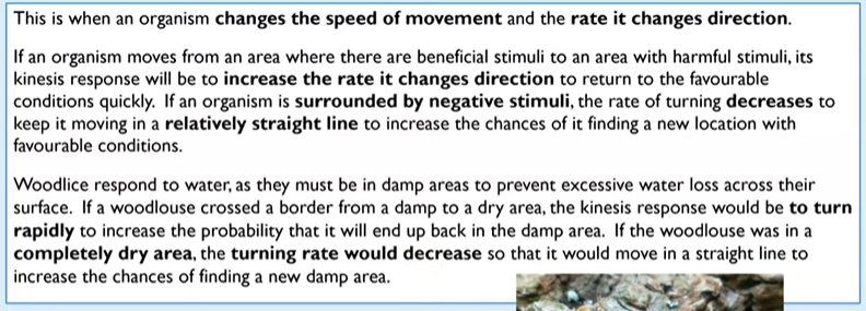
What two types of photoreceptors does the retina contain?
Rods and cones
They detect light
How is the amount of light that enters the eye controlled?
By the muscles of the iris.
How are light rays focused?
By changing the nature of the lens through the ciliary muscle and suspensory ligaments.
Focused onto retina so it can be detected by photoreceptors there.
Mostly focuses on the fovea (specialised area of retina containing a dense number of photoreceptors- mainly cone cells).
Retina is a photosensitive layer at the back of the eye.
What does sensitivity of light refer to?
The amount of light required to stimulate the receptor.
Receptors hit by light are stimulated and if threshold level is reached, then an action potential will be triggered.
Summary of structure of rod and cone cells
Once a receptor is stimulated, it can send impulses to the brain.
The brain interprets the pattern of impulses to form an image.
There are synapses connecting the rods and cones to bipolar neurones.
The bipolar neurones connect to ganglion cells via synapses.
Ganglion cells have axons that extend to the optic nerve which is directly connected to the brain.
Due to the high number of receptors, it is not possible for there to be individual connections between each receptor and the brain.
The way that rods and cones are connected to the optic nerve affects visual acuity.
Rods
Have rod-like shapes
Cannot distinguish different wavelengths of light, so it processes images in black and white.
Can detect light even at very low light intensity due to retinal convergence- survival mechanism
What is retinal-convergence?
Means there are multiple rod cells connected to a single bipolar cell- e.g. of summation.
Does mean that the brain cannot distinguish between the separate sources of light that stimulated it as only one impulse from the bipolar cell is sent.
Two light sources close together cannot be seen as separate- rod cells give low visual acuity, meaning one can’t see as well in lower light intensity.
The human retina- rods
To create the generator potential, the pigment of rod cells (rhodopsin) must be broken down by light energy.
There is enough energy from low light-intensity to cause the breakdown of this pigment →can trigger action potential (next flashcard)
If enough pigment is broken down, then threshold is met (in bipolar cells) and the action potential is triggered.
This threshold can be reached even in low light because so many rod cells are connected to a single bipolar cell- example of summation.
Why does the breakdown of iodopsin trigger an action potential if threshold is met?
Iodopsin ⇌ Opsin and retinal (forward reaction is called bleaching and backwards is dark adaptation).
Presence of opsin causes a change in the permeability of the rod cell to sodium
Describe what happens when light is detected
Rod
Rhodopsin broken down → Generator potential if threshold met → Release of transmitter in the rod cell → nerve impulse impulse carried across the synapse to bipolar cells → synapse to ganglion cells → optic nerve to the brain if generator potential is large enough.
Cone
Iodopsin broken down → Generator potential if threshold met → Release of a transmitter in the cone cell → nerve impulse carried across the synapse to bipolar cells → synapse to ganglion cells → optic nerve to the brain ig generator potential is large enough.
Summation/ Spatial summation
The rhodopsin broken down by all the rod cells connected to bipolar cell are collected and hopefully it can collectively trigger a large enough stimulus to result in generator potential.
What is rhodopsin?
A protein pigment found within the rod cells.
Breaks down when enough energy from light is absorbed.
This breakdown of pigment can go on to trigger a generator potential.
Why is rhodopsin unstable in bright light?
Rhodopsin is broken down more quickly than it can reform, disabling the rod cell.
Cone cells
3 types of cone cells: red green and blue
They contain different types of iodopsin pigment which all absorb different wavelengths of light.
Depending on the proportion of each cone cell that is stimulated, we perceive colour images and a whole range of different colours.
Iodopsin is only broken down if there is a high light intensity, so action potentials can only be generated with enough light.
This is why we can’t see very well in the dark because there isn’t enough energy to break down iodopsin and trigger an action potential.
Why can cone cells only respond to high light intensity?
Usually only one cone cell connects to a bipolar cell.
So no retinal convergence so no spatial summation.
However, because there is only one cone cell connected to one bipolar cell, the brain can distinguish between separate sources of light detected -cone cells give high visual acuity, sharper clearer vision in colour.
Distribution of rods and cones in the retina
Rods found across most of retina but less in fovea whereas cone mainly in fovea.
Light is focused by the lens on the part of the retina opposite the pupil, the fovea, which will receive the highest intensity of light.
Therefore, most cone cells are located near the fovea as they only respond to high light intensities and rod cells further away as these can respond at lower light intensities.
What is blind spot?
Spot in retina with no cone or rod cells.
No photoreceptors and no light to be detected there.

Pacinian corpuscle
Responds to pressure changes AS ITS A RECEPTOR
Receptors occur deep in skin, mainly in fingers and feet.
Consists of a single sensory neurone, end wrapped with layers of connective tissue (lamellae) with gel in between (containing K+ and Na+).
Sensory neurone in the Pacinian corpuscle has special channel proteins in it’s plasma membrane called stretch-mediated sodium ion channels.
Only responds to mechanical pressure.
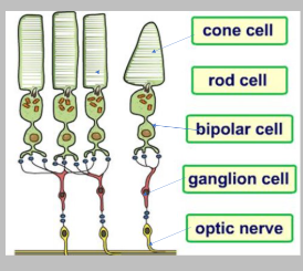
The structure of a neurone
What does the cell body of a neurone contain?
The organelles found in a typical animal cell, including the nucleus.
Proteins and neurotransmitter chemicals are made here.
What do dendrites do?
Receive impulses from other neurones to pass to other neurones.
What is the axon and what does it do?
Long conductive fibre
Connected to cell body and axon.
Carries nervous impulses along a neurone, away from cell body.
Myelin sheath
A lipid layer, insulating the axon (made of Schwann cells).
Because it’s lipid, charged ions cannot pass through.
There are gaps in the myelin sheath, called nodes of Ranvier.
What do Schwann cells do?
Wrap around the axon to form the myelin sheath.
What are the gaps between the myelin sheath called?
Nodes of Ranvier
Impulses can ‘jump’ here.
What is resting potential?
The difference in electrical charge between the inside and outside of the neurone when it is not conducting an impulse.
It’s -70mV because the inside of the axon is more negative than the outside- said to be polarised.
At resting potential, the neurone is not transmitting an impulse.
How are resting potentials maintained?
By the sodium-potassium pump, involving active transport and therefore ATP.
The pump moves 2 K+ ions in and 3 Na + ions out.
Both of these are transported against concentration gradient (more K+ inside and more Na+ outside).
This creates an electrochemical gradient → facilitated diffusion
K+ diffuses out and some Na+ diffuses in, but membrane is more permeable to K+ so more of that moves out and resting potential is maintained.
Why is the membrane more permeable to K+?
More potassium ion protein channels.
Potassium ion channels are mainly opened whereas sodium ion channels are usually closed and open after an increase in pressure
Sequence
Stimulus: If generator potential was large enough action potential can be started.
Depolarisation: If threshold is reached (-55mV), voltage-gated sodium ion channels open and lots of Na+ enters via facilitated diffusion. The inside of the axon becomes more positive, until +30mV.
Repolarisation: Sodium ion channels and voltage-gated Na+ channels close. Voltage-gated K+ channels open and K+ leaves the axon, making the inside negative again.
Hyperpolarisation: K+ channels are slow to close and so too much leaves and the inside has become more negative than resting potential, -80mV is when they begin to close.
After it has closed, the sodium-potassium pump restores the original ion balance, and resting potential is restored.
All-or- nothing principle
If the stimulus isn’t large enough, not enough energy will be provided to open enough sodium ion channels, so the voltage won’t exceed -55mV and an action potential and impulse isn’t produced.
Any stimulus that does trigger depolarisation to -55mV will always peak at the same maximum voltage.
Bigger stimuli doesn’t increase the peak but the frequency of action potentials.
Why is the all-or-nothing principle important?
As it makes sure that animals only respond to large enough stimuli, rather than responding to every slight change in the environment which would overwhelm them (and instead hinder survival).
Refractory period
After an action potential is generated, the membrane enters the refractory period when it can’t be stimulated, because ion channels are recovering and can’t be opened.
Why is the refractory period important?
Ensures that discrete impulses are produced, meaning an action potential cannot be generated immediately after another one to make sure each is separate from another.
It ensures that action potentials travel in one direction. This stops the action potential from spreading out in two directions, which would prevent a response.
It limits the number of impulse transmission. This is important to prevent over reaction to a stimulus and therefore overwhelming the senses.
Transmission of the impulse
In the non-myelinated neurone, the action potential moves as a wave along the axon→ one region depolarises the next.
In a myelinated neurone, the impulses ‘jump’ from node to node- saltatory conduction.
Much faster as depolarisation only happens at the nodes, and not at every part of the membrane.
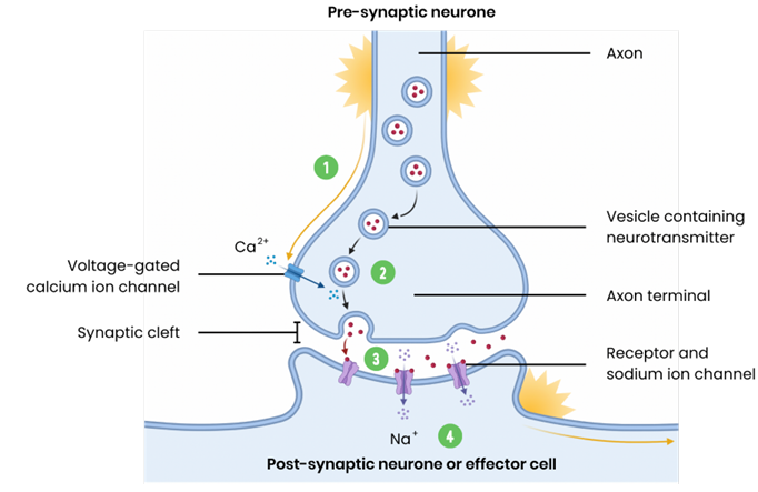
After action potential reaches the axon terminal, it triggers Ca2+ channels to open. Ca2+ moves in and causes vesicles (containing neurotransmitters) to fuse with the presynaptic membrane.
These neurotransmitters diffuse across the synaptic cleft and bind to receptors on the postsynaptic membrane, opening ion channels.
Inhibitory opens Cl- so they can enter, making inside more negative. When it opens K+ channels, they leave. Both of these make it harder to trigger an action potential.
Excitatory neurotransmitters make it easier to trigger an action potential by opening Na+ channels.
Extra stuff on the heart structure
What is the rate of heart contraction controlled by?
Wave of electrical activity/ the nervous system
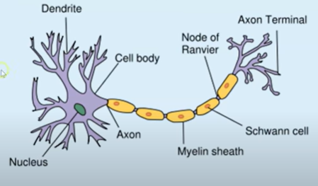
What is the sinoatrial node?
SAN
Pacemaker located in the right atrium.
Tissue which releases a wave of electricity (depolarisation).
When this hits cardiac muscle, it causes it to contract.
What is the atrioventricular node?
AVN
Located near the border of the right and left ventricle within the atria still.
What is the bundle of His?
Conductive tissue that runs through the septum of the heart.
What are the Purkyne fibres?
Conductive tissue that go all the way through the walls of the ventricle.
Control of the heart
Sinoatrial node (SAN) acts as pacemaker → releases regular waves of electrical activity across atria
○ Causing atria to contract simultaneouslyNon-conducting tissue between atria / ventricles prevents impulse passing directly to ventricles
○ Preventing immediate contraction of ventriclesWaves of electrical activity reach atrioventricular node (AVN) which delays impulse
○ Allowing atria to fully contract and empty before ventricles contractAVN sends wave of electrical activity down bundle of His, conducting wave between ventricles to apex where it branches into Purkyne tissue
○ Causing ventricles to contract simultaneously from the base up
The medulla oblongata- coordination centre
Found in the brain
Controls heart rate, via the autonomic nervous system. It does this by controlling how quickly the SAN releases the wave of depolarisation.
Two parts that it is linked to
Sympathetic and parasympathetic nervous system.
What does autonomic mean?
Automatic and done without thought
What does the sympathetic nervous system do?
Increase heart rate by increasing rate of depolarisation.
What does the parasympathetic nervous system do?
Decreases the heart rate by decreasing the rate of depolarisation.
Why does the heart rate change in response to pressure?
If blood pressure is too high, this can cause damage to walls or arteries → heart attack or stroke
Too low could mean there is an insufficient supply of oxygenated blood to respiring cells and removal of waste.
How does heart rate change in response to changes in pH and why?
The pH of blood will decrease during times of high respiration (e.g. exercise) due to the production of carbon dioxide or lactic acid.
Excess acid must be removed from the blood rapidly to prevent enzymes denaturing. This is achieved by increasing the heart rate, so carbon dioxide can diffuse out into the alveoli more quickly.
How does the heart rate change when pressure is increased?
Stimulus: Increased pressure
Receptor: Pressure receptors/ Baroreceptors in the wall of aorta and carotid artery are stretched if high blood pressure.
Coordination centre: More electrical impulses sent to medulla oblongata and then more sent via parasympathetic nervous system to SAN to decrease frequency of electrical impulses/ depolarisation waves.
Effector: Cardiac muscle-/SAN tissues release fewer waves of depolarisation
Response: Reduced heart rate
How does heart rate change when pressure is decreased?
Stimulus: Decreased pressure
Receptor: Pressure receptors/ baroreceptors in the wall of aorta or carotid artery are not stretched in low blood pressure.
Coordination centre: More electrical impulses sent to medulla oblongata and then more impulses sent via sympathetic nervous system to SAN to increase the frequency of electrical impulses/ waves of depolarisation.
Effector: Cardiac muscle/ SAN tissues then depolarised more frequently.
Response: Increased heart rate
How does the heart rate change in response to decreased pH?
Stimulus: Decreased pH
Receptor: Chemoreceptor in wall of aorta and carotid artery.
Coordination centre: More electrical impulses sent to medulla oblongata and then more impulses sent via sympathetic nervous system to SAN to increase frequency of depolarisation.
Effector: Cardiac muscle/ SAN tissues to release waves of depolarisation more frequently.
Response: Increased heart to deliver blood to lungs rapidly to remove carbon dioxide.
Factors that affect the speed of conductance
Myelination and saltatory conduction
Axon diameter
Temperature
Myelination and saltatory conduction
The axon is the conductive, long fibre that carries the nervous impulse along the motor neurone.
Schwann cells wrap around the axon to form the myelin sheath, which is a lipid and therefore does not allow charge ions or the impulse to pass through it. There are gaps between these myelin sheath called the nodes of Ranvier.
The action potential jumps from node to node (Saltatory conduction), which means the action potential travels along the axon faster as it doesn’t have to generate an action potential along the entire length of the axon, just at the nodes of Ranvier.
Axon diameter
With a wider diameter, the speed of conductance increases.
A wider diameter means that there is less leakage of ions and therefore action potentials travel faster- threshold met faster.
Temperature
A higher temperature increases the speed of conductance for 2 reasons:
Ions diffuse faster
Enzymes involved in respiration work faster. Therefore there is more ATP for active transport in the Na+/K+ pump.
What are synapses?
Gaps between the axon of one neurone and the dendrite of another one.
Here the action potential is transmitted as neurotransmitters that diffuse across the synapse.
Function of a synapse
An action potential arrives at the synaptic knob. Depolarisation of synaptic knob leads to the opening of voltage-gated Ca 2+ channels and Ca2+ diffuses into the synaptic knob/neurone.
Vesicles containing neurotransmitters move towards and fuse with the presynaptic membrane. Neurotransmitter is released to the synaptic cleft.
Neurotransmitter diffuses down concentration gradient (no neurotransmitter in post-synaptic neuron), to post-synaptic membrane; neurotransmitter binds by complementary of shape to receptors on the surface of the post-synaptic membrane (receptors only on post-synaptic neurone).
This causes Na+ ion channels on the post-synaptic membrane to open and Na+ diffuse in; if enough Na+ diffuses in, above threshold, then post-synaptic neurone becomes depolarised.
Neurotransmitter is hydrolysed (by acetylcholinesterase) and released from the receptor where it can then be reabsorbed by the presynaptic neurone, and recycled., the Na+ channels close and the post-synaptic neuron can re-establish resting potential.
This stops overstimulation as if it wasn’t removed, then depolarisation would continue as neurotransmitter would continue binding the complementary receptors on post-synaptic membrane.
Cholinergic synapse
Exactly the process above but you must know the name of neurotransmitter and the enzyme that breaks it down.
Neurotransmitter- Acetylcholine
Enzyme- Acetylcholinesterase- hydrolyses into choline and acetate.
Explain how synapses result in unidirectional nerve impulses.
Neurotransmitter only released/ made in the pre-synaptic neurone.
Receptors are only on post-synaptic membrane.
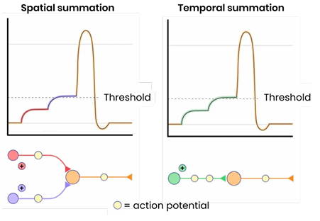
What is summation?
The rapid build up of neurotransmitters in the synapse caused by the addition of a number of impulses converging on a single post-synaptic membrane.
Makes action potential more likely.
Two methods: spatial and temporal
This is needed because some action potentials do not result in sufficient concentrations of neurotransmitter being released to generate a new action potential.
Spatial summation:
Many different pre-synaptic neurones collectively trigger a new action potential by combining the neurotransmitter they release, to exceed the threshold value.

Temporal summation:
One neurone releases neurotransmitter repeatedly over a short period of time to add up to enough to exceed the threshold value.
It does this by releasing action potentials multiple times in presynaptic neurone.
Inhibitory synapses
Neurotransmitter causes for Cl- channels to open instead.
Causes chloride ions to move into the postsynaptic neurone and potassium ions to move out.
These both cause the postsynaptic membrane potential to decrease to -80mV, hyperpolarisation, and therefore an action potential is highly unlikely
Not a disadvantage- don’t want to respond to every single stimulus in environment- overstimulate the senses.
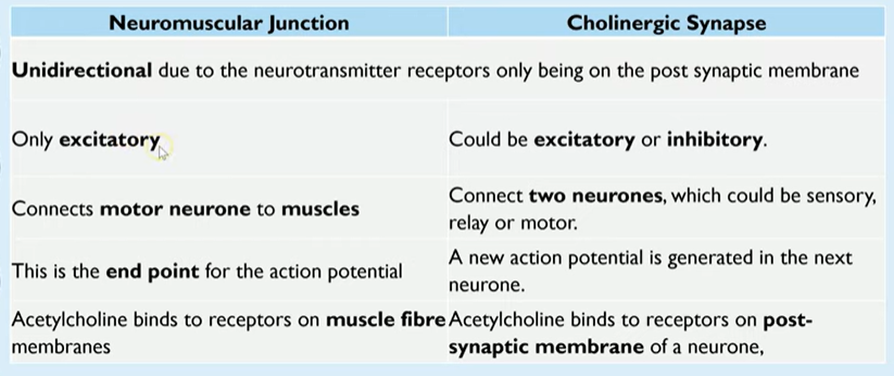
Neuromuscular junction
This is a synapse that occurs between a motor neurone and a muscle and is very similar to a synaptic junction.
Neurotransmitter still involved.
Instead of action potential, muscles are caused to contract.
Comparison of neuromuscular junction and cholinergic synapse
Muscle fibre sarcolemma
Cholinergic can also be connected to glands.
Action potential MAY be initiated in postsynaptic neurone but it propagates along sarcolemma down T-tubules.
Structure of a synapse
What are cholinergic synapses?
Synapses that use the neurotransmitter acetylcholine.
Use examples to explain the effect of drugs on a synapse.
Some drugs stimulate the nervous system, leading to more action potentials, eg.
Similar shape to neurotransmitter
Stimulate release of more neurotransmitter
Inhibit enzyme that breaks down neurotransmitter → Na+ continues to enter
Some drugs inhibit the nervous system, leading to fewer action potentials, eg.:
Inhibit release of neurotransmitter eg. prevent opening of calcium ion channels
Block receptors by mimicking shape of neurotransmitter.
Overview of muscle contraction
Myosin heads slide actin along myosin, causing the sarcomere to contract/shorten.
When all the sarcomeres in all the myofibrils shorten, the entire muscle fibre shortens.
When many fibres shorten together, the whole muscle shortens/contracts, pulling on the tendon which pulls on the bone and causes movement.
H and I bands shorten, bringing Z lines closer together.
Describe how muscles work
In antagonistic pairs → pull in opposite direction:
One contracts (agonist), pulling on bone/producing force.
One muscle relaxes (antagonist).
Skeleton is incompressible so muscle can transmit force to bone, creating movement.
Advantage of this is that because the second muscle is able to reverse movement caused by first muscle, posture can help be maintained.
Can be automatic as part of a reflex response or controlled by conscious thought.
Structure of skeletal muscle
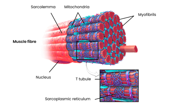
Made of bundles of muscle fibres/cells packaged together.
Attached to bones by tendons.
Muscle fibre contains:
Sarcolemma (cell membrane) which folds inwards (invagination) to form T (transverse) tubules.
Sarcoplasm
Multiple nuclei
Many myofibrils
Sarcoplasmic reticulum
Many mitochondria.
Myofibril= one piece of spaghetti and a sarcomere is a unit within myofibril.
Structure of a myofibril
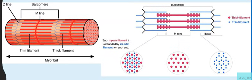
Muscle fibres are made of millions of myofibrils which collectively bring about the force to cause movement.
Myofibril are made of two key types of proteins: myosin and actin, that form a sarcomere.
Myosin is a thicker protein filament.
Arrange in parallel in functional units called sarcomeres.
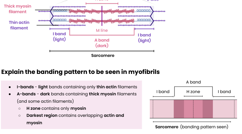
Bands and stuff
The curvy wall of sarcomere represents the bands.
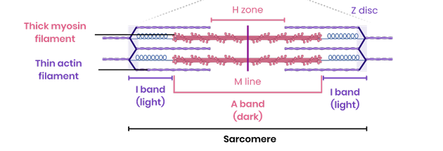
Ends- Z line
Middle- M line
H zone
Myosin heads slide actin along myosin, causing the sarcomere to contract/shorten
When this happens: H zones and I bands shorten.
Allowing Z lines to become closer.
A bands stay the same
Sliding filament theory
When an action potential reaches a muscle, it stimulates a response.
Depolarisation spreads down sarcolemma via T-tubules causing Ca2+ release from sarcoplasmic reticulum and diffuse into myofibrils.
Calcium ions cause movement of tropomyosin in actin.
This exposes binding sites on actin.
Allowing myosin heads to attach to binding sites.
The ATP on myosin head is hydrolysed into ADP and Pi by ATP hydrolase, causing myosin heads to bend as tension is created, pulling and sliding the actin filament across myosin, shortening sarcomere.
A new ATP molecule can then bind to the myosin head and cause it to change shape slightly, and as a result it detaches from the actin.
Myosin reattaches to a different actin binding site further along actin. Process is repeated as long as calcium ion concentration is high and ATP is available and until the actin has slid towards the centre of the sarcomere ( M line).
What happens during muscle relaxation?
Ca2+ is actively transported back into the sarcoplasmic reticulum using energy from ATP.
Tropomyosin moves back to block myosin binding site on actin again → no actinomyosin cross bridges.
Explain the banding pattern to be seen in myofibrils
I bands are light because they contain only thin actin filaments.
A bands are dark because they contain thick myosin filaments and some actin filaments
H zone only contains myosin
Darkest region contains overlapping actin and myosin.
ATP significance
Active muscles require a high concentration.
That’s why there are so many mitochondria in myofibril.
In times when aerobic respiration cannot create enough ATP to meet this demand, anaerobic respiration occurs.
The chemical phosphocreatine, which is stored in muscles, assists this by providing phosphate to regenerate ATP from ADP.