Test #2 - common sonographic findings
1/22
There's no tags or description
Looks like no tags are added yet.
Name | Mastery | Learn | Test | Matching | Spaced | Call with Kai |
|---|
No analytics yet
Send a link to your students to track their progress
23 Terms
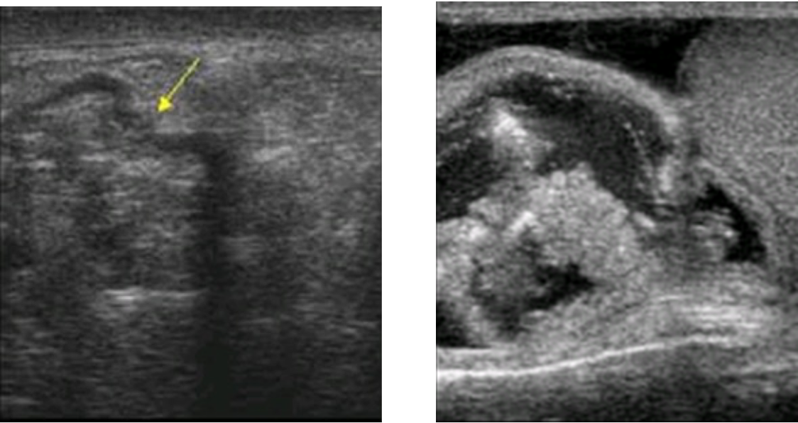
Tubular ectasia of rete testis
hypoechoic, small cystic spaces
Dilated tubules appear avascular and fluid filled on Col Dop
Yolk sac tumor
inhomogeneous
echogenic foci
Choriocarcinoma
heterogenous mass
calcifications
acute epididymitis
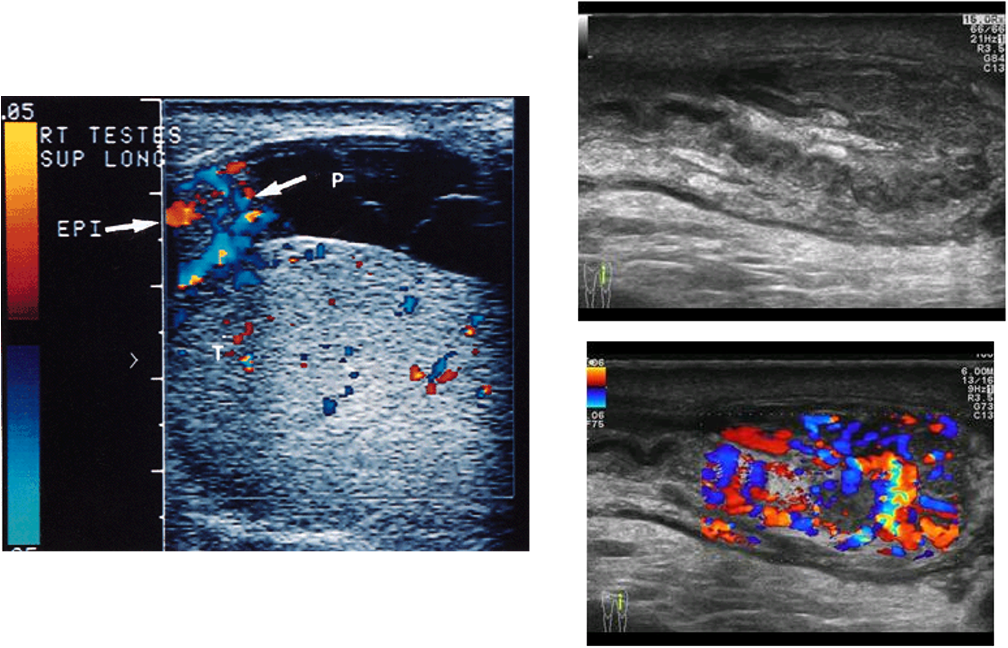
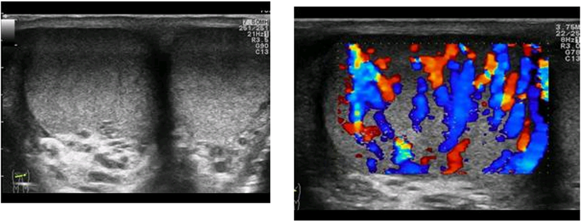
enlarged, hypoechoic epididymis head, increased flow
Chronic Epididymitis
thickened echogenic epididymis, calcifications
epididymitis
Cryptorchidism (Undescended testis)
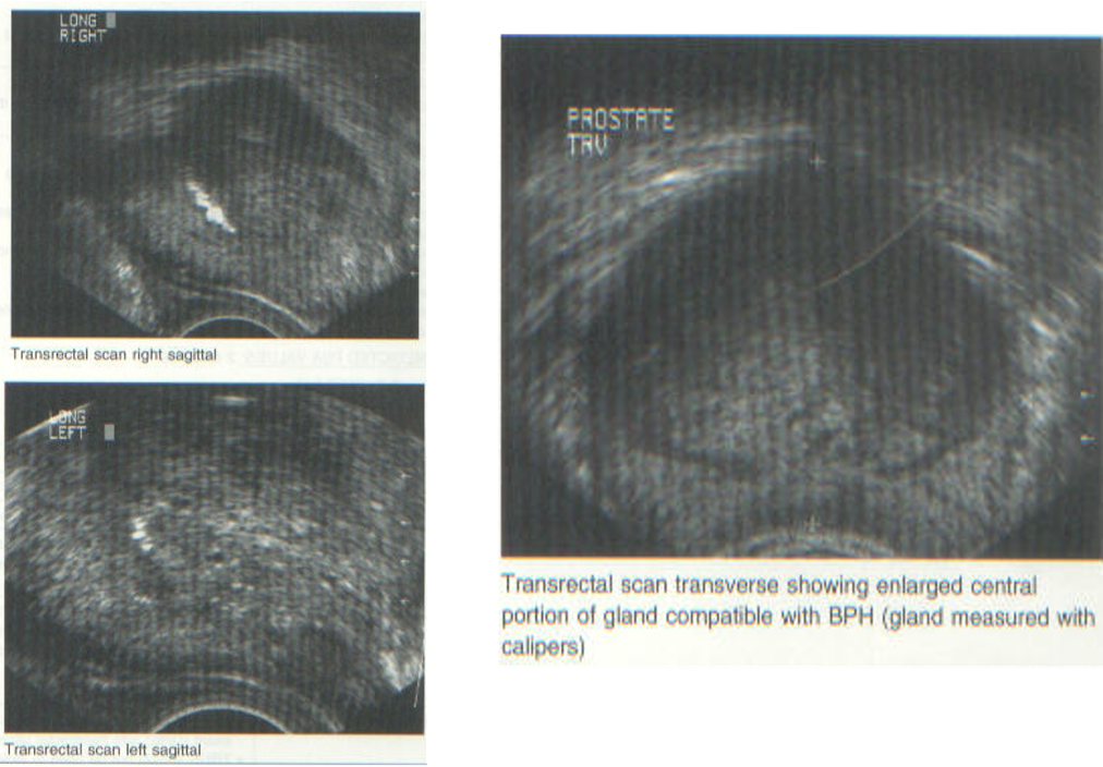
Prostate carcinoma (Adenocarcinoma)
‘beak” where seminal vesicles enters central zone
70 % Peripheral zone, 20% transition, 1-5% central
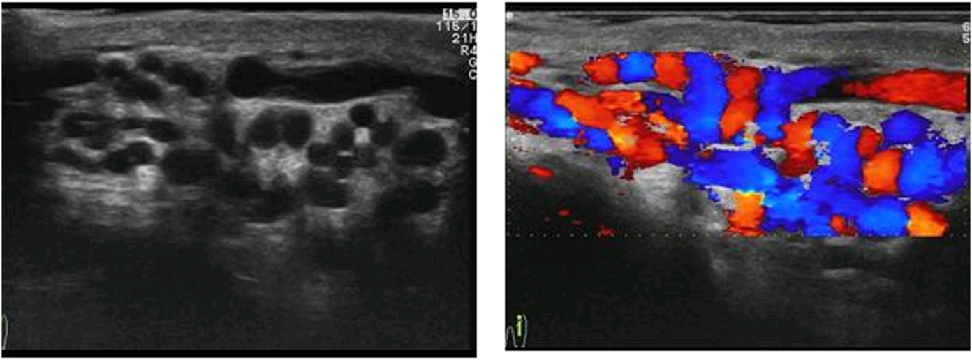
varicocele
greater than 2 mm
Preform valsalva for increased flow in prominent veins
Scrotal hernia
Look for peristalsis
fluid-filled loops
Benign prostatic hyperplasia (BPH)
transitional zone
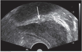
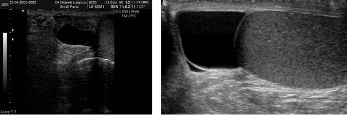
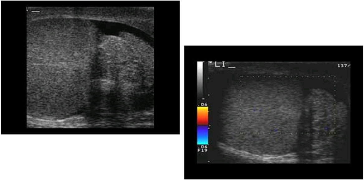
Hydrocele
anechoic fluid, calcifications with posterior shadowing

hematocele
focal mural calcification
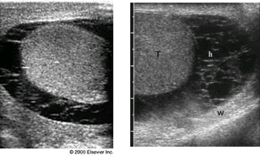
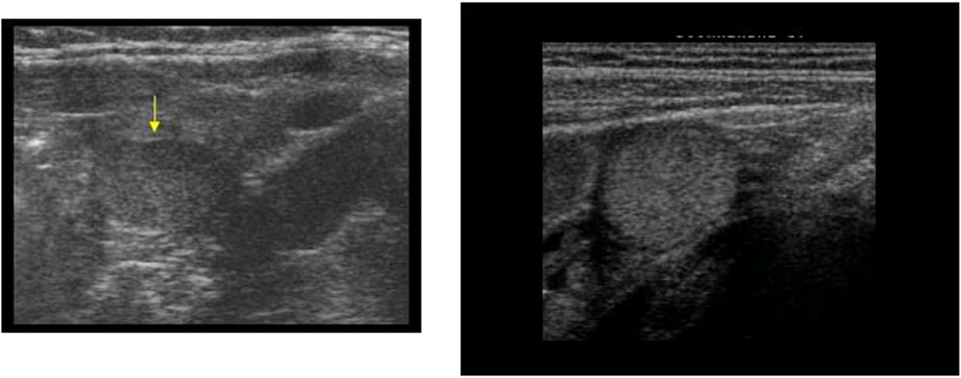
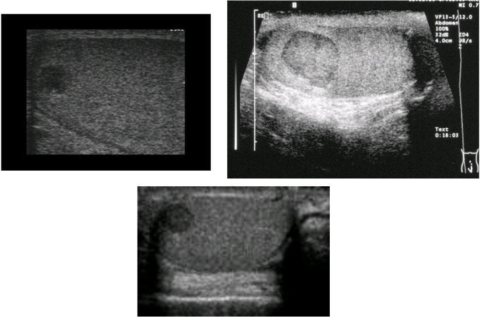
spermatocele
round or oval anechoic mass, no posterior enhancement
orchitis
Torsion “bell clapper deformity”
enlarged epididymis and testicle
No blood flow
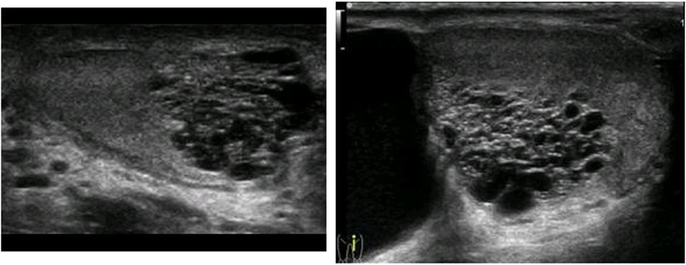
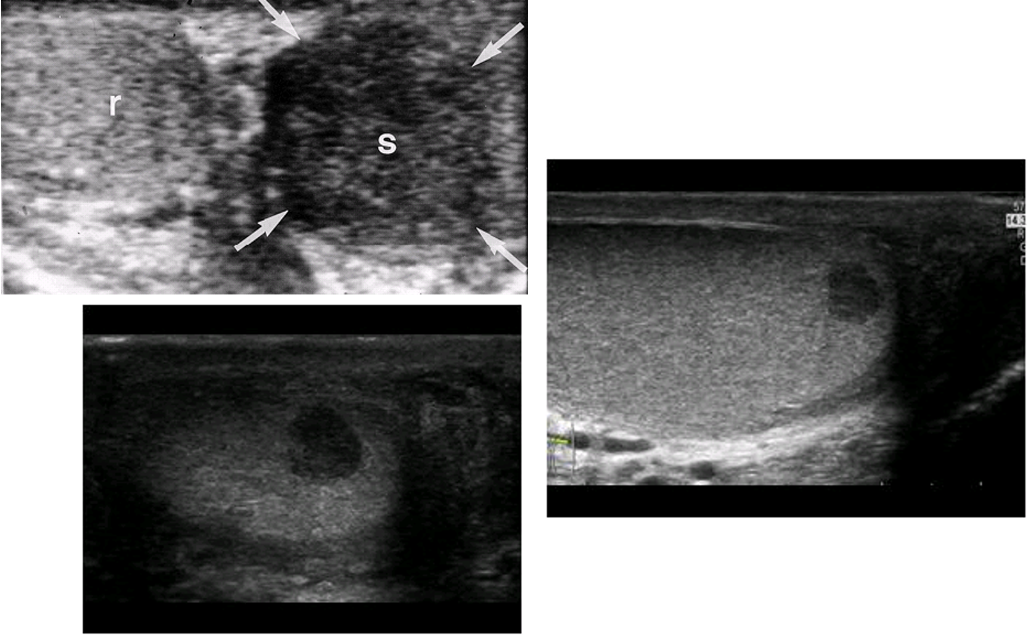
Leydig Cell tumor
non-germ cell
benign leydig cell tumor
solid, well-circumscribed intratesticular mass
Less than 1 cm
Peripheral flow
malignant leydig cell tumor
hypoechoic
greater than 5 cm
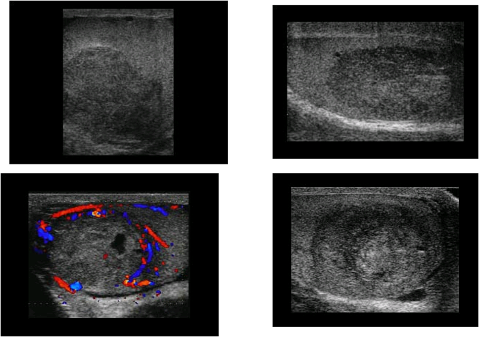
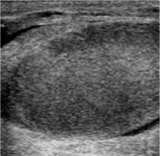
seminoma
Well-defined, solid, hypoechoic mass
Homogeneous echotexture
Hypervascularity on color Doppler in tumors larger than 1.6 cm
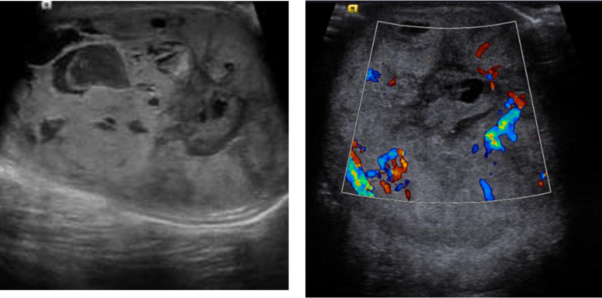
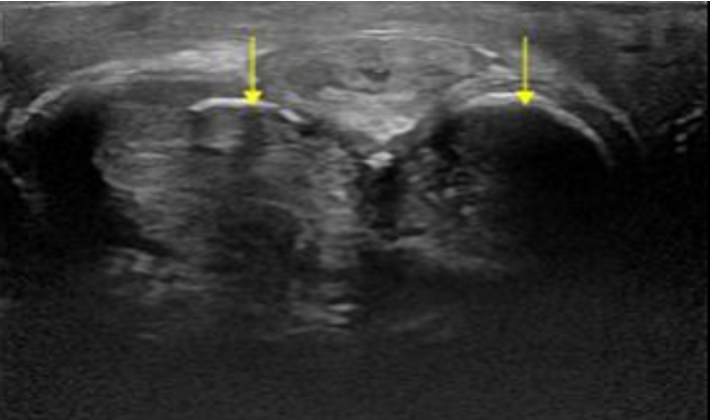
lymphoma
enlargement of testes
Decreased echogenicity diffuse or focal
Increased vascularity
Peyronie’s Disease
Plaque is dense hyperechoic area near peripheral margin
Calcifications with or w/o shadow
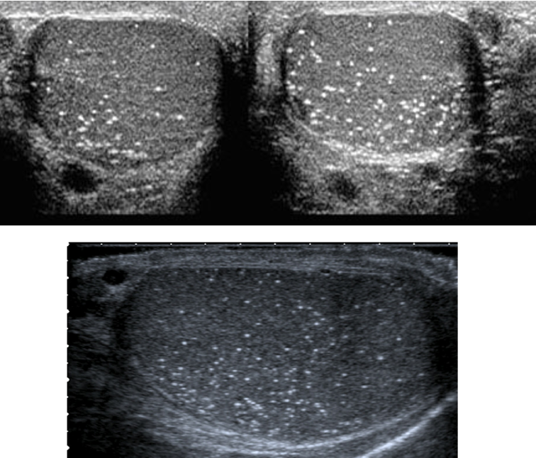
Microlithiasis
1-3 mm echogenic foci