times on slide 38 not important, just know general process
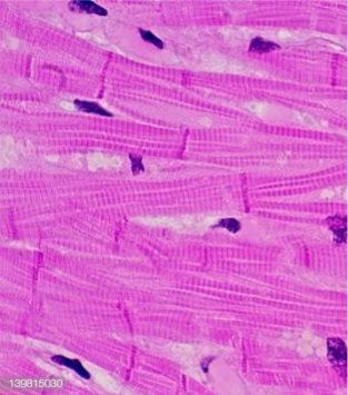
Cardiac Muscle - Characteristic
cardiac muscles cells are _____
striated
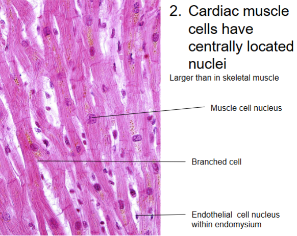
Cardiac Muscle - Characteristic
cardiac muscle cells have ____ located _____
larger than in skeletal muscle
centrally, nuclei
Cardiac Muscle - Characteristic
Y-shaped ____: cells are _____
allows muscle fibers to ______ in more complicated arr w/i ______ to make an efficient _____ mechanism for emptying the heart
fibers, branched
interweave, fascicles, contraction
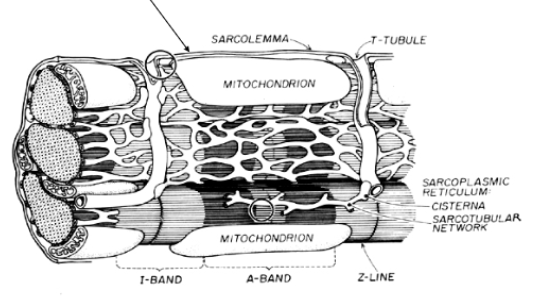
Cardiac Muscle - Characteristic
myofibrils are ____ dense and organized than those of skeletal muscle
alternate w/ abundant _______
may take up to 40% of _____ (abt 2% in skeletal)
heart relies almost exclusively on _____ respiration
less
mitochondria
cytoplasm
aerobic
Cardiac Muscle - Characteristic
intercalated discs: contact b/w cells is accomplished by ______ in the _______ region
T tubules are ______ and _____ numerous in cardiac muscles than skeletal muscles
cardiac muscle has _____ sarcoplasmic _____
interdigitation, transverse
larger, more
reduced, reticulum
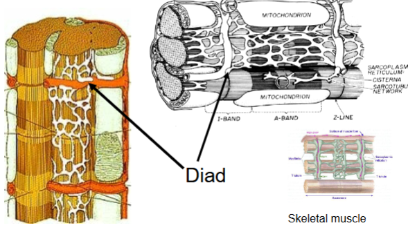
Cardiac Muscle - Characteristics
cardiac muscles have _____ rather than _____
SR touches it in ____ rather than _____ length
cardiac muscle makes up the _____ of the heart
cardiac muscle is capable of generating ______ action potentials at ______ intervals
diads, triads
spots, whole
myocardium
endogenous, periodic
Cardiac Muscle - Characteristics
cardiac muscles can make a membrane potential that is ____ _____ nerve membrane potential
diff from
Cardiac Muscle - Characteristics
has cardiac cycle
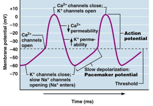
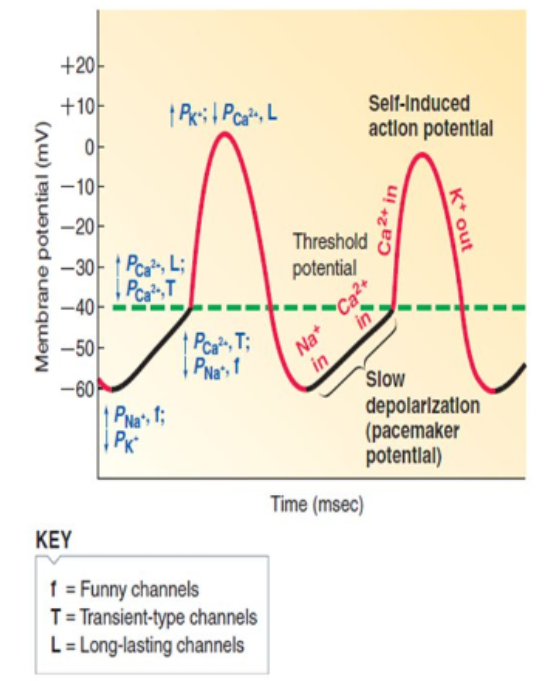
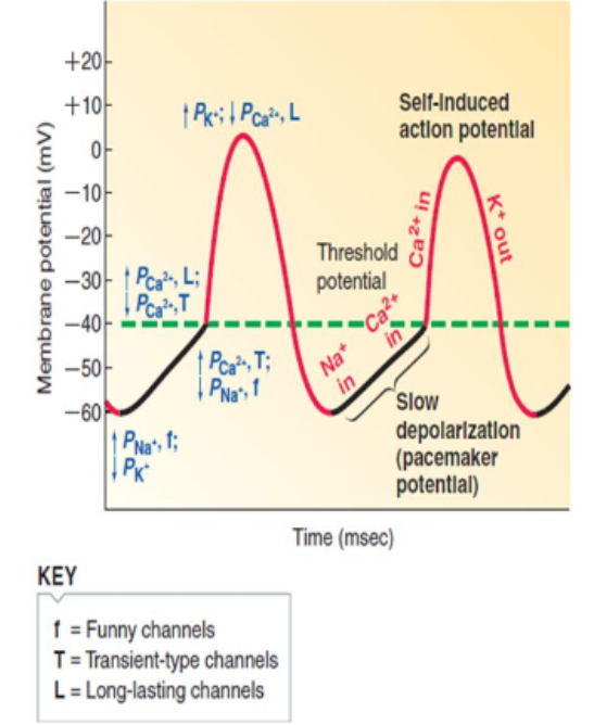
autorhythmic cell membranes show a _____ drift to threshold = ______ potential
What is rest membrane potential at?
slow, pacemaker
-55
Pacemaker potential process
1) _____ depolarization/pacemaker potential
2) ______ ______
3) ______ potential
4) _____ _______
slow
rapid depolarization
action
rapid repolarization
Ionic basis of AP of Autorhythmic cells - Phase 1: Pacemaker potential
_______ of voltage gated ____ channels (_____ ______) and voltage gated transient ______ channels
_______ of voltage gated _____ channels
opening, Na+, funny channels, Ca2+
closure, K+
Ionic basis of AP of Autorhythmic cells - Phase 2: Rising Phase/Depolarization
______ of long-lasting voltage-gated _____ channels (______ Ca2+ channels)
_____ influx of _____
opening, Ca2+, L-type
large, Ca2+
Ionic basis of AP of Autorhythmic cells - Phase 3: Falling Phase/Repolarization
______ of voltage gated ____ channels
______ of long lasting voltage gated _____ channels
Potassium _____
opening, K+
closing, Ca2+
efflux
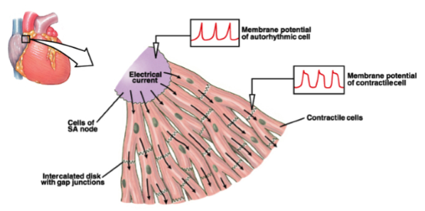
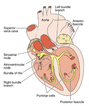
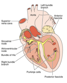
Intrinsic cardiac conduction system
made of ________ cardiac cells specialized to ____ and distribute ______ t/o the heart
noncontractile, initiate, impulses
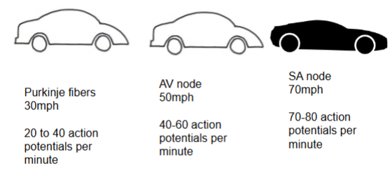
Sinoatrial/SA Node
located in right ____ wall just inferior to ______ _____ _____ & generates 75 AP/min
_______ (has sinus rhythm)
no ______ elements, but connected directly to ____ fibers which makes it spread ____
atrial, superior vena cava
pacemaker
contractile, atrial, faster
Propagation
more ______ thru 3 specialized bundles of atrial muscle called _____ pathways
nodal pathways have specialized ______ cells + ________
rapid, internodal
conductive, cardiomyocytes
Internodal Pathway
___ to ___ node
Atria/Ventricles separate by ___-_____ fibrous tissue
AV is only ______ connection
AV nodal ____ is abt 0.1 sec
net effect of transporting _____ impulse to ___ node w/i 30 msec; _____ of 130 msec in AV node and ____ system during which atria contract, filling the ______
SA, AV
non-conductive
electrical
delay
conductive, AV, delay, bundle, ventricles
Interatrial pathway
___ → ___ atrium
thru ____ junctions
really ____
right, left
gap
fast
Atrioventricular/AV node
located in ____ part of ______ septum
uses _______ pathway to spread _______
upper, interarterial
internodal, depolarization
Bundle of His
AV bundle
only _____ connection b/w atria/ventricles
____ & ____branch
______ abt 30 times per sec
(nothing)
electrical
left, right
depolarize
Purkinje fibers
modified _____ fibers with few _____
_____ 30 times per sec
controls _____ and ______ muscles
____ chordae tenineae
____ tricuspid/mitral valve
muscle, myofibrils
depolarize
ventricles, papillary
tighten
open
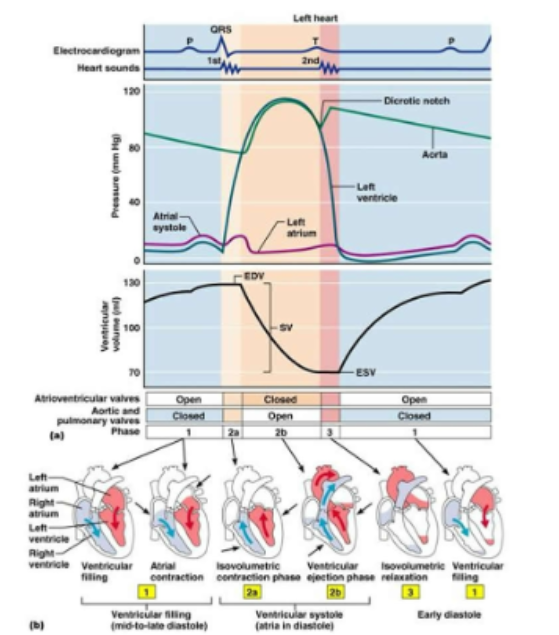
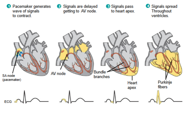
Control of heart rhythm
1) pacemaker (__ ____) generates wave of signals to _____
2) signals _____ getting to __ node
3) signals pass to ____ ____
4) signals spread thru-out ______
SA node, contract
delayed, AV
heart apex
ventricles
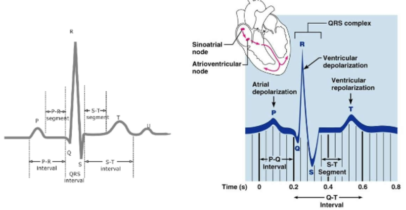
P-wave
depolarization of SA node → atria
QRS wave
ventricular depolarization/covers atrial repolarization
T-waves
ventricular repolarization
Cardiac Muscle Refractory Period
____
period which heart _____ make AP
_____ go into ____/_____
long
can’t
can’t, tetany/summation
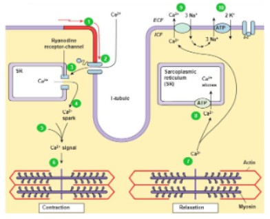
AP travels down T Tubules
bulk of this req ____ for ______ which origin from the ______ ______ (SR)
____-____ voltage gated _____ channels in T tubule _____
Ca2+ binds to _____ receptors
Ca2+ released from ___ thru ____ channel
Ca2+ is actively _____ _____ into __
_____ & ____ antiport exchangers (NCX) make more minor contributions
ca2+, contraction, sarcoplasmic reticulum
L-type, Ca2+, opens
Ryr
SR, Ryr
pumped back, SR
Na+, Ca2+
Regulation of Stroke Volume
Regulated by: Preload, contractility, and afterload
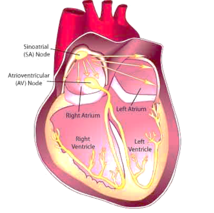
Explain the process happening in this image
fluids flow from the SA node into the right and left atrium
it flows faster into the left atrium and slower into the right
the fluids then make contact with the AV node, travels quickly into the left and right ventricles, and spreads out