Stage 2 Biology everythingwegot! herewego! 🪄
1/94
Earn XP
Description and Tags
Starting off with Cells as the Basis of Life (Topic 2)! I SKIPPED LIKE A BUNCH OF THIS TOPIC, HENCE THE EMPTY CARDS: JUST SCROLL DOWN. Secondly! Topic 1! DNA & Proteins! Green: Need to be able to detail and represent as highlighted by the SACE Curriculum. Highlighted in green: The key terms that need to be hit and known. Yellow: Expected to know/assumed knowledge. Don't need to know off by heart, but need to know/be able to distinguish and recognise. Highlighted in yellow: The key terms/phrases that need to be known. Expected/assumed knowledge. Everything else: Also don't need to know off by heart, but need to know/be able to distinguish and recognise (maybe slightly less urgent than yellow, but still crucial to understand these concepts). Assumed knowledge i guess. If it's in bold it is important. If it's in italics, it is either fluff information to enhance understanding, or does not need to be memorised FOR THAT QUESTION. crucial. for that question of flash card, we absolutely need to know the information otherwise. Italics may also be information i've dumbed down or added extra information too so that it makes sense. Cue word: Is a word/term/phrase used to help me remember (like a recognition cue), it may also be a key term used by SACE (describe/state/represent/compare/etc). Ok update, since its changed a little. Even if non-highlighted its assumed information, but it may also be similar to italics, where its fluff info to enhance understanding, but also maybe not, so just use brain to connect the dots... or double check with supervising professor.
Name | Mastery | Learn | Test | Matching | Spaced | Call with Kai |
|---|
No analytics yet
Send a link to your students to track their progress
95 Terms
What is cell theory?
.
.
.
.
Section: Cells as the Basis of Life
SACE Terms:
.
.
Cue word:
All living organisms are made of one or more cells (prokaryotic/eukaryotic).
All cells contain hereditary information (DNA) which is passed onto daughter cells.
All cells arise from pre-existing cells via division.
Cells carry out the life processes of organisms.
What is the controversy surrounding the cell theory and viruses?
.
.
.
.
Section: Cells as the Basis of Life
SACE Terms:
.
.
Cue word:
The cell theory states that all living things arise from pre-existing cells.
This does not hold true for viruses.
What is the cell membrane?
.
.
.
.
Section: Cells as the Basis of Life
SACE Terms:
.
.
Cue word:
The cell membrane is responsible for the exchange of materials into and out of the cell, including the entry of nutrients and exit of waste products.
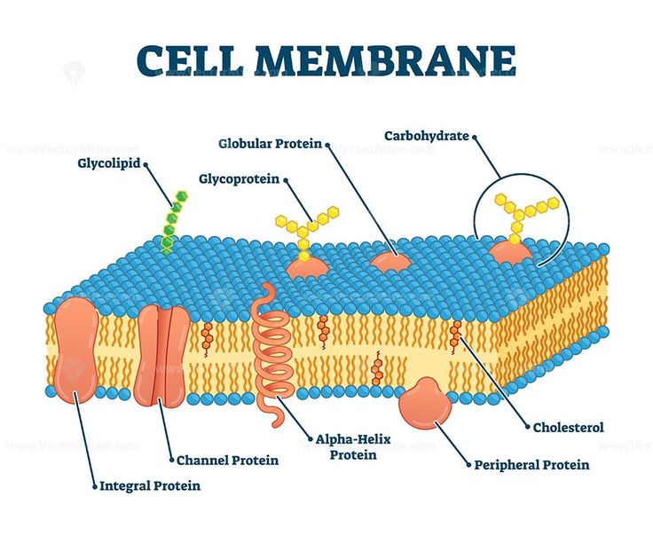
What is the fluid mosaic model?
.
.
.
.
Section: Cells as the Basis of Life
SACE Terms:
.
.
Cue word:
The fluid mosaic model describes the cell membrane which comprises of several phospholipids, with a polar phosphate head and a non-polar hydrophilic tails which face inwards towards each other to form a phospholipid bilayer.
Throughout the membrane are several integral proteins embedded and peripheral proteins atop the bilayer which are responsible for processes like transport and cell communication/recognition.
The fluidity of the phospholipids, which are in constant motion, provide the membrane with its permeability, allowing the entry of some substances but not others.
The membrane relies on its fluid nature to carry out its functions, and the proteins which facilitate transport or catalysations/catalysis.
What features do prokaryotes and eukaryotes share in common?
.
.
.
.
Section: Cells as the Basis of Life
SACE Terms:
.
.
Cue word:
Prokaryotes and eukaryotes both have DNA, ribosomes, cytoplasm, and a cell membrane.
Compare the eukaryotic and prokaryotic cell types.
.
.
.
.
Section: Cells as the Basis of Life
SACE Terms:
.
.
Cue word:
Prokaryotic cells are much smaller than eukaryotic cells, and contain a singular circular chromosome. Eukaryotes are much larger and possess several linear chromosomes. Unlike prokaryotes, eukaryotes have a nucleus and membrane bound organelles, providing a much more complex internal organisation.
Describe the nucleus.
.
.
.
.
Section: Cells as the Basis of Life
SACE Terms:
.
.
Cue word:
The nucleus controls and regulates all cellular activities, and is the site of transcription.
Describe the nucleolus.
.
.
.
.
Section: Cells as the Basis of Life
SACE Terms:
.
.
Cue word:
The nucleolus synthesises RNA molecules.
Describe the mitochondrion.
.
.
.
.
Section: Cells as the Basis of Life
SACE Terms:
.
.
Cue word:
The mitochondrion is responsible for carrying out the latter stages of aerobic respiration.
It has a double membrane, inner and outer. The inner membrane is referred to as the cristae and is folded within the mitochondria providing a high surface area for the
Describe the chloroplast.
.
.
.
.
Section: Cells as the Basis of Life
SACE Terms:
.
.
Cue word:
The chloro
Describe the vacuole.
.
.
.
.
Section: Cells as the Basis of Life
SACE Terms:
.
.
Cue word:
Describe the Golgi body.
.
.
.
.
Section: Cells as the Basis of Life
SACE Terms:
.
.
Cue word:
The Golgi body is responsible for the packaging and secretion of materials via vesicles.
Note that: This includes mention of the vesicles.
Describe the Endoplasmic Reticulum.
.
.
.
.
Section: Cells as the Basis of Life
SACE Terms:
.
.
Cue word:
Describe the ribosomes.
.
.
.
.
Section: Cells as the Basis of Life
SACE Terms:
.
.
Cue word:
Describe the lysosomes.
.
.
.
.
Section: Cells as the Basis of Life
SACE Terms:
.
.
Cue word:
Describe the cytoskeleton.
.
.
.
.
Section: Cells as the Basis of Life
SACE Terms:
.
.
Cue word:
What is the difference between Heterotroph and Autotroph?
.
.
.
.
Section: Cells as the Basis of Life
SACE Terms:
.
.
Cue word:
What is photosynthesis?
.
.
.
.
Section: Cells as the Basis of Life
SACE Terms:
.
.
Cue word:
What is aerobic respiration?
.
.
.
.
Section: Cells as the Basis of Life
SACE Terms:
.
.
Cue word:
What is anaerobic respiration?
.
.
.
.
Section: Cells as the Basis of Life
SACE Terms:
.
.
Cue word:
Compare the energy/ATP release of aerobic and anaerobic respiration.
.
.
.
.
Section: Cells as the Basis of Life
SACE Terms:
.
.
Cue word:
During anaerobic respiration, the net release of ATP is 2ATP during glycolysis per molecule of glucose.
During aerobic respiration, the net release of ATP is 36ATP during glycolysis and the Kreb’s cycle/electron transport chain which continues through the mitochondria.
Hence aerobic respiration provides more energy to a cell to fuel cellular processes as more ATP is available to be broken down, thus releasing the large magnitudes of energy useable by the cell.
What is the formation of ATP from ADP and Pi?
.
.
.
.
Section: Cells as the Basis of Life
SACE Terms:
.
.
Cue word:
ASHUICASBCOANFCA TO COME BACK TO IM JUST SKIPPING TO CELL METABOLISMMM EHIEHIEJEKEK
How do the inputs and outputs of autotrophs and heterotrophs differ?
.
.
.
.
Section: Cells as the Basis of Life
SACE Terms: Compare
.
.
Cue word: idunnouseyourbrainforthisone
In order to survive, cells require an input of matter, including gases, simple nutrients, and ions, and the removals of wastes.
[seriously don’t memorise this next section, its in italics for a reason, its just some background info which may help if i’ve forgotten all this, and am coming back to it months later].
Metabolic processes in cells produce wastes that may become toxic to the cell in high concentrations, and thus, it must be removed.
Depending on the cell, and the circumstances, substances which need to be removed include (or may include):
Oxygen, carbon dioxide, lactic acid, ethanol, urea, and various ions.
Even water may harm the cell if its been provided time to accumulate.
Autotrophic cells do not need to be supplied with organic compounds, such as glucose, since they can produce it from carbon dioxide and water (can synthesise it themselves).
In eukaryotic autotrophs, the synthesis of organic compounds occurs in the chloroplast, and the glucose produced in this way can be used to form other organic molecules such as lipids, amino acids, nitrogen bases, and nucleotides.
However, input of inorganic substances containing elements such as nitrogen, phosphorus, and sulphur is also required for this.
These elements are usually acquired in the form of inorganic substances like nitrite or nitrate ions, phosphate ions, and sulphate ions.
Heterotrophic cells also require these inorganic inputs, as well as an input of the organic compounds that they are unable to produce themselves.
(simply: Autotrophic cells do not need require an input of organic substances, such as glucose, since they are capable of synthesising such molecules themselves. However, they do need to be supplied with an input of inorganic molecules, usually in the form of inorganic substances like specific ions. Heterotrophs also require an input of inorganic substances, as well as an input of the organic compounds they cannot produce themselves).
As a result of their different metabolisms, autotrophic and heterotrophic cells have different outputs.
An autotrophic cells produce a net output of oxygen when the rate of photosynthesis exceeds the rate of its respiration. However, when its rate of respiration and fermentation exceed its rate of respiration (e.g., in a low light-intensive environment), it will have a net output of carbon dioxide.
(simply: Autotrophic cells produce a net output of oxygen when the rate of photosynthesis exceeds the rate of respiration, and a net output of carbon contrariwise).
Heterotrophic cells that are respiring aerobically will have a net output of carbon dioxide.
The outputs from fermentation also differ from one type of cell to another.
Autotrophs produce an output of ethanol and carbon dioxide during fermentation, while heterotrophs produce lactic acid.
Refer to attached table for more summary.
INPUTS:
Substance | Autotrophs | Heterotrophs |
|---|---|---|
oxygen | for aerobic respiration when the rate of respiration exceeds rate of photosynthesis | for aerobic respiration |
carbon dioxide | for photosynthesis when the rate of photosynthesis exceeds the rate of respiration | not required |
nitrates, nitrites | source of nitrogen for amino acid synthesis | source of nitrogen for amino acid synthesis |
phosphates | source of phosphorus for nucleotide synthesis | source of phosphorous for nucleotide synthesis |
calcium | a component of plant cell walls | an enzyme cofactor |
other inorganic nutrients | required for synthesis reactions | required for synthesis reactions |
organic compounds | not required since they are manufactured by cell | some are required (glucose, some amino acids, lipids), as not all can be manufactured by cell |
OUTPUTS:
Substance | Autotrophs | Heterotrophs |
|---|---|---|
oxygen | from photosynthesis when the rate of photosynthesis exceeds the rate of respiration | no output |
carbon dioxide | from respiration and fermentation when their rate exceeds the rate of photosynthesis | from aerobic respiration |
lactic acid | not normally produced | a waste from lactic acid fermentation |
ethanol | a product of fermentation | mainly yeasts (product of fermentation) |
urea | not normally produced | a nitrogenous waste product from the breakdown of excess amino acids |
![<p><mark data-color="yellow">In order to survive, cells require an input of matter, including gases, simple nutrients, and ions, and the removals of wastes.</mark></p><hr><p><em>[seriously don’t memorise this next section, </em><strong><em><u>its in italics for a reason</u></em></strong><em>, its just some background info which may help if i’ve forgotten all this, and am coming back to it months later].</em></p><p><em>Metabolic processes in cells produce wastes that may become toxic to the cell in high concentrations, and thus, it must be removed.</em></p><p><em>Depending on the cell, and the circumstances, substances which need to be removed include (or may include):</em></p><ul><li><p><em>Oxygen, carbon dioxide, lactic acid, ethanol, urea, and various ions.</em></p></li><li><p><em>Even water may harm the cell if its been provided time to accumulate</em>.</p></li></ul><hr><p><mark data-color="green">Autotrophic cells do not need to be supplied with organic compounds</mark>, such as glucose, since they can produce it from carbon dioxide and water <mark data-color="green">(can synthesise it themselves)</mark>.</p><p>In eukaryotic autotrophs, the synthesis of organic compounds occurs in the chloroplast, and the glucose produced in this way can be used to form other organic molecules such as lipids, amino acids, nitrogen bases, and nucleotides.</p><p><mark data-color="green">However, input of inorganic substances</mark> containing elements such as nitrogen, phosphorus, and sulphur is also required for this.</p><p>These elements are usually acquired in the form of inorganic substances like nitrite or nitrate ions, phosphate ions, and sulphate ions.</p><p><mark data-color="green">Heterotrophic cells also require these inorganic inputs, as well as an input of the organic compounds that they are unable to produce themselves</mark>.</p><p>(simply: <em>Autotrophic cells do not need require an input of organic substances, such as glucose, since they are capable of synthesising such molecules themselves. However, they do need to be supplied with an input of inorganic molecules, usually in the form of inorganic substances like specific ions. Heterotrophs also require an input of inorganic substances, as well as an input of the organic compounds they cannot produce themselves</em>).</p><p>As a result of their different metabolisms, autotrophic and heterotrophic cells have different outputs.</p><p>An autotrophic cells produce a net output of oxygen when the rate of photosynthesis exceeds the rate of its respiration. However, when its rate of respiration and fermentation exceed its rate of respiration <em>(e.g., in a low light-intensive environment)</em>, it will have a net output of carbon dioxide.</p><p>(simply: <mark data-color="green">Autotrophic cells produce a net output of oxygen when the rate of photosynthesis exceeds the rate of respiration, and a net output of carbon contrariwise</mark>).</p><p><mark data-color="green">Heterotrophic cells that are respiring aerobically will have a net output of carbon dioxide</mark>.</p><p>The outputs from fermentation also differ from one type of cell to another.</p><p><mark data-color="green">Autotrophs produce an output of ethanol and carbon dioxide during fermentation, while heterotrophs produce lactic acid</mark>.</p><p><strong>Refer to attached table for more summary</strong>.</p><p><u>INPUTS</u>:</p><table style="minWidth: 75px"><colgroup><col><col><col></colgroup><tbody><tr><th colspan="1" rowspan="1"><p>Substance</p></th><th colspan="1" rowspan="1"><p>Autotrophs</p></th><th colspan="1" rowspan="1"><p>Heterotrophs</p></th></tr><tr><td colspan="1" rowspan="1"><p><strong>oxygen</strong></p></td><td colspan="1" rowspan="1"><p>for aerobic respiration when the rate of respiration exceeds rate of photosynthesis</p></td><td colspan="1" rowspan="1"><p>for aerobic respiration</p></td></tr><tr><td colspan="1" rowspan="1"><p><strong>carbon dioxide</strong></p></td><td colspan="1" rowspan="1"><p>for photosynthesis when the rate of photosynthesis exceeds the rate of respiration</p></td><td colspan="1" rowspan="1"><p>not required</p></td></tr><tr><td colspan="1" rowspan="1"><p><strong>nitrates, nitrites</strong></p></td><td colspan="1" rowspan="1"><p>source of nitrogen for amino acid synthesis</p></td><td colspan="1" rowspan="1"><p>source of nitrogen for amino acid synthesis</p></td></tr><tr><td colspan="1" rowspan="1"><p><strong>phosphates</strong></p></td><td colspan="1" rowspan="1"><p>source of phosphorus for nucleotide synthesis</p></td><td colspan="1" rowspan="1"><p>source of phosphorous for nucleotide synthesis</p></td></tr><tr><td colspan="1" rowspan="1"><p><strong>calcium</strong></p></td><td colspan="1" rowspan="1"><p>a component of plant cell walls</p></td><td colspan="1" rowspan="1"><p>an enzyme cofactor</p></td></tr><tr><td colspan="1" rowspan="1"><p><strong>other inorganic nutrients</strong></p></td><td colspan="1" rowspan="1"><p>required for synthesis reactions</p></td><td colspan="1" rowspan="1"><p>required for synthesis reactions</p></td></tr><tr><td colspan="1" rowspan="1"><p><strong>organic compounds</strong></p></td><td colspan="1" rowspan="1"><p>not required since they are manufactured by cell</p></td><td colspan="1" rowspan="1"><p>some are required (glucose, some amino acids, lipids), as not all can be manufactured by cell</p></td></tr></tbody></table><p><u>OUTPUTS</u>:</p><table style="minWidth: 75px"><colgroup><col><col><col></colgroup><tbody><tr><th colspan="1" rowspan="1"><p>Substance</p></th><th colspan="1" rowspan="1"><p>Autotrophs</p></th><th colspan="1" rowspan="1"><p>Heterotrophs</p></th></tr><tr><td colspan="1" rowspan="1"><p><strong>oxygen</strong></p></td><td colspan="1" rowspan="1"><p>from photosynthesis when the rate of photosynthesis exceeds the rate of respiration</p></td><td colspan="1" rowspan="1"><p>no output</p></td></tr><tr><td colspan="1" rowspan="1"><p><strong>carbon dioxide</strong></p></td><td colspan="1" rowspan="1"><p>from respiration and fermentation when their rate exceeds the rate of photosynthesis</p></td><td colspan="1" rowspan="1"><p>from aerobic respiration</p></td></tr><tr><td colspan="1" rowspan="1"><p><strong>lactic acid</strong></p></td><td colspan="1" rowspan="1"><p>not normally produced</p></td><td colspan="1" rowspan="1"><p>a waste from lactic acid fermentation</p></td></tr><tr><td colspan="1" rowspan="1"><p><strong>ethanol</strong></p></td><td colspan="1" rowspan="1"><p>a product of fermentation</p></td><td colspan="1" rowspan="1"><p>mainly yeasts (product of fermentation)</p></td></tr><tr><td colspan="1" rowspan="1"><p><strong>urea</strong></p></td><td colspan="1" rowspan="1"><p>not normally produced</p></td><td colspan="1" rowspan="1"><p>a nitrogenous waste product from the breakdown of excess amino acids</p></td></tr></tbody></table>](https://knowt-user-attachments.s3.amazonaws.com/511b8db9-daf2-4a49-9d3b-af02b024b8ca.png)
How does the structure of a membrane facilitate different processes of movement through it?
.
.
.
.
Section: Cells as the Basis of Life
SACE Terms: Explain
.
.
Cue word: Phospholipid bilayer, polarity and characteristics of substances, polarity and semi-permeability.
It consists of a phospholipid bilayer, with polar phosphate heads which surround the non-polar lipid tails facing inwards.
The bilayer has fluid properties; allowing it break, reform, and enclose materials (e.g., proteins in vacuoles/vesicles for bulk transport).
With respect to its polarity, the cell membrane is semi-permeable, but not equally permeable to all substances. Through a narrow range, it restricts the entry and exit of substances, and ensures to seperate the cells intracellular and extracellular fluids.
The concentration of substances, and the pH of the cell must be maintained within a narrow range in order for it to function normally.
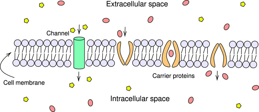
Channel and carrier proteins are embedded throughout the bilayer, which assist in the transport of molecules.
The way a substance is transported depends on its characteristics (like polarity, size, lipid-soluble, charge), the presence or absence of a concentration gradient, and the cell’s SA:V ratio.
What are the roles of transport proteins?
.
.
.
.
Section: Cells as the Basis of Life
SACE Terms: Explain
.
.
Cue word: Including channel proteins (such as aquaporins), and carrier proteins.
Transport proteins are embedded throughout and across the cell membrane, and function to speed up the movement of substances into and out of the cell.
Channel proteins do not bind to the substances that move through them passively, with the concentration gradient. Channel proteins are involved in diffusion and osmosis, and it is a rapid process.
An aquaporin is an example of a specialised channel protein; involved in the movement of H2O molecules.
Carrier proteins bind to specific molecules, and some assist them to move passively across the membrane with the concentration gradient. This process is facilitated diffusion, which does not require energy, and is considerably slower than movement through channel protein.
A glucose transporter protein (GLUT) is an example of a specialised carrier protein.
Other carrier proteins are protein pumps which bind to selected substances, particularly ions, and move them against the concentration gradient, using energy from the breakdown of ATP. This is active transport.
Compare active and passive transport
.
.
.
.
Section: Cells as the Basis of Life
SACE Terms: Explain
.
.
Cue word: Including channel proteins (such as aquaporins), and carrier proteins.
Passive transport
Does not require any additional input of energy.
Passive movement across a phospholipid bilayer membrane relies on the inherent energy of the molecules.
The specialised proteins involved in passive transport also do not require energy from the breakdown of ATP.
Substances are moved with the concentration gradient (move from areas of high concentration to low).
Active transport
Requires the additional input of energy.
Active transport uses specific transport proteins (carrier protein pumps).
Energy from the breakdown of ATP is required for the conformational change in shape of the protein to pump molecules into and out of the cell.
Solutes are moved against the concentration gradient (move from areas of low concentration to high)
What is diffusion?
.
.
.
.
Section: Cells as the Basis of Life
SACE Terms: None
.
.
Cue word: None
All particles have kinetic energy. This kinetic energy is why they are in constant motion. Due to this random motion, the particles of a substance have a tendency to diffuse until they take up all of the available space. When the substance is evenly dispersed, equilibrium is met.
Even when equilibrium is reached, the particles do not cease to move just because they have stopped diffusing. The particles moving in one direction will be balanced by the particles moving in the opposite direction. (The net movement is zero; no concentration gradient).
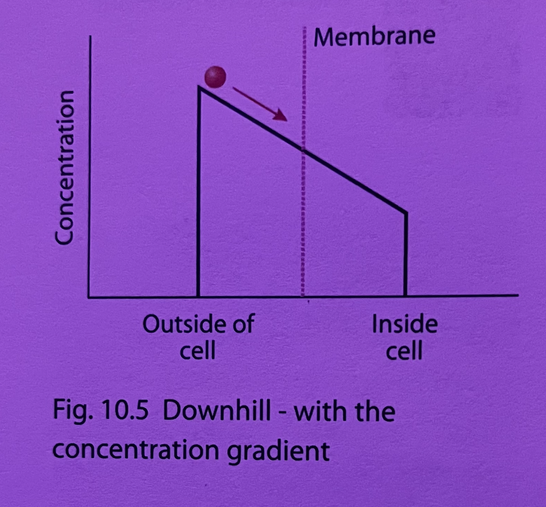
Diffusion (aka. simple diffusion) is defined as the net random movement of molecules that results in their uniform distribution.
Diffusion is the movement of a substance in a fluid from a region of high concentration, towards a region of lower concentration (e.g., oxygen, carbon dioxide, . ethanol, small uncharged lipids). The particles move with, and follow the concentration gradient. Diffusion is a passive process, which does not require the input of energy.
(in case you forgot, concentration gradient refers to a region of space over which the concentration of a substance changes. It is a measurement of how the concentration of something changes from one place to another. And in biology, the concentration gradient is high→low).
The relevance of diffusion for cells, is that a substance will diffuse across a membrane if a concentration gradient exists.
(Refer to attached image: It can be thought of as a ball at the top of a hill. Think of the hill as the concentration gradient, and the ball as the substance. The ball naturally wants to roll down the hill because of gravity. Hence, a substance has a tendency to diffuse when there is a concentration gradient.)
Examples include the diffusion of oxygen into cells, and the diffusion of carbon out of cells.
These molecules are capable of penetrating through the hydrophobic core of the phospholipid bilayer since they hydrophobic and non-polar in nature.
Small polar molecules can also cross the membrane by simple diffusion.
What is facilitated diffusion?
.
.
.
.
Section: Cells as the Basis of Life
SACE Terms: None
.
.
Cue word: None
Like simple diffusion, facilitated diffusion also occurs with a concentration gradient; from high concentration to low concentration. However, it requires the presence of specific channel or carrier proteins.
Channel and carrier proteins assist molecules to move more passively through the concentration gradient.
The semi-permeability of the cell membrane makes it difficult for the molecules of many substances to diffuse across it.
Facilitated diffusion is still a form of diffusion since it moves molecules from regions of high concentration to regions of lower concentration; it also does not require the input of energy.
Substances which enter the cell by facilitated diffusion often that contain hydrophilic molecules, and some ions.
This includes glucose, some amino acids, certain drugs, and potassium/sodium ions.
What is osmosis?
.
.
.
.
Section: Cells as the Basis of Life
SACE Terms: None
.
.
Cue word: Plant and animal cells
The movement of water differs to the diffusion processes, because the movement of water is also affected by the concentration of solutes.
Osmosis is the passive diffusion of water from an area of low solute concentration (high [water]) to an area of high solute concentration (low [water]).
(i.e., from a more watery solution to a less watery one).
Water is a small polar molecule, and can therefore slowly move directly through the phospholipid bilayer.
Aquaporins are specialised protein channels which facilitate the efficient diffusion of water molecules.
Only water molecules can use aquaporins.
Aquaporins are commonly found in the membranes of nephron and kidney cells, and respond to anti-diuretic hormone, which controls how much water is excreted in urine; thereby contributing to homeostatic processes for regulating blood volume.
Osmosis is significant to the cell since the osmotic pressure maintains cell shape in animal cells, and provides support in plant cells.
However, the pressure must be just right.
Human red blood cells burst when placed in a distilled water solution; and shrivel in a concentrated salt solution.
A solution of 0.9% sodium chloride will exactly balance the solute concentration of solutes in the cell’s cytoplasm.
Which results in no net movement of water (no osmosis), known as a “physiological saline” solution.
The result of excess uptake of water is less severe in plant cells (due to presence of cell wall) than animal cells.
When placed in distilled water, plant cells will not burst, but will completely swell with water, and are said to be fully turgid.
If plant cells lose water, they become flaccid or limp. Under conditions when the cell becomes flaccid, the whole plant will wilt, and if the water is not replaced, it will eventually die.
Isotonic:
When two solutions have the same solute concentration on either side of the membrane.
Hypotonic:
When there are more free water molecules (low solute concentration outside the cell), and hence water tends to move into the cell.
(SO, there are more water molecules outside of the cell, and its more watery to less watery, so if theres more outside, the water is gonna want to go inside the cell.
in other terms, there is low solute concentration outside the cell, and high solute concentration inside the cell. And its high solute concentration to low solute concentration.
anyways in hypotonic solutions, water is going in because theres more water outside than inside.
think of hypO… like O the cell is yk.. O its swellin up, water is going in).
Hypertonic:
There are higher solute concentrations inside the cell, and thus less free water molecules. Hence, water moves out of the cell.
What is active transport?
.
.
.
.
Section: Cells as the Basis of Life
SACE Terms: None
.
.
Cue word: None
Active transport is the opposite of diffusion., since substances are moved across the membrane against the concentration gradient.
Therefore, substances are moved from regions of low concentration to regions of high concentration.
Active transport requires carrier protein pumps, which require energy for the conformational change in shape of the protein to pump molecules into or out of the cell.
The cell is selective about what it transports into and out of the cell. It may transport ions and substances into the cytoplasm, while transporting others out.
What is Endocytosis?
.
.
.
.
Section: Cells as the Basis of Life
SACE Terms: None
.
.
Cue word: Pinocytosis, phagocytosis
Endocytosis is the movement of a substance into the cell by causing invagination of the cell membrane, forming a vesicle which facilitates entry into the cell.
To ensure that only required substances are transported into the cell through endocytosis, the process may involve specific receptors, where the substance must bind to its receptor, and be recognised before it can enter the cell.
Substances which enter the cell through endocytosis include: iron, cholesterol, bacteria, phagocytes capable of engulfing bacterium, and cells of the small intestine ingesting lipid droplets.
Simply, endocytosis is described as the ingestion of particles (such as bacteria) and the uptake of fluids or macromolecules in small vesicles. A process; requiring ATP.
Material (solids or fluids) that are brought into the cell are engulfed by an invagination of the cell membrane.
Vesicle buds off from the plasma membrane.
The vesicle carries molecules into the cell, where they can then be digested by lysosomes containing enzymes.
Endocytosis (phago- and pino-) require lots of ATP.
Phagocytosis is the intake of particles, and often referred to as cell eating.
The membrane invaginates in the vicinity of the particle, and encloses it in a vacuole which then breaks away from the cell membrane and enters the cytoplasm.
Lysosomes (containing digestive enzymes) can then fuse with the vacuole, which breakdown the particle.
Examples of phagocytic cells are unicellular amoebae, which feed in this way, and certain cells of the human immune system involved in engulfing and destroying foreign particles (bacteria).
Cells that carry out phagocytosis are selective, and will not engulf just any particle.
Pinocytosis is the intake of liquids, and often referred to as cell drinking.
Pinocytosis is similar to phagocytosis, however, on a smaller scale.
Pinocytosis may be non selective, and involve the intake or certain large molecules, such as fat droplets in the small intestine (takes in samples of any solutes in the surrounding extra cellular fluid).
What is exocytosis?
.
.
.
.
Section: Cells as the Basis of Life
SACE Terms: None
.
.
Cue word: None
Exocytosis is the secretion of materials produced by the cell, and usually involves the packaging of the material into a vesicle,
which then migrates to the plasma membrane,
fuses with the membrane,
and then secretes its contents to the external cellular environment.
The manufacture of the material often occurs in the endoplasmic reticulum, and the packaging is a function of the Golgi body.
The cells of the salivary gland are a prime example of exocytosis; which produce and secrete saliva.
This card could maybe maybe not use more Information? I cant tell if its actually this long, or if i accidentally made it short because i am in a hurry…
How is the exchange of materials across membranes affected by various factors?
.
.
.
.
Section: Cells as the Basis of Life
SACE Terms: Explain
.
.
Cue word: SA:V of cell, concentration gradient, the physical and chemical nature of the materials being exchanged
I half assed this card so bad, so uh, we may needa revisit this…
Surface area to volume ratio (SA:V):
As the size of the cell increases, its surface area to volume ratio decreases.
(this is because there is less space available for “things” to react with it… think of a bath bomb, it fizzles and melts slower as one big than, compared to when you beak it up into smaller parts. or idk, a berocca tablet).
Cells obtain their nutrients from their surroundings, and taking them in through their membranes. Cells also remove waste products by passing them through the membrane.
As the cell gets bigger, there is proportionally less surface area, and as a result, there is a decrease in the efficiency with which the cell can exchange materials with its surroundings.
Theres also the notion that DNA (which controls cellular activity) can only exert an influence over a finite volume, which limits the size to which most cells can grow.
Concentration gradients:
The direction of movement of a substance across a membrane depends on the difference in concentration of the substance on either side of the membrane.
When there is no input energy, movement is with the concentration gradient from a region of higher concentration towards a region of lower concentration, until equilibrium is reached.
If particles move across membranes against the concentration gradient from a region of low concentration towards a region of higher concentration, energy is required.
Nature of the exchange materials:
Small, uncharged particles diffuse easily through cell membranes, while particles that are large or charged require assistance from channel proteins, carrier proteins, or even whole sections of the membrane (endo- and exocytosis).
TO COME BACK TO AND ADD THE THNGS IN BETWEEN BECAUSE IM SKIPPING AHEAD, REMEMBER IF YOU HOVER YOUR MOUSE THING IN BETWEEN TWO FLASHCARDS TEHRES AN OPTION TO ADD IN BETWEEN, YEA
DO THAT
What is cell metabolism?
.
.
.
.
Section: Cells as the Basis of Life
SACE Terms: None
.
.
Cue word: None
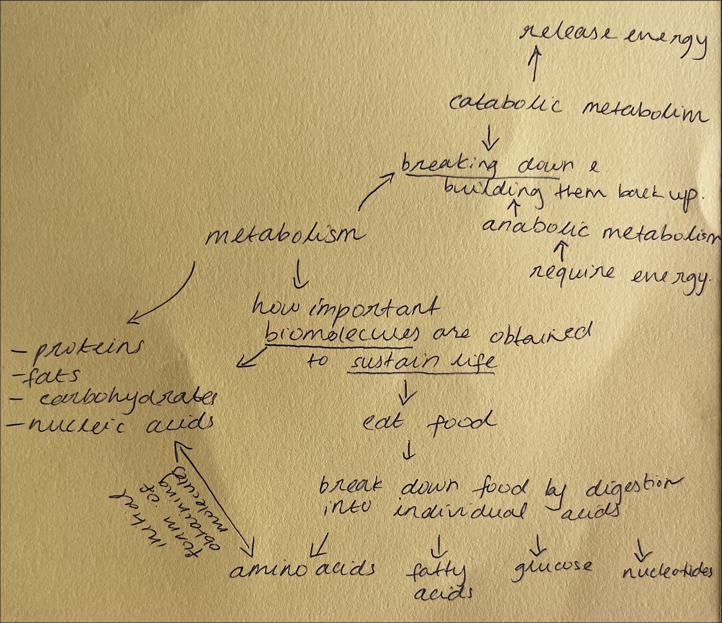
Metabolism refers to all of the biochemical reactions occurring in cells. It is critical to the survival of cells, and hence, to organisms as a whole.
Metabolism involves a series of controlled, complex chemical reactions, which are sequenced into metabolic pathways. This ensures efficient functioning of cells.
However, these biochemical processes may be influenced by the nature and arrangement of internal membranes, and the presence of specific enzymes.

What are metabolic pathways?
.
.
.
.
Section: Cells as the Basis of Life
SACE Terms: Explain
.
.
Cue word: None
Metabolic pathways refer to most of the chemical reactions that occur in cells.
A metabolic pathway consists of several regulated steps, where each step of the pathway requires a specific enzyme to catalyse it (as each new substrate has its own specific shape and can only bind the active site of its own enzyme).
Such that, the substrate for the first reaction is converted into a product by the first enzyme. This product is then the substrate for the second reaction, and so forth. The sequence of chemical conversions of substrates into products continues until the end-product is formed.

What are the advantages of metabolic pathways?
.
.
.
.
Section: Cells as the Basis of Life
SACE Terms: None
.
.
Cue word: None
As a result of the many regulated steps, the cell can exert a greater level of control of the reaction with a different enzyme at each step.
Other substrates can ‘feed in’ at different points of the pathway, allowing cells to use other resources.
(simply: this means that if the primary substrate used in the pathway is in limited supply or not available, the cell can use alternative substrates to maintain the pathway).
Some steps produce intermediate compounds that are often essential molecules needed by the cell.
The cell can release energy more gradually, a portion (of energy) of which can be trapped in energy coupling processes, such as ATP synthesis
(simply: this means that the cell can capture more of the energy released, and can therefore put more of it towards the synthesis of ATP rather than it being wasted. This maintains the cell’s energy balance, allowing energy to be more efficiently used in cellular processes).During each step in the metabolic pathway, some chemical energy is lost, and transformed into heat, which can be used to maintain stable body temperatures in many organisms - including humans.
How does the mitochondria facilitate biochemical processes?
.
.
.
.
Section: Cells as the Basis of Life
SACE Terms: Explain
.
.
Cue word: Internal membrane
The latter stages of aerobic respiration occur in the mitochondrion (Kreb’s cycle and the electron transport chain).
The mitochondrion has (an outer membrane and) an inner membrane which is folded to form structures called cristae.
The cristae provides a large surface area for the attachment of enzymes such as ATP synthase, which are crucial in aerobic respiration.
The inner membranes of the mitochondrion are the main site of ATP production - it is here where much of the energy is released and used to make ATP.
How does the chloroplast facilitate biochemical processes?
.
.
.
.
Section: Cells as the Basis of Life
SACE Terms: Explain
.
.
Cue word: Internal membrane
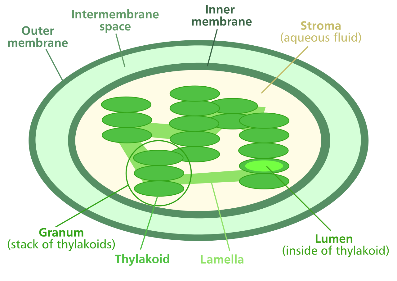
Chloroplasts have two outer membranes, and an internal system of membraneous flattened sacs called thylakoids.
The light-dependent reactions of photosynthesis occur in the thylakoids, which contain chlorophyll and the enzymes that catalyse them. These thylakoids are stacked in piles called “grana,” where inside are stacks of chlorophyll molecules, called “lamellae.”
The internal system of membranous flattened sacs in chloroplast increase the surface area available for light absorption, thereby facilitating photosynthesis by increasing the frequency of light being successfully absorbed by chlorophyll molecules within the chloroplast.
(simply: by increasing the chance that light passing through a chloroplast will be absorbed by chlorophyll molecules.)
The light-independent stages of photosynthesis make up a pathway termed the ‘Calvin Cycle.’ This occurs in the stroma; it is here where the organic molecule, glucose, is formed.
How does temperature influence metabolic reactions?
.
.
.
.
Section: Cells as the Basis of Life
SACE Terms: None
.
.
Cue word: None
Biochemical processes in the cell are influenced by environmental factors.
Temperature has a significant effect on metabolic rate.
Increased temperatures increase the kinetic energy of the molecules. This makes the particles move faster, allowing them to collide more frequently, and thus, increasing the chances of more successful enzyme-substrate binding (thereby increasing reaction rate).
This will be true within the tolerance range for any given organism (simply: increased reaction rate and enzyme activity holds true only within the temperature-tolerance-range of the organism).
However, at temperatures too high, the enzymes often denature, causing them to lose their 3D structure necessary for enzyme-substrate binding. Most human enzymes work best between temperatures 35-40℃.
Since enzymes are biological catalysts, any change to the function of these proteins directly affect metabolic rate.
How does pH influence metabolic reactions?
.
.
.
.
Section: Cells as the Basis of Life
SACE Terms: None
.
.
Cue word: None
Biochemical processes in the cell are influenced by environmental factors.
Each enzyme has an optimum pH.
Proteins (enzymes) function within a defined pH range, where in this range, their structure is maintained.
If the pH range is not met, this may result in changes to the enzyme’s 3D structures causing them to denature. Consequently, leading to a reduction in metabolic rate.
Each enzyme has an optimum pH range, generally ranging from pH6-8. However, some digestive enzymes favour acidic conditions (pH=2), and others in the intestines favour alkaline conditions (pH=10).

How does nutrient availability and location influence metabolic reactions?
.
.
.
.
Section: Cells as the Basis of Life
SACE Terms: None
.
.
Cue word: This is not substrate concentration.
Biochemical processes in the cell are influenced by environmental factors.
Metabolic pathways require substrates (reactants) to process.
Lack of nutrient availability decreases metabolic rate, since there are less molecules for enzymes to catalyse.
Conversely, increased nutrient availability simulates increased metabolic rate.
Some inorganic (copper, iron, zinc ions) act as co-factors for enzymes, and can bind to the enzyme or with the substrate. Organic molecules (vitamins) that are co-factors are called co-enzymes. These co-factors and co-enzymes help maintain metabolic rates, by directly participating in enzymatic reactions in the pathway.
(note that co-factors don’t replace substrates, they just help in the binding of substrates to the active site - e.g., correct orientation - hence, less of these may lead to less substrates being able to successfully bind).
Reactions will only occur if all components are available (enzymes/substrates/co-factors), therefore, location may limit the speed and progression of some reactions.

How do chemical inhibitors influence metabolic reactions?
.
.
.
.
Section: Cells as the Basis of Life
SACE Terms: None
.
.
Cue word: This is a looooong cue card.
Poison, cyanide, malonate, herbicides, medicines, insecticides.
Biochemical processes in the cell are influenced by environmental factors.
Chemicals can interfere with cell metabolism.
General: Chemical inhibitors can prevent or block the action of enzymes, thereby decreasing the cell’s metabolic rate.
Specific: The binding of chemical inhibitors to enzymes can be reversible or irreversible. Irreversible inhibitors often react with, and chemically alter the structure of the individual animo acids which make up the precise shape of the enzyme. Consequently, altering its functionality. Many drugs are chemical inhibitors of enzymes, and as such, this is an important field of medical research.
The following is in yellow as you do not need to know this off by heart, but are expected to be able to apply this knowledge. For instance, if you were to be asked: Discuss possible benefits and/or harmful effects of chemicals that human beings use.
Poisons:
Substances that are harmful to organisms are called poisons. Many poisons work by inhibiting enzymes involved in metabolic pathways. (e.g., mercury, lead).Cyanide:
Potassium cyanide is an irreversible inhibitor of cytochrome c oxidase (complex IV) (key enzyme in electron transport chain of aerobic respiration).
Cyanide competitively inhibits the enzyme by preventing oxygen from binding at the active site.
The poison binds to iron in the 4th complex of the electron transport chain, and stops electron transport to oxygen. ATP production is severely limited, often resulting in death.Malonate:
Malonate binds to the active site of succinate dehydrogenase, and acts as a competitive inhibitor of its substrate succinate.
This enzyme is key during the Kreb’s cycle and the electron transport chain. Its inhibition results in cell death.Herbicides:
Herbicides are used to kill or greatly slow the growth of undesired plants (weeds). Glyphosate is a herbicide used to competitively inhibit an enzyme which catalyses a metabolic process that produces substances that plants need to grow. As a result of being sprayed by glyphosate, the plants die shortly after.Medicines:
Enzyme inhibitors to the viral enzyme, protease, act to slow and stop viral growth by preventing them from building new protein coats.
The antibiotic, penicillin, inhibits the bacterial enzyme, transpeptidase, which is necessary for the development of cell walls in new bacteria. In the pathway, penicillin is accepted as a substrate, and irreversible binds to the active site. Bacteria subjected to penicillin cannot form effective cell walls during binary fission, and thus cannot reproduce or survive.Insecticides:
Insecticides kill insect pests. Many plants secrete naturally-occurring insecticides. Humans use both naturally occurring and synthetic insecticides, as well as genetically modifying organisms (GMO) to produce insecticides to produce productivity in agriculture or assist in reducing the spread of disease.
DDT (1944) was an insecticide which disrupted the transmission of nerve impulses by opening sodium channels in an insects nerve cells. Nowadays, however, DDT is banned from use in many countries due to its toxic tendencies towards many types of animals, including humans.

How do internal membranes influence metabolic reactions?
.
.
.
.
Section: Cells as the Basis of Life
SACE Terms: None
.
.
Cue words: None
Biochemical processes in the cell are influenced by environmental factors.
Internal membranes increase the surface area available for the enzymes to attach, hence, increasing metabolic rate.

Why does the amount of DNA in a cell double before division?
.
.
.
.
Section: Cells as the Basis of Life
SACE Terms: Explain
.
.
Cue word: None
[ Cells and organisms function efficiently for a finite period of time. The continuity of life therefore requires new cells to be produced.
This involves the replication of the genetic material of pre-existing cells (DNA replication), and the transfer of replicated DNA to the next generation (cell division: binary fission, mitosis, meiosis, fertilisation).
Cell division in somatic cells is different from the cell division that produces gametes from germ-line cells. ]
In multi-cellular organisms, cells need to be continually replaced, and each of the new cells must receive a copy of the genetic information stored in the DNA. For this reason, the DNA in a cell is replicated before the cell divides. This ensures that when a cell divides, each new cell will receive exactly the same amount of DNA containing identical information, given no mutations occur. It’s important to note that while the DNA doubles during this process, the number of chromosomes do not double.
What is binary fission?
.
.
.
.
Section: Cells as the Basis of Life
SACE Terms: Recognise, describe, represent
.
.
Cue word: the phases
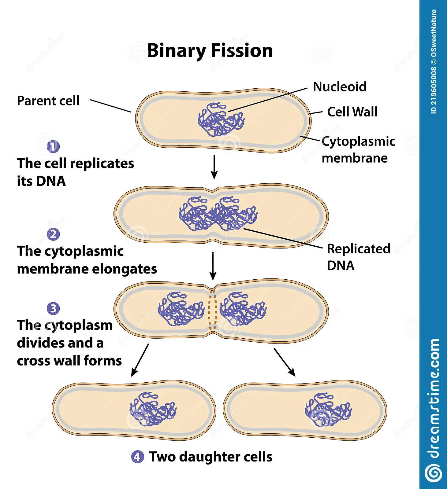
Binary fission is the asexual reproduction of prokaryotes.
Just prior to the process, DNA replication occurs, resulting in two identical DNA double helices in circular loops.
The DNA is in the form of a single, circular double helix, so it has to be cut and untwisted to allow enzymes to get to the twin strands. Enzymes (helicase, gyrase, swivelase, topoisomerase) facilitate this replication. After the strands of the double helix have separated, both of the newly formed single-strands then act as templates for the new strand to form. The result is two DNA double helices (two chromosomes) in circular loops.Condensation of the cell’s DNA occurs to form two identical circular chromosomes.
Both chromosomes attach to the inside of the cell membrane.
The chromosomes are attached separately by proteins to the cell membrane near the middle of the cell.The cell elongates; separating the two chromosomes.
The separated chromosomes are moved to the opposites ends of the parent cell as the cell elongates.
As the cell grows, the cell membrane between the attached chromosomes, expands, and moves the attached chromosomes in opposite directions.A new cell wall and cell membrane form, and the cells divides into two daughter cells, each containing one circular chromosome.
The circular chromosomes are well apart before the cell pinches across its equator to give new cells. The division into two daughter cells is completed by the synthesis of new wall material.
There exists no genetic variation in binary fission. The daughter cells are identical to its parent cell.
Binary fission can produce enormous numbers of prokaryotes in very little time. In optimal conditions, bacteria divide every 20 minutes, doubling their numbers each time. The rapid increase of cells (or organisms) is referred to as exponential growth.
What is mitotic division?
.
.
.
.
Section: Cells as the Basis of Life
SACE Terms: Recognise, describe, represent
.
.
Cue word: the phases
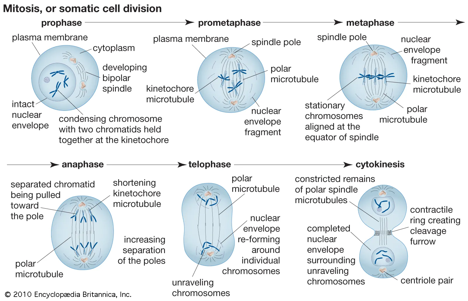
Mitotic division (aka. somatic cell division) is the asexual reproduction of somatic cells in eukaryotes.
Prior to mitosis, interphase occurs (G1, S, G2).
When a cell is not dividing, its genetic material is present in a form known as chromatin, which consists of long thin threads of DNA and protein.
During interphase, the DNA is replicated.
In interphase, the chromatin becomes slightly unwound (confirm this first part). Each DNA strand is replicated, so that each of the chromosomes consist of two identical sister chromatids, joined together at the centromere.
The first appearance of chromosomes in the cell is an indication that prophase has commenced.
Prophase:
In prophase, the chromatin condenses, causing the DNA strands to wind up, and the chromosomes to shorten and thicken (making them visible under a light microscope). The nuclear envelope disintegrates, and the nucleoli disappear, resulting in the chromosomes temporarily residing in the cytoplasm. The spindle apparatus then begins to form. In animal cells, the centrioles divide to form two pairs, and migrate towards the poles of the cell. The spindle fibres of the spindle apparatus, and the centrioles are both made of microtubules (proteins) which are components of the cytoskeleton. Midway between the poles, is a region of the spindles called the equator. Plant cells do not contain centrioles.Metaphase:
During metaphase, the microtubules in the cytosol radiate from the poles, and attach to the chromosomes; moving the chromosomes so that they line up at the equator (aka. metaphase plate). The spindle fibres from the opposite poles are attached to either sides of the centromere. This is important to ensure that both sister chromatids do not end up at one pole of the cell.Anaphase:
Anaphase is characterised when the centromeres separate, and the pairs of sister chromatids are drawn apart by the spindle fibres towards opposite poles. Each chromatid (of the set at opposite poles) is considered to be an identical daughter chromosome.Telophase:
After the daughter are moved to opposite poles, new nuclei begin to form, each one complete with a nucleolus. The chromosomes then decondense into chromatin; signalling the end of mitosis. The spindle apparatus is also broken down.
Mitosis is complete with telophase, and cytokinesis ensures that the cytoplasmic contents are divided between the two new daughter cells.
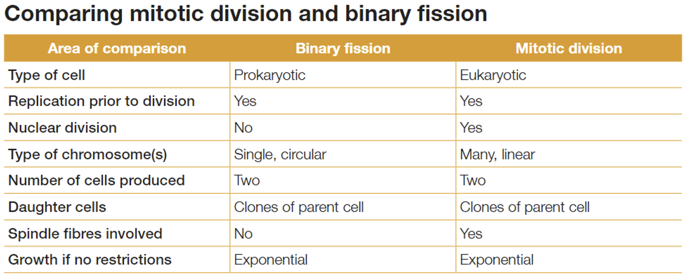
What is the difference between the products of binary fission and mitotic division?
.
.
.
.
Section: Cells as the Basis of Life
SACE Terms: Compare
.
.
Cue word: None
The cells formed as a result of binary fission and mitotic division are genetically identical to each other and to their parent. This is because the genetic material is replicated, and each daughter cell receives an identical copy. Thus, the number and type of chromosome in each daughter cell, is the same as that of the parent.
In binary fission, nuclear division does not occur, while it does in mitotic division.
Spindle fibres are only involved in mitotic division.
The chromosome involved in binary fission is single, circular one, whereas in mitotic division there are many, linear chromosomes.
What is meiotic division?
.
.
.
.
Section: Cells as the Basis of Life
SACE Terms: Recognise, describe, represent
.
.
Cue word: the phases
Each individual phase of meiosis was not written in Biology: Levels of Life, so I don’t know how much of it needs to be locked-in-memorised. However, certain aspects of the process did have a section - which have their own cue card anyway. Maybe each individual step is more of a yellow field than green? Hence, don’t bend over backwards memorising the stages word-for-word. I could also be wrong though… After writing this card, this is kind of like an information card.
Meiosis functions to reduce the number of chromosomes to half (by dividing twice), and is the process which forms gametes.
Gametes are haploid cells (half the amount of chromosomes as somatic cells).
The overall result of meiosis, is that from one diploid germ-line cell, four haploid cells are formed; either male or female gametes. While all cells in mature male animals can develop into sperm; in mature females, usually only one of them develops into a mature ovum (or egg cell).
The chromosome number of a sexually reproducing species is kept constant from one generation to the next. In summary: meiotic division is the reduction division; from diploid to haploid, and then, if fertilisation occurs, the diploid state is restored.
MEIOSIS PROCESS:
Prior to meiosis, interphase occurs, where the DNA is replicated. In meiosis, the chromatin is less unwound, and less visible compared to the interphase of mitosis. (This is because the chromosomes undergo events specific to meiosis, such as crossing over).
Meiosis is divided into two stages.
Meiosis I
Meiosis II
Meiosis I
Prophase I:
In prophase I, the chromatin condenses, the DNA is unwound, and the chromosomes become visible, each consisting of identical sister chromatids. The homologous chromosomes pair up to form a bivalent. Crossing over then occurs, where fragments of chromatids are exchanged between the paired homologous chromosomes, forming new combinations of maternal and paternal genes. The nucleolus disappears, and the nuclear membrane breaks down, resulting in homologous pairs moving into the cytoplasm. The spindles form, and radiate towards the equator of the cell.Metaphase I:
The pairs of homologous chromosomes (bivalents) are moved to the equator of the cell by the spindle fibres. The homologous chromosomes line up next to each other (either side of the equator). The side of the equator that a chromosome lines up on is entirely random, and is known as independent assortment. This results in the random assortment of maternal and paternal genes in the resulting haploid cells (genetic variation~).Anaphase I:
The homologous pairs, randomly arranged at the equator, are then separated by the spindle fibres, pulling each homologous chromosome to opposite poles of the cell. The sister chromatids are not separated during anaphase I, only the homologous pairs are.Telophase I & Cytokinesis:
Chromosomes arrive at the poles of the cell. In some organisms, the chromosomes decondense into chromatin, and nuclei from around the two haploid sets of chromosomes. In others, cells skip this step, skipping into prophase II.
Cytokinesis divides the cytoplasm of the parent cell, forming two daughter cells. The result of meiosis I, is two haploid cells with only one set of chromosomes; however, both cells still have their sister chromatids, and further division is required.
Meiosis II
Prophase II:
The haploid cells produced from meiosis I do not require DNA replication (sister chromatids are already present).
If required, the chromatin condenses into chromosomes, and the nucleolus and nucleus break down. Chromosomes in cytoplasm. (Note that homologous pairs do not form in prophase II since they were separated during meiosis I). A new spindle apparatus beings to form.Metaphase II:
The spindle fibres from opposite poles attach to the centromeres of the chromosomes, each consisting of identical sister chromatids, and move them to where they align on the equator.Anaphase II:
The sister chromatids of each chromosome are separated and move to opposite poles.Telophase II & Cytokinesis:
During telophase II, a nuclear membrane forms around each of the four sets of chromosomes, and the nucleolus for each cell becomes visible. The chromosomes decondense, forming the chromatin, and are no longer visible as separate structures (the chromosomes).
Cytokinesis then follows for both cells, dividing each cytoplasm. The result is four genetically variable haploid cells.
Why are the products of meiosis haploid cells containing only a single set of chromosomes?
.
.
.
.
Section: Cells as the Basis of Life
SACE Terms: Explain
.
.
Cue word: None
Remember that chromosomes are “counted” by the number of centromeres. There could be 46 chromatids, or 92 chromatids, but still 46 chromosomes.
During anaphase I, whole chromosomes are separated by the spindle fibres (sister chromatids still attached at the centromere), thus halving the diploid - forming the haploid.
At metaphase II, the chromosomes migrate to the equator and have become attached to the spindle fibres by their centromeres.
This single line of chromosomes, with each sister chromatid facing opposite poles, may appear similar to metaphase of mitosis, however in meiosis only one member of each homologous pair is present, whereas in mitosis (involving diploid cells), two of each type of chromosome are present.
During anaphase II, the centromeres divide, the spindle fibres contract, and the sister chromatids are drawn towards the opposite poles. In telophase II, the nuclear membrane reforms, nucleoli reappear, and cytokinesis occurs.
Hence, this process results in four haploid cells, each containing a single set of chromosomes, as a product of one diploid cell undergoing mitosis.
Additionally, as a result of crossing over (prophase I) and independent assortment (metaphase I), each of the four daughter cells possess a unique combination of genetic material.
What is the importance of crossing over and independent assortment in meiosis?
.
.
.
.
Section: Cells as the Basis of Life
SACE Terms: Explain
.
.
Cue words: None
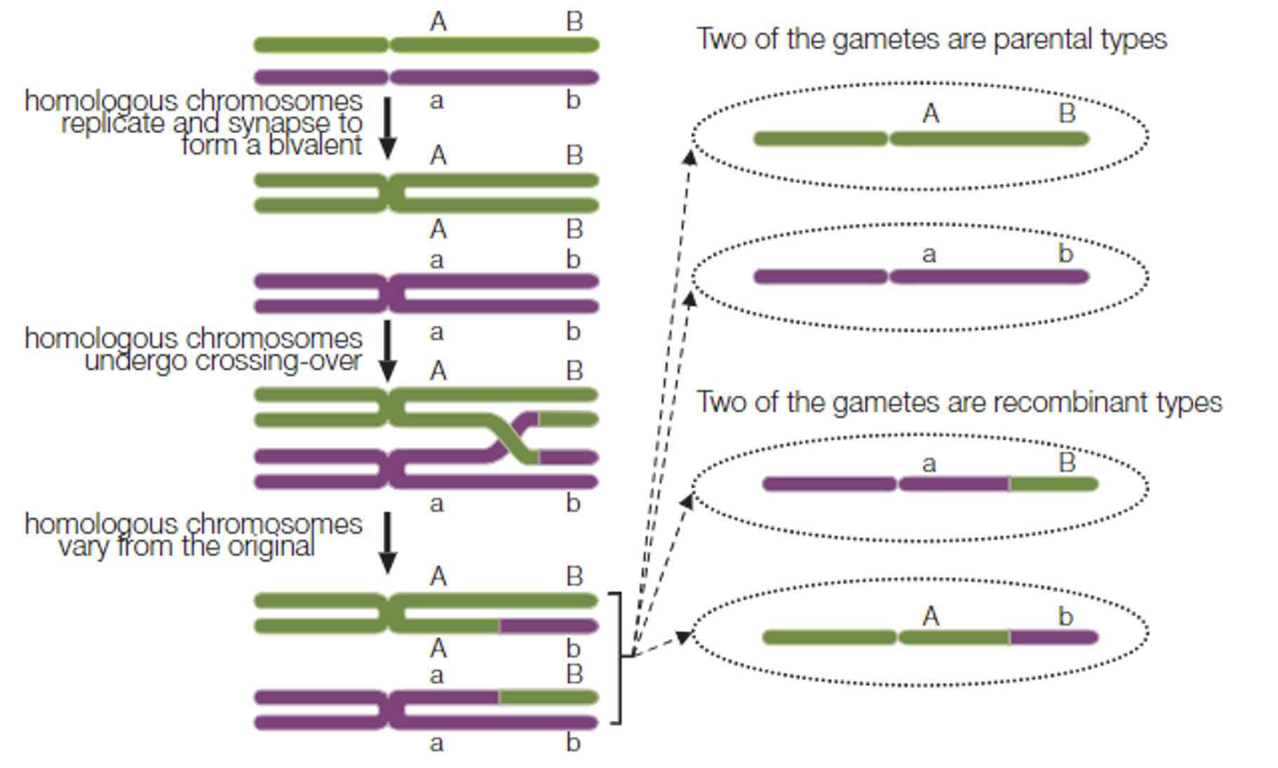
In prophase I, synapsis occurs, where the homologous chromosomes (each consisting of two identical sister chromatids) lie next to each other.
In this position, corresponding sections of non-sister chromatids may touch, break, and then rejoin, thereby exchanging chromosomal material. The positions at which crossing over occurs are referred to as chiasmata.
(in other words: When synapsis is in effect, crossing over may occur. This is when sections of non-identical chromatids come into contact causing an exchange of chromosomal material. The positions at which crossing over occurs are referred to as chiasmata).
Chromosomes with unique gene combinations as a result of crossing over are referred to as recombinants (recombinant chromosomes). Crossing over forms new combinations of maternal and paternal genes.
Independent assortment:
During metaphase I, the maternal and paternal chromosomes of each homologous pair line up on the equator of the spindle independently of the other pairs.
This greatly increases the number of unique genetic combinations by randomly distributing the maternal and paternal chromosomes.
Hence, crossing over and independent assortment are important in increasing the genetic variability of offspring, by altering the combination of genes that is passed on from one generation to the next.
How does fertilisation restore diploid number?
.
.
.
.
Section: Cells as the Basis of Life
SACE Terms: Explain
.
.
Cue words: None
The gametes formed in meiosis are haploid cells, and therefore contain half the chromosomes of a diploid cell.
When two gametes fuse during fertilisation, the normal diploid condition is restored, forming a zygote (fertilised cell).
Since fertilisation is random, any male gamete may fuse with any female gamete of the same species, As a process of sexual reproduction, this randomisation contributes to the genetic variation present in the offspring.
What is the difference of product of mitotic and meiotic cell division?
.
.
.
.
Section: Cells as the Basis of Life
SACE Terms: Compare
.
.
Cue words: None
Where does the process occur?
Mitosis occurs in somatic cells, while meiosis involves cells in the sexual cycle.Are the processes capable of further division?
Mitosis is a singular cell division, forming two daughter cell; While, meiosis involves two cell divisions, forming four daughter cells.Is there change in the chromosome number?
The chromosome number is maintained in mitosis, diploid (2n); whereas the chromosome number is halved in meiosis, haploid (n).Is there change in the composition of chromosomes?
The composition of chromosomes is identical in mitosis, but different in meiosis.When does variation occur?
Variation in mitosis only occurs due to mutation, whereas in meiosis, variation occurs as a result of crossing over, and independent assortment.Are the cells formed capable of fusion?
Cells formed in mitosis are not capable of fusion, while cells formed in meiosis are.How many daughter cells are formed?
Mitosis forms two daughter cells, while meiosis forms four daughter cells.
What are the sources and degree of genetic variation of the products of asexual and sexual reproduction?
.
.
.
.
Section: Cells as the Basis of Life
SACE Terms: Compare
.
.
Cue words: None
In asexual reproduction, which involves mitotic cell division, the only source of genetic variation is mutation.
In sexual reproduction, genetic variation results not only from mutation, but also in the crossing over and independent assortment processes of meiosis, and the random fertilisation of gametes.
Thus, there is much greater variation in the products of sexual reproduction than in the products of asexual reproduction.
What are the stages of the cell cycle?
.
.
.
.
Section: Cells as the Basis of Life
SACE Terms: Describe
.
.
Cue words: Include checkpoints
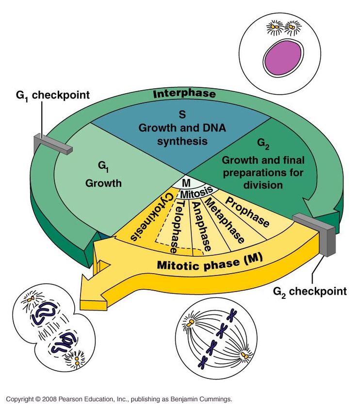
There are two main stages of the cell cycle.
Interphase: Where the cell lives most of its life; where DNA replication, development, and growth occur.
Mitosis: Where cell division occurs.
Interphase has three stages, G1, S, and G2. M represents mitosis, which is followed by cytokinesis.
The timing for the cell cycle varies in each cell, and hence, it needs to be regulated.
In the cell cycle, there are three checkpoints. The first in G1 (aka. restriction point, R), the second in G2, and the third in M.
At these checkpoints, the cell cycle is halted, and cannot proceed until a specific signal is received by the cell cycle control mechanism.
G1 (growth stage 1):
Longest stage, where the cell grows, performs protein synthesis and its natural functions.Checkpoint 1: Restriction point R
Here, the cell checks whether the internal and external conditions are ready for division. Conditions include its size, energy levels, and if there is enough external space.
The first check point is where the cell decides if it is ready to divide. Once the cell passes the G1 phase and enters S, it becomes irreversibly committed to division.S (synthesis):
After the first checkpoint, synthesis begins; categorised as the time when DNA replication occurs.G2 (growth stage 2):
Shortest stage, where the cell finalises its growth and development, and prepares the organelles before dividing.Checkpoint 2:
The cell checks to ensure that DNA replication occurred correctly. If faults are identified, the cell stops the cycle to repair the DNA if necessary. Subsequently, the cell proceeds into mitosis.Mitosis.
Checkpoint 3: M checkpoint (at metaphase)
Cell checks if chromosomes aligned correctly.
In the cell cycle, there is also a G0 phase. When a cell exits the cell cycle and stops dividing, it enters this phase. Cells in G0 are still metabolically active, however, are not actively preparing to divide. Cells in G0 may remain there temporarily or indefinitely, depending on signals from their environment or internal cues.
When a cell differentiates, it undergoes cellular changes to become a specialised cell. Cells often enter the G0 phase as they differentiate, as they no longer divide, and can focus on their specialised functions.
(G0 may also be referred to as the ‘resting phase,’ since the cell has exited the cell cycle and can ‘rest’ while it carries out its specialised functions).
So what happens if the cell does not fulfil the conditions for the checkpoint?
As previously mentioned in Checkpoint 2, the cell will try to repair itself, to restore the conditions and prerequisites required to move onto the next stage. However, in the case the cell cannot do so, the cell self-destructs in a process of apoptosis.
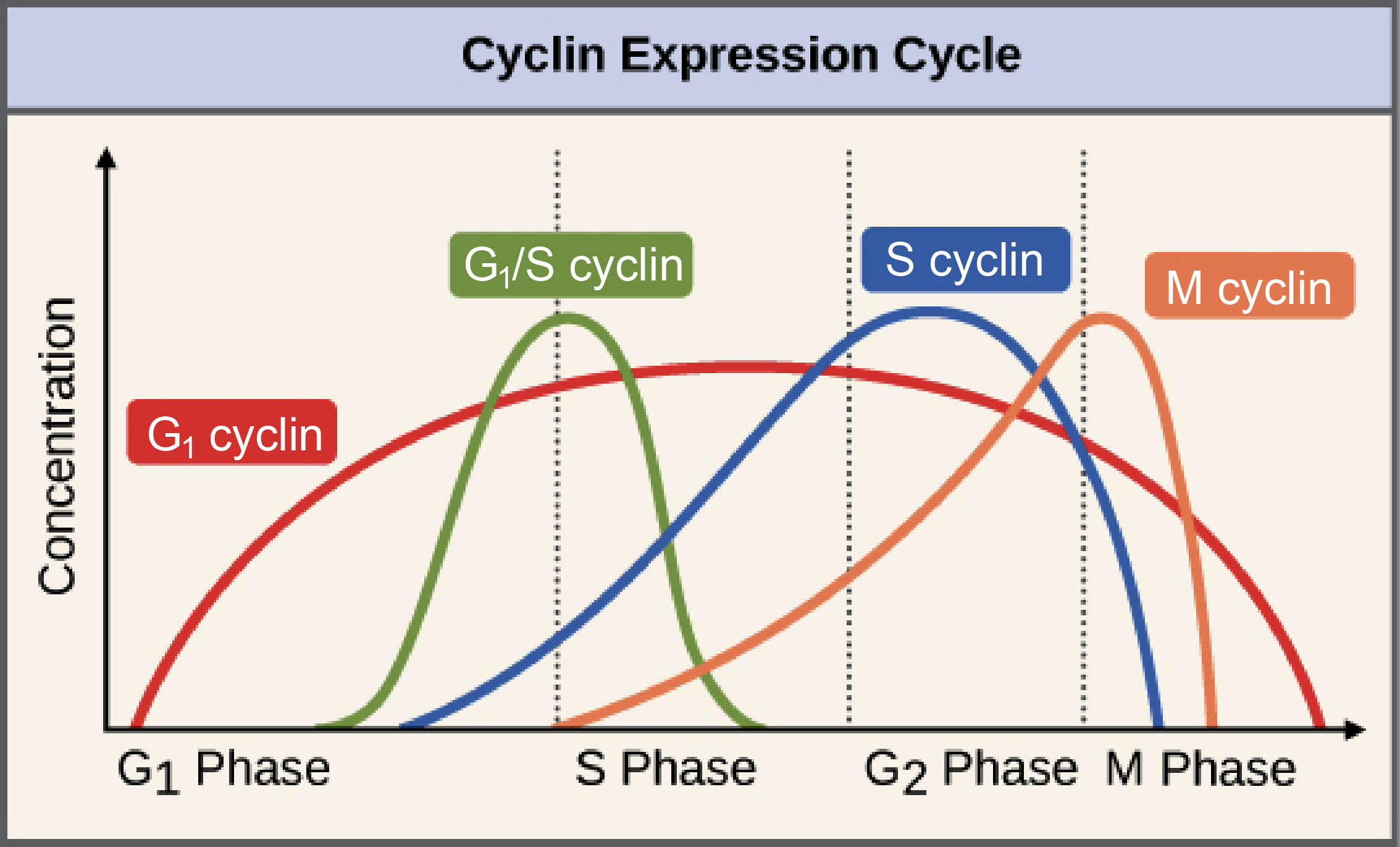
What are the cyclins?
.
.
.
.
Section: Cells as the Basis of Life
SACE Terms: None
.
.
Cue words: For each phase
G1: Cyclin D | CDK4/6
S: Cyclin E | CDK2
G2: Cyclin A | CDK2
M: Cyclin B | CDK1
(bolded: most important cyclins).
Maybe add more information to this card one day? This information isn’t written itself is not included in biology levels of life, but it might be handy? Anyways don’t bend over backwards trying to memorise all this.
What are the internal factors that may regulate cell division?
.
.
.
.
Section: Cells as the Basis of Life
SACE Terms: None
.
.
Cue words: Gene products/proteins produced by cell
Internal and external factors trigger the production of proteins that drive the cell through the checkpoints.
Regulators may be positive or negative (to either speed up or slow down the process).
Internal factors are often proteins which act to regulate the cycle.
The cell produces gene products (proteins) that regulate the cell cycle, and cyclins are the most important ones (regulators of these gene products).
These help to drive the cell to reach the threshold level at each checkpoint.
Its main types include G1 cyclin, G1/S cyclin, S cyclin, and M cyclin.
Each cyclin is associated with a specific stage of the cell cycle (as their names suggest), and helps to drive the cell through that stage.
For instance, the M cyclin promotes the events of mitosis, such as the breakdown of the nuclear membrane and the condensation of the chromosomes.
Cyclins operate by binding to cyclin-dependent kinases (Cdks) (enzymes), which thereby activates the enzyme, and forms a complex called the maturation promoting factor (MPF) (aka. mitosis promoting factor).
Cdk + cyclin → MPF
Typical cyclins are present at low levels for most of the cycle, and increase strongly when required, binding to Cdks shortly before the checkpoint (refer to figure attached).
It is the levels of the Cdk/cyclin complexes which drive the cell through the checkpoints.
When MPF levels reach the threshold, that is when it signals/triggers the cell to move through the checkpoint.
(remember: that the checkpoints halt the cell cycle, and it cannot continue until the cell receives a signal from the cell cycle control mechanism).
Subsequently, MPF is quickly broken down, allowing the cycle to proceed.
What are the external factors that may regulate cell division?
.
.
.
.
Section: Cells as the Basis of Life
SACE Terms: None
.
.
Cue words: None
Internal and external factors trigger the production of proteins that drive the cell through the checkpoints.
Regulators may be positive or negative (to either speed up or slow down the process).
External factors may be physical or chemical in nature, and provide the cell with information about the cell’s condition, and location.
External factors mainly influence decisions at the G1 checkpoint.
Growth factors:
Hormones that bind to receptors, and stimulate the cell to produce certain proteins, which act to stimulate cell division (or the production of internal factors which help the cell move through the checkpoint).SA/V ration:
Large cells have a low SA/V ratio, which acts to stimulate cell division.Cell density:
Crowded cells will not divide, but if gaps form, this stimulates cells to divide.
How do hormones regulate cell division?
.
.
.
.
Section: Cells as the Basis of Life
SACE Terms: Explain
.
.
Cue words: None
NOTE: PAGE 119 IN BIOLOGY LEVELS OF LIFE SECTION SHOULD BE ADDED TO THIS, BUT I AM IN A RUSH, SO I AM ONLY ADDING HUMAN RELATED HORMONE THINGS HERE/MENTIONS IN PPT. BUT ONE DAY I WILL ADD MORE, HOPEFULLY.
Hormones, produced by glands such as the pituitary and thyroid, regulate cell division by interacting with receptors on target cells.
They trigger signalling pathways that influence gene expression and protein synthesis, impacting the cell cycle.
For instance, growth hormones stimulate cell division and growth, while reproductive hormones like estrogen and progesterone regulate cell proliferation in reproductive tissues.
Thyroid hormones indirectly affect cell division by controlling metabolism and energy production.
Overall, hormones play crucial roles in coordinating cellular processes, and maintaining homeostasis.
I have no idea where the homeostasis part came from either considering we are talking about regulating the cell cycle, but yes, okay, yes.

How do carcinogens upset the normal controls of cell division by causing mutations in key regulatory genes?
.
.
.
.
Section: Cells as the Basis of Life
SACE Terms: None
.
.
Cue words: Cancer
There is a strong correlation between carcinogenesis, the onset of cancer, and mutagenesis, a change in the DNA sequence. Three main classes of agent are responsible for mutagenesis.
Chemical carcinogens causing changes to the nucleotide sequence in DNA.
Radiation, especially X-rays, which cause chromosome breakage.
Viruses that can cause an addition of foreign DNA sequences into the host DNA.
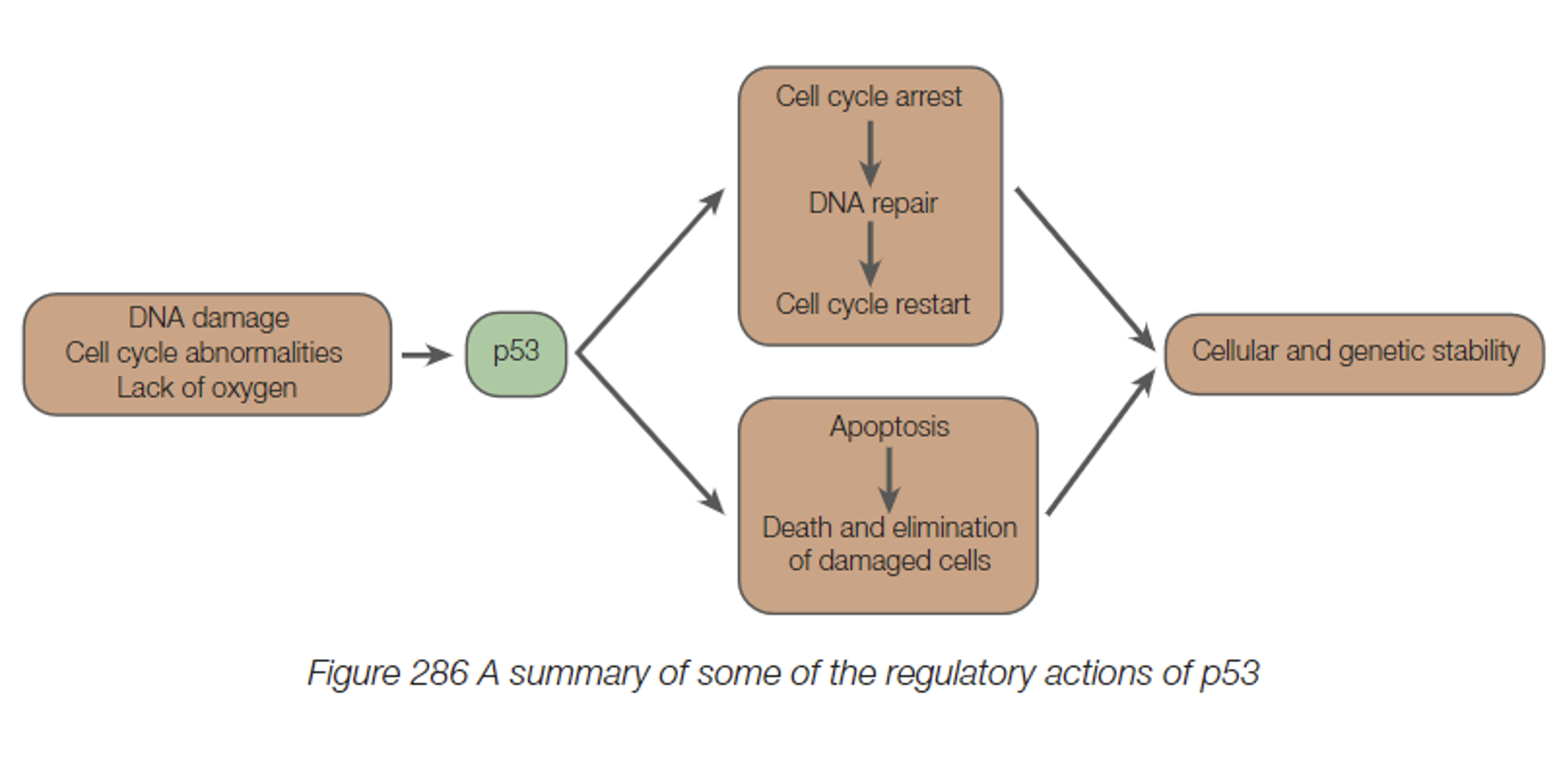
Cancer is a disease caused by uncontrolled cell division, leading to tumour formation. Cancers occur as a result of factors which disrupt the normal controls of the cell cycle.
Carcinogens, physical or chemical, induce gene mutations, giving rise to cancer, by causing changes to the nucleotide sequence in DNA.
Oncogenes (aka. tumour genes) arise from gene mutations, and are capable of over activating the cell cycle, leading to uncontrolled cell growth.
DNA repair genes produce products which can repair DNA that has been damaged by X-rays or mutagenic chemicals. If the normal DNA repair processes malfunction, this may facilitate the continuation of faulty cells through the cycle.
Tumour suppression genes, such as p53, produce proteins which prevent the production and proliferation of cells with damaged DNA, by binding to specific DNA sequences, and halting the cycle. Once stopped, the p53 protein activates the gene expression of proteins involved in DNA repair.
It’s important that such genetic alterations are not passed onto future generations. If the DNA cannot be repaired, DNA repair genes activate the gene expression of proteins involved in apoptosis.
In many types of cancers, the p53 gene is mutated or otherwise dysfunctional, leading to a loss in its tumour-suppressive functions, allowing cells with damaged DNA to continue and divide.
It has been found that p53 is inactive in almost 50% of all cancers.
What are the techniques of cell culture?
.
.
.
.
Section: Cells as the Basis of Life
SACE Terms: Explain
.
.
Cue words: Process and its requirements. Culture~cultivation
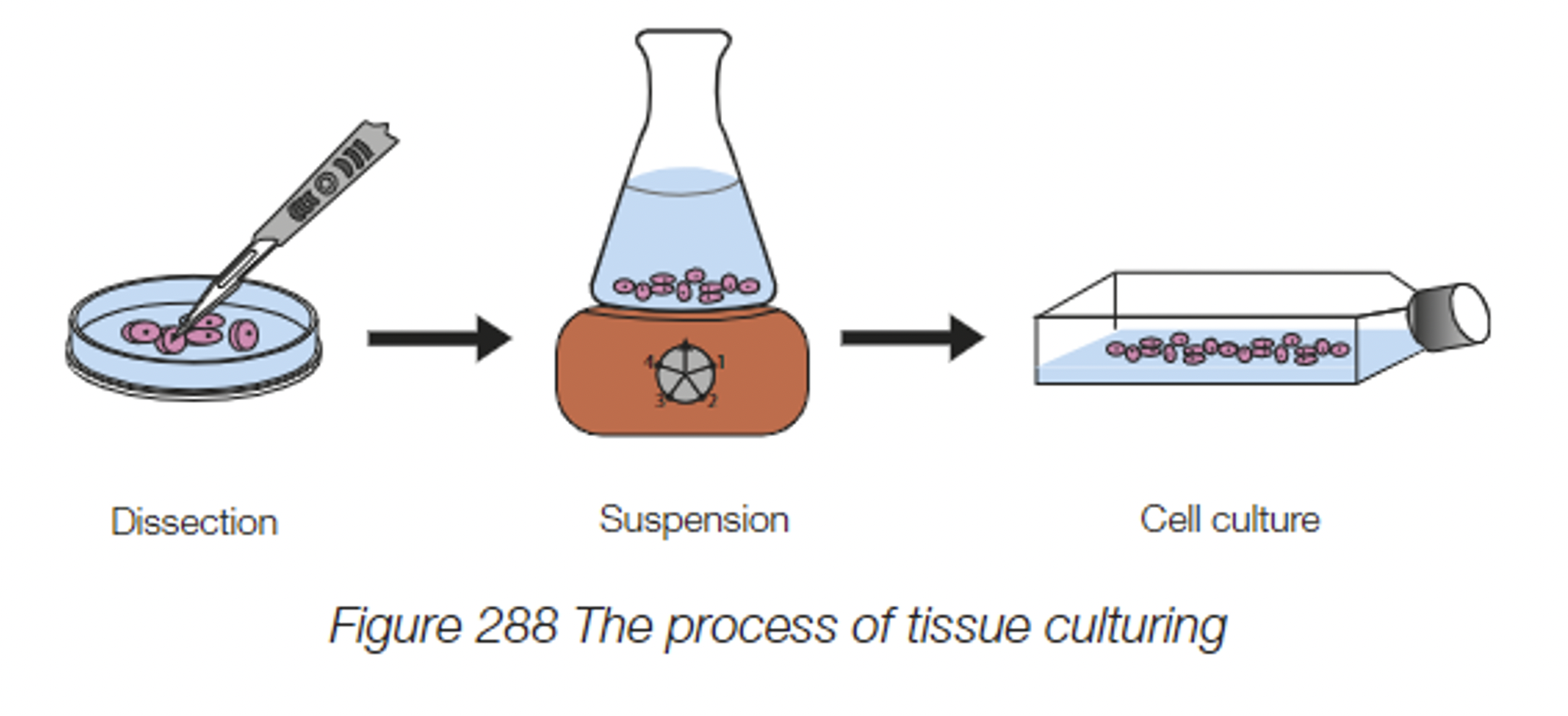
Cell culture is the process of growing, and cloning cells within a controlled environment of a lab.
The requirements for cell culturing include:
Nutrients such as glucose, and oxygen (for respiration)
Growth factors (to stimulate division)
DNA nucleotides (to facilitate DNA replication)
Correct and consistent temperature and pH conditions (so enzymes do not denature) (37℃ is optimal temperature in humans)
Amino acids (for protein synthesis)
A sterile environment (prevent infection caused by unwanted microorganisms)
(Think of all the things a cell needs in order to grow and divide).
The process for cell culturing involves the following.
Firstly, the tissue (containing the desired cell) is dissected to expose the cells.
The dissected tissue is then placed in a suspension medium of protein digesting enzymes, to release the cells.
Thirdly, the cells. are placed into a dish (or sterile environment) with all of the requirements they need (mentioned above), and left to culture and grow.
What are the applications and limitations of cell culture?
.
.
.
.
Section: Cells as the Basis of Life
SACE Terms: Explain
.
.
Cue words: None
Science as a Human Endeavour moment~
Applications of cell culturing include:
Manufacturing vaccines.
Bacterial cultures:
Testing the effectiveness of antibiotics on bacterial infections before application (to determine which antibiotics the species are not resistant to, and can therefore be applied in treatment). As well as for genetic engineering.IVF (in vitro fertilisation):
To allow people to have children;
To culture embryos to implant into expecting mothers, or surrogate mothers through IVF.Culturing human stem cells for scientific research and differentiation into specific somatic cells.
Manufacturing anti-cancer agents:
Use of biotechnology, and recombinant DNA to manufacture a range of critical products such as enzymes, hormones, and monoclonal antibodies and anti-cancer agents.A key process in cellular agriculture is the production of transgenic organisms with targeted genes, e.g., for herbicide resistance.
Food industry:
Improving the techniques of food production and in the food industry (producing cell-cultured meat; yeast cells in brewing industry).Product (medical and cosmetic) testing and Skin grafts:
Growing human tissue, and culturing skin cells, for drug testing (studying effects on human cells before application, this is instead of animal testing too), and reconstructive surgery (skin grafts for skin burns)
While cell culture is advantageous in many ways, its application is not without limitations, and thus more research is required in the field.
Limitations of cell culturing include:
Hard to maintain the sterile conditions required for cell culturing.
There is a finite number of times in which most cells can divide. Most tend to stop at after 50 divisions.
After time, toxins and dead cells begin to accumulate.
Changes may occur in the pH of the growth media.
Genetic and epigenetic changes may lead to cultured-adapted cells with changed capacities (the cells have adapted and are used to the cultured environment, and hence real-world application may vary slightly to when it is in its cell-cultured environment).
Contact inhibition: As the cells begin to come into contact with one another, they slow their rate of division. This limits the cell culture’s application in researches which require large populations of actively dividing cells.
As the cells fill the bottom of the dish, division slows further and is balanced by the rate of cell death, so that the total cell number remains constant. This state is called confluence (“confluence reached”). This confluence ensures that cells remain as a monolayer. However, (drifting off topic here) in cancer cells, even when confluence is reached, their growth continues to divide in an unregulated manner. Anyways, remember that, an external factor which regulates cell division is cell density; a crowded cell will not divide.
Despite limitations, cell culture is invaluable for biomedical research and biotechnology.

Is CO2 an organic compound?
.
.
.
.
Section: DNA and Proteins
SACE Terms: None
.
.
Cue words: None
No.
While it contains carbon, and is produced by and associated with living things, it is not a complex molecule.
Hence, carbon dioxide is an inorganic molecule.
What is DNA?
.
.
.
.
Section: DNA and Proteins
SACE Terms: None
.
.
Cue words: None
DNA stores and transmits genetic information; it functions in the same way as all living things.
DNA is a helical, double-stranded molecule.
Deoxyribonucleic acid (DNA) is a macromolecule found in all living cells, and is the chemical that controls virtually everything that happens in cells.
DNA stores and transmits the information that is necessary to direct the cell’s complex processes.
DNA in cells is found in structures called chromosomes.
(Chromosomes are known as the structural unit of information in cells).
It is the most fundamental chemical of life, believed to have formed ~3 billion years ago when the first forms of life arose.
The below will be further brushed upon in other cards.
DNA is a unique macromolecule, capable of self-replication.
Its sequence of nucleotides allows it to store information that can be copied and passed onto daughter cells.
DNA provides the link between one generation and the next.
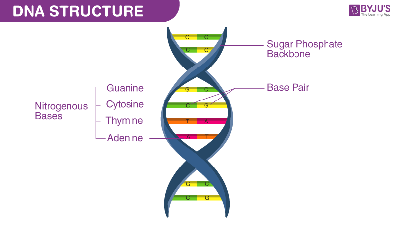
Where is DNA unbound and circular?
.
.
.
.
Section: DNA and Proteins
SACE Terms: None
.
.
Cue words: None
DNA is unbound and circular in the cytosol of prokaryotes, and in the mitochondria and chloroplasts of eukaryotes.
Prokaryotic cells contain a single circular chromosome, which is double-stranded, made of DNA with no protein attached to it (unbound to protein molecules).
The DNA in a prokaryotic cell is not enclosed in a nucleus, but floats freely in the cytosol. (The cytosol is the fluid part of the cytoplasm, not including organelles such as ribosomes). The location of DNA is called the nucleoid region.
Mitochondria and chloroplasts have their own DNA, separate from the DNA in the nucleus.
Their DNA resembles prokaryotic DNA as it is circular, and has no protein attached.
This genetic information contains all the information needed for the mitochondria and chloroplasts to undertake their respective functions, and for their own division (by binary fission).
The presence of these circular DNA molecules in these organelles is hypothesised to be due to their prokaryotic origins.
(additional information: nucleic acid known as RNA and proteins are also found in the nucleoid region. Also found in many prokaryotes, are plasmids, which are small amounts of circular, double-stranded DNA that are separate from the cell’s chromosomal DNA).
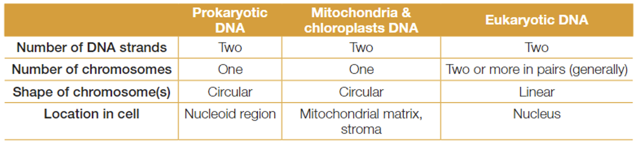
Where is DNA bound to proteins and linear?
.
.
.
.
Section: DNA and Proteins
SACE Terms: None
.
.
Cue words: None
In eukaryotic cells, DNA is bound to proteins (histones) in linear chromosome, which are found in the nucleus.
In eukaryotic cells, the strands of DNA in the nucleus are coiled around proteins (histones) to form structures called chromosomes.
These chromosomes are linear.
Typically, chromosomes are not visible, unless the cell is dividing (where they condense, shorten, and thicken).
(im not sure how much of the rest of this information we need, but i shall proceed to add).
Characteristics of DNA in eukaryotes.
The chromosomes are arranged into condensed, linear strands, consisting of a double-stranded DNA molecules and histone proteins.
The ends of chromosomes have short lengths of DNA called telomeres. These protect the tips of the chromosomes from breakdown, and prevent the chromosomes from binding to each other.
Chromosomes are only visible under a light microscope during cell division when the DNA is tightly coiled around the histone proteins (condensed).
The de-condensed form of chromosomes is called the chromatin.
Chromatin is present in the nucleus of cells throughout the cell cycle (excluding cell division).
It is important that the genetic code is accessible during the growth and development of a cell so that DNA synthesis, RNA synthesis, and protein synthesis can occur.
If the DNA molecule is condensed, the genetic code is very hard to access, and these processes are unlikely to occur.
However, it is still important for the chromatin to condense into chromosomes prior to cell division to protect the genetic code, and ensure identical copies are transferred to both daughter cells.
Each human somatic cell contains ~2-3 meters of DNA when completely de-condensed to linear form.
If chromatin was present during cell division, it is likely that the de-condensed DNA would get tangled; resulting in DNA breakages.
The chromosomes in prokaryotic cells do not contain histones.
It is the DNA of the chromosomes that contain the vast store of information, in the form of base sequences, that is needed by the cell and the organism.
The information coded in the DNA determines what proteins are made within a cell, and hence it influences the structure and function of the cell.
A segment of DNA on a chromosome that contains the complete sequence of bases required to direct the manufacture of a polypeptide or an RNA molecule is called a gene.
Each chromosome will carry particular genes which will be the same for all individuals of the species, except for any chromosomal mutations which may have occurred.
Each chromosome will have a homologous pair, where each carries the same set of alleles, encoding variants of the same gene.
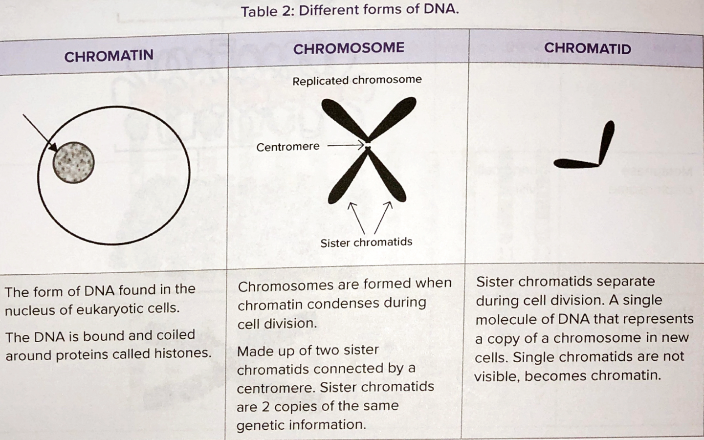
Why is DNA replicated?
.
.
.
.
Section: DNA and Proteins
SACE Terms: None
.
.
Cue words: What does it allow for?
Replication of DNA allows for genetic information to be inherited.
Every time a cell divides, its DNA is copied, and each new (daughter) cell receives a complete copy.
Thus, the information on the DNA is passed from one generation of cells to the next.
The process of DNA replication is the same in prokaryotes and eukaryotes, and is considered to be a universal process in all organisms.
The only difference between prokaryotes and eukaryotes is the location where it occurs.
(prokaryotes - nucleoid region; eukaryotes - nucleus).
Similarly, when organisms reproduce, they pass genetic information on to their offspring via DNA.
In sexual reproduction an individual receives half of each parent’s genetic material.
In humans, this genetic material is carried in egg cells and sperm cells.
However, this still involves copying and passing on DNA.
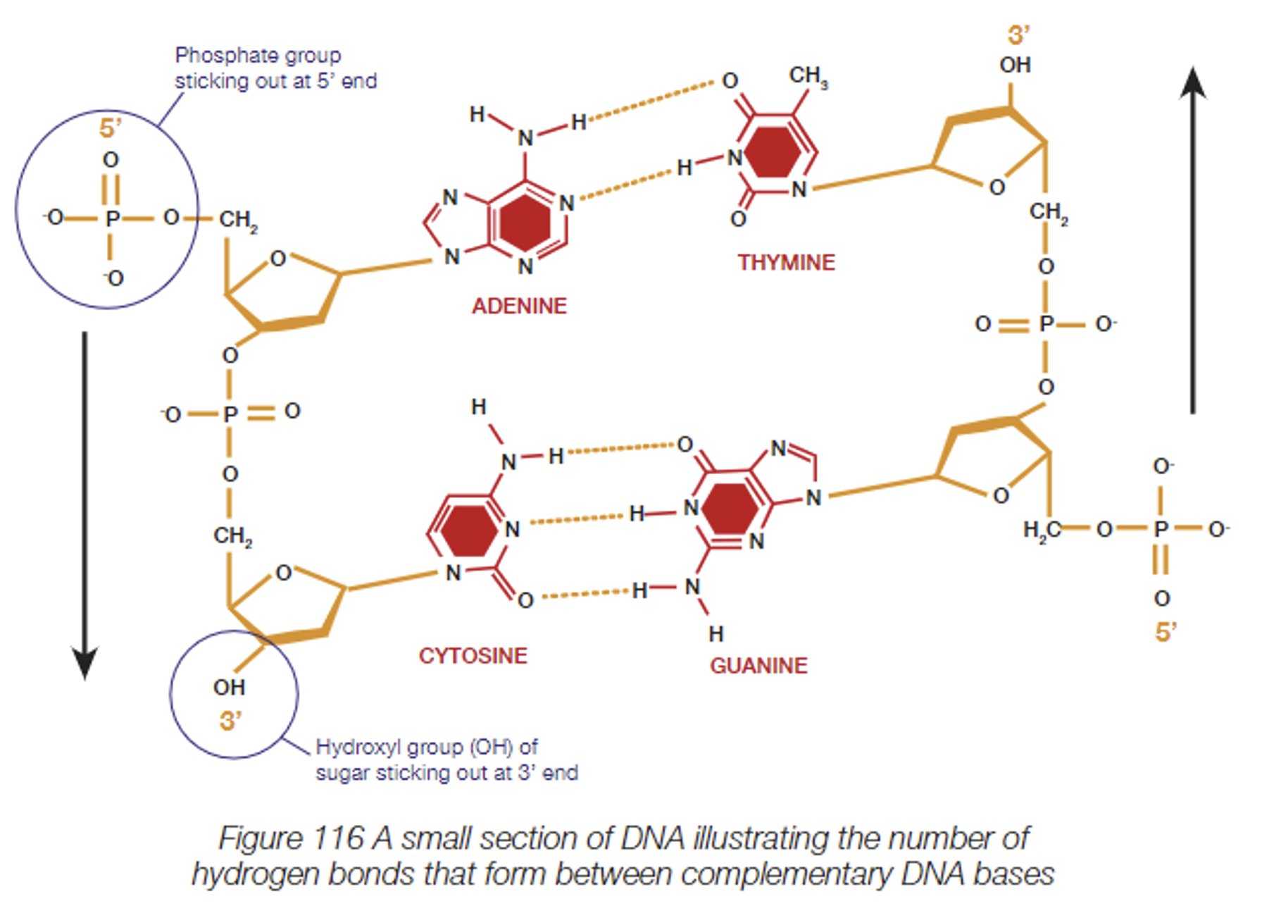
What is the function of DNA, and what are the structural properties of the DNA molecule?
.
.
.
.
Section: DNA and Proteins
SACE Terms: Describe
.
.
Note: This is kind of a brief/unspecified section, since it overlaps with other seahorses, but feel free to go into as much depth as pleased (double check on google if unsure/or using other cards).
.
.
Cue words: Nucleotide composition and pairing, and the weak bonds between strands of DNA that allow for replication.
DNA stores and transmits genetic information.
The structure of DNA enables it to perform two vital functions necessary for life.
Make identical copies of itself so that genetic information can pass from cell to cell, and can be inherited or passed on to the next generation.
Provide a code that can be used by cells to manufacture protein molecules.
Structure of DNA
DNA is a large macromolecule made up of repeating units called DNA nucleotides.
Nucleotides are complex molecules made up of three smaller molecules that are linked together by covalent bonds (refer to attached figure).
A pentose (5-carbon) sugar called deoxyribose.
a phosphate group, and
one of the four organic bases: adenine (A), thymine (T), guanine (G), or cytosine (C).
Each base forms weak hydrogen bonds with its complementary base (located at the same position on the other strand).
Weak hydrogen bonds between complementary base pairs in DNA are an advantage to cells since they are easy to break and re-form.
(simply: Weak hydrogen bonds form between the complementary base pairs, which are advantageous to the cell since they can be easily broken and reformed.)
This is important in DNA replication, which involves breaking apart the DNA double helix, to expose its organic bases to base pairing.
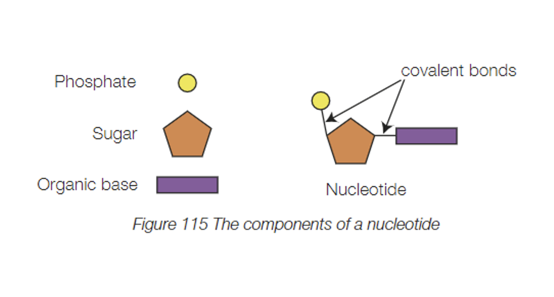
How are DNA strands read?
.
.
.
.
Section: DNA and Proteins
SACE Terms: Recognise
.
.
Note: This is kind of a brief/unspecified section, since it overlaps with other seahorses, but feel free to go into as much depth as pleased (double check on google if unsure/or using other cards).
.
.
Cue words: directional
DNA strands are directional and are read 5’ to 3’.
The phosphate of one nucleotide is attached to the sugar of the next nucleotide.
This results in a backbone of alternating phosphates and sugars.
The structure of the phosphate-sugar backbone resulting in one end of the DNA strand ending in a phosphate, and the other ending in a sugar, gives the strand directionality.
The beginning of the DNA strand has a phosphate (PO43—) sticking out, and is known as the 5’ (“five prime”) end.
At the opposite end of the chain, a sugar molecule exposed with a hydroxyl group (OH) sticking out. This is called the 3’ (“three prime) end of the DNA strand.
The DNA sequence is therefore written in the 5’ to 3’ direction.
The phosphate group has a negative charge, giving the DNA molecule an overall negative charge.
This charge can be used to help separate DNA molecules.

What is the important of complementary base pairing?
.
.
.
.
Section: DNA and Proteins
SACE Terms: Explain
.
.
Note: This is kind of a brief/unspecified section, since it overlaps with other seahorses, but feel free to go into as much depth as pleased (double check on google if unsure/or using other cards).
.
.
Cue words: Include the base-pairing rules
The base pairing rules, and the method of DNA replication are universal.
The possible nitrogenous bases include:
Adenine (A)
Thymine (T)
Guanine (C)
Cytosine (C).
The base-pairing rules are that:
Adenine only bonds with thymine through a double hydrogen bond (A=T); and cytosine always bonds with guanine through a triple hydrogen bond (C≡G).
Evidence for complementary base pairing is supported by DNA analysis which reveals that species have identical amounts of adenine and thymine, as well as identical amounts of cytosine and guanine.
For example, if there’s 20% thymine in a DNA molecule, there would be 20% adenine, and therefore 30% guanine, and 30% cytosine.
When DNA is assembled, it consists of two complementary strands linked by the complementary base pairs.
As the strands are complementary, they are read in opposite directions. Such that the DNA strand is read from top to bottom in the 5’ to 3’ direction, and vice versa for the other side (in the 5’ to 3’ direction).
This consistent pairing of bases (aka. complementary base-pairing) is important since it allows for the accurate replication of DNA. It also allows DNA to be transcribed accurately into RNA and then translated from RNA into amino acids.
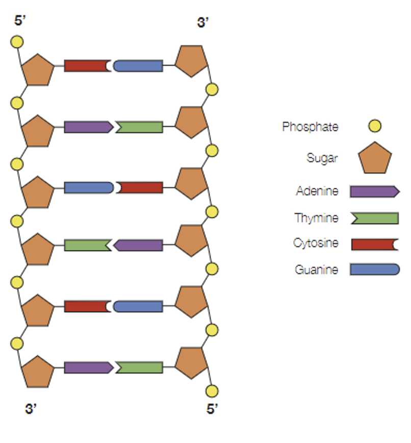
What are chromosomes comprised of?
.
.
.
.
Section: DNA and Proteins
SACE Terms: None
.
.
Cue words: How species on earth differ
Chromosomes consist of several functional units of information known as genes.
Each gene contains a unique sequence of nitrogen bases on one strand of the DNA (the coding strand).
Hence, the sequence of bases varies from one DNA molecule to another, and it is this variation in the base code which enables DNA to be such a versatile and diverse molecule.
Different species have different characteristics, and therefore a different number of genes and chromosomes.
Different species on earth differ in:
The number of chromosomes found in cells (and therefore, number of genes)
The base sequences of genes found in their DNA
The length of the DNA molecule, and hence the number of bases.
What is the process of semi-conservative replication of DNA?
.
.
.
.
Section: DNA and Proteins
SACE Terms: Describe and represent
.
.
Cue words: None
The enzyme, helicase, breaks the hydrogen bonds between the complementary (nucleotide) bases. This unwinds and leaves the bases exposed.
Each strand serves as a template for the new complementary strand.
DNA polymerases (enzymes) bond single, free nucleotides with the exposed bases (according to the base pairing rules), and other enzymes join together the sugar-phosphates of the free nucleotides to form a new DNA strand. Hence, it is synthesises in the 5’ to 3’ direction.
Each new double-stranded DNA molecule rewind to form its double helix structure, and join at the centromere (forming the chromosome).
The result is two new DNA double helices that are identical to each other and to the original molecule; where each new DNA molecule consists of one ‘old’ strand and one ‘new’ strand. This process is therefore called semi-conservative replication.
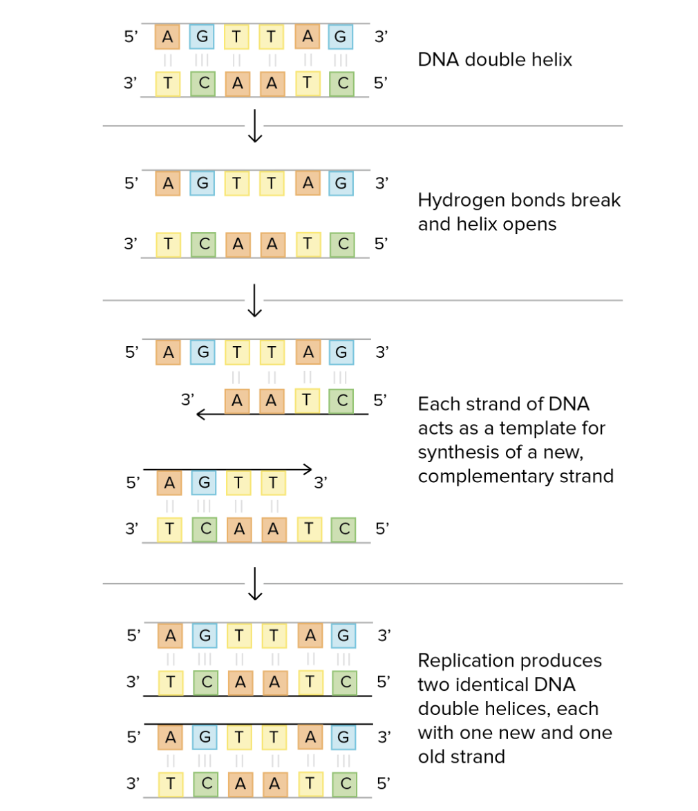
Why is the number of times DNA replication can occur limited?
.
.
.
.
Section: DNA and Proteins
SACE Terms: None
.
.
Cue words: None
DNA replication can only occur a finite number of times since the telomeres (at the ends of the chromosomes) shorten with each replication.
Eventually, the telomeres are too short for further replication to occur.
What is the involvement of genes with RNA molecules?
.
.
.
.
Section: DNA and Proteins
SACE Terms: None
.
.
Cue words: None
A gene consists of a unique sequence of nucleotides that codes for a functional protein, or an RNA molecule.
A gene is a particular segment of a chromosome which contains the genetic code (specific nucleotide sequence) required to direct the synthesis of a particular polypeptide (protein) or RNA molecule.
Genes function to make polypeptide chains, proteins, and RNA molecules.
What does an RNA nucleotide consist of?
.
.
.
Section: DNA and Proteins
SACE Terms: None
.
.
Cue words: None
A pentose (5-carbon) sugar called ribose
A phosphate group
One of the four organic bases
Adenine (A), Uracil (U), Guanine (G), Cytosine (C).
Same as DNA, except for the presence of Uracil in place of Thymine.
What are the 4 major types of RNA?
.
.
.
Section: DNA and Proteins
SACE Terms: None
.
.
Cue words: None
Messenger RNA (mRNA)
Encodes proteins.
Transfer RNA (tRNA)
Acts as an adaptor between mRNA and amino acids.
Ribosomal RNA (rRNA)
Forms the ribosomes.
Micro RNA (miRINA)
Regulates gene expression.
Describe the translation and transcription in the context of introns and exons.
.
.
.
Section: DNA and Proteins
SACE Terms: Distinguish between exons and introns as coding and non-coding segments of DNA found in genes in eukaryotes; Describe how both exons and introns are transcribed but only exons are translated to form a polypeptide in eukaryotes.
.
.
Cue words: None
In the nucleus, introns and exons are transcribed into pre-mature mRNA by RNA polymerase (as apart of protein synthesis).
The introns are then spliced out of the premature mRNA by RNA-splicing endonuclease inside the spliceosome (an RNA-protein complex), forming mature mRNA.
Introns are spliced out prior to translating since they interfere with the sequence of the gene product (e.g. proteins). After splicing they can be used to form other RNA (e.g. tRNA). mRNA splicing ensures that the correct sequence of amino acids are synthesised.Mature mRNA moves out of the nucleus towards a ribosome, where the ‘code’/information is translated into amino acids, forming proteins, using codons and tRNA.
What are the roles of DNA, mRNA, tRNA, and rRNA in transcription and translation?
.
.
.
Section: DNA and Proteins
SACE Terms: Describe the role of DNA, mRNA, tRNA, and rRNA in transcription and translation.
.
.
Cue words: None
DNA:
The non-coding strand of the DNA is used as a template in the transcription of pre-mature mRNA (pre-mRNA).
The coding strand is not used since it contains the correct sequence of genes for producing the specific products. Hence, using the coding strand as a template would lead to the wrong sequence being transcribed.
mRNA:
mRNA acts as a working copy of the gene; enabling the synthesis of the protein encoded by that gene. Mature mRNA carries codons which are complementary to anticodons on tRNA molecules, which then correspond to a specific amino acid.
mRNA is a working copy of the DNA coding strand, and transfers this information to the ribosomes where it can be translated into a specific sequence of amino acids (which form proteins useable by the cell).
tRNA:
The role of tRNA is to carry the specific amino acid to the correct placement on the polypeptide being synthesised—which is specified and directed by the codons on the mRNA. Once complete, the tRNA will collect another of the same amino acid in preparation for its future involvement in translation.
rRNA:
rRNA and proteins make up the ribosomes; the site of translation. It is the rRNA which facilitates protein synthesis as the ribosome moves along the mRNA strand, translating the mRNA code into a specific sequence of amino acids.
SACE: Protein synthesis involves the transcription of a gene into mRNA, and translation of mRNA into an amino acid sequence at the ribosomes. In eukaryotic cells, transcription occurs at the nucleus.
What is the relationship between DNA codons, RNA codons, anticodons, and amino acids?
.
.
.
Section: DNA and Proteins
SACE Terms: Describe
.
.
Cue words: None
The DNA coding strand codes for genes of interest (which codes for the gene product). The coding strand determines the correct mRNA sequence, but does not directly take part in transcription.
The coding strand consists of triplets of bases called DNA codons.
The DNA template strand is complementary to the coding strand, and serves as a genetic code template for synthesising the mRNA strand.
The template strand consists of triplets of bases called DNA anti-codons.
Hence, it follows that the mRNA strand consists of RNA codons, and the tRNA molecules have complementary anti-codons.
Where these RNA codons code for a specific amino acid, and the RNA anti-codons allow the amino acid to be brought to the correct position in the polypeptide chain.
What are the factors that determine the primary, secondary, and tertiary, and quaternary structure of proteins?
.
.
.
Section: DNA and Proteins
SACE Terms: Describe
.
.
Cue words: None
Primary structure:
The primary structure is shaped by the polypeptide strand formed at the completion of translation, where the amino acids are held together by peptide bonds. This structure thus determines and affects all of the subsequent levels of structure.
Secondary structure:
The secondary structure is the localised coiling or folding of the polypeptide, which forms as a result of hydrogen bonding between the amino acids. This stage acts as an intermediate between the primary structure and its three-dimensional shape established in the tertiary structure. The two most common ways the secondary structure changes are through alpha helices (coiling), or beta pleated sheets (folding).
Tertiary structure:
The tertiary structure is the ‘result’ of how the polypeptide is coiled or folded in the secondary stage.
The tertiary shape forms spontaneously due to forces of attraction between amino acids (interactions such as ionic bonding, hydrogen bonding, and strong covalent disulphide bonds), resulting in its unique 3D structure.
Some proteins may be fully functional and complete at this stage (since proteins can comprise of one or more polypeptide).
Proteins that have a folded shape are called globular proteins.
Quaternary structure:
If the protein consists of more than one polypeptide, the quaternary structure is formed by the polypeptides chemically bonding together.
Why is the three-dimensional shape of a protein critical to its function?
.
.
.
Section: DNA and Proteins
SACE Terms: Explain
.
NOTE: THIS CARD NEEDS MAD REFINING FOR SACE EXAM
Cue words: None
Questions surrounding this often refer to a specific scenario, but generally (how I do it at least) you want to talk about the following, and then LINK it to the given scenario.
The three-dimensional structure of proteins is crucial to its function, since the folding, coiling, grooves and clefts formed by the forces of attraction between the amino acids produce a shape complementary to the receptor or molecule which it is supposed to bind to.
When binding to its complementary molecule/receptor, it will elicit a specific response inside the cell, correlated with its specialised function.
As such, damage to this three-dimensional structure (particularly to the bonds between the amino acids) means that the protein cannot (or at least, not as efficiently) carry out is intended function—and thus, three-dimension shape is critical for function.
You’re going to want to link the following points to your given scenario.
For instance, in the context of adrenaline binding with the cell membrane of the liver cells and not other cells of the organism:
This is because the receptor proteins on the cell membrane of liver cells are specifically complementary to the shape of adrenaline hormones. Not all cells will have this particular receptor, hence it follows that adrenaline is not complementary to those receptors, and thus will not bind to it.
Or in the context of antigens and antibodies:
Bacterium and viruses are characterised by specific antigens or antigenic markers on their membranes. The antibodies are complementary to these antigens, and bind to the bacteria and viruses, which enables it to neutralise and inactivate it.
What is the induced-fit model of enzyme-substrate binding?
.
.
.
Section: DNA and Proteins
SACE Terms: Explain
.
.
Cue words: None
The active site of an enzyme is a groove or cleft formed by the unique folding (enzymes are usually globular proteins; hence folding) of the polypeptide chain; it is where the substrate binds to the enzyme.
The induced fit model, proposed in the 1950s, explains how complementary binding works (between enzyme-substrate).
It was originally theorised that substrates bind to enzymes as per the lock and key model; suggesting that only the correct ‘key’ (substrate) will fit and work the lock (enzyme).
However, the induced-fit model explains that instead of a lock and key, the substrate and enzyme bind together through weak bonds, and modify their shape in the process to create an ideal fit for catalysis - refer to attached figure.
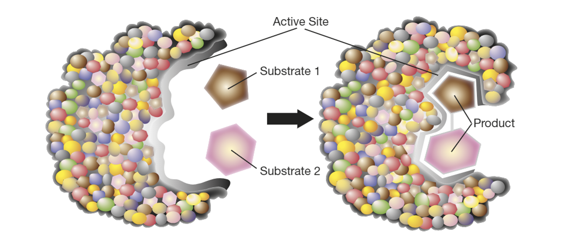
How do changes in DNA methylation and histone modification alter gene expression?
.
.
.
Section: DNA and Proteins
SACE Terms: Recognise
.
.
Cue words: None
Recognise that changes in DNA methylation and histone modification can alter gene expression
DNA Methylation:
Methylation involves the addition of a methyl group (CH3) onto the DNA strand; often on a cytosine base.
Methylation does not alter the DNA sequence, but changes how the gene is read and expressed.
Cytosine methylation inhibits RNA polymerase from binding to the promotor sequence of the DNA strand (hence, inhibiting transcription).
(remember that the ‘promotor sequence’ is essentially nucleotide bases upstream/prior to the gene being encoded… it is the site where the RNA polymerase binds to).
After methylation, genes usually retain the methyl group during cell division. As such, this methylation can be passed on and inherited by future generations.
Histone Modification:
DNA is package with histones to form the chromatin, where these histones may undergo modification.
Acetylation: Acetylation causes the chromatin to de-condense, resulting in the loose packaging of histones. This promotes gene expression since the RNA polymerase can access the gene more easily, and bind to the DNA.
Deacetylation: Deacetylation causes the chromosome to condense, resulting in the tighter packaging of histones. Consequently, RNA polymerase cannot bind to the DNA, which inhibits gene expression.
Methylation: Histones may also undergo methylation, which may increase or decrease gene expression. The consequence of histone methylation depends on the amino acids in the methylated histone, and the amount of methyl groups added to that amino acid.
How would epigenetic modifications in genes that control cell division lead to cancer?
.
.
.
Section: DNA and Proteins
SACE Terms: Explain
.
.
Cue words: Such as changes in DNA methylation
Explain how epigenetic modifications in genes that control cell division, such as changes in DNA methylation, can lead to cancer.
Cancer is caused by uncontrolled cell division, therefore, genes associated with cell division are tightly regulated.
Proto-oncogenes:
Proto-oncogenes normally code for genes which promote cell division.
Therefore, decreased methylation promotes cell division, where uncontrolled division increases the risk of cancer formation.Tumour suppression genes:
Normally, tumour suppression genes code for proteins that suppress or inhibit cell division. This helps to regulate the number of cells in a tissue, which suppresses tumour formation.
One example is the p53 gene that expresses the p53 protein. The p53 protein can stop the cell cycle, and therefore, cell division. It can also activate proteins involved in DNA repair; and in extreme scenarios, activate apoptosis.
Consequently, increased DNA methylation of tumour suppression genes may lead to uncontrolled cell division due to the lack of cell division suppressors (which are normally present).DNA repair genes:
DNA repair genes produce proteins involved in repairing damaged DNA. Hence, increased methylation to the DNA repair genes result in less damaged DNA being repaired, causing damaged DNA to accumulate. This increases the risk of uncontrolled cell division, and cancer formation.
What are the different consequences of mutations in germ cells and somatic cells?
.
.
.
Section: DNA and Proteins
SACE Terms: Compare
.
.
Cue words: None
Mutations in genes can lead to changes in the characteristics of the descendants. Hence, its important to distinguish two types of cells in which mutations can occur.
Germline cells:
Germline cells refer to cells in the testes and ovaries which give rise to gametes; sperm and egg cells.Germline cell mutations can be passed on and inherited by future generations. During DNA replication for the new growing embryos, the mutated gene is also replicated, and appears in all new cells.
Somatic cells:
Somatic cells refer to non-gamete cells. While somatic cell mutations can cause serious disease, they are not inheritable by future generations (only affect that specific individual).
Many cancers are caused by somatic cell mutations
DO A COUPLE SLIDES ON SICKLE/CYSTIC FIBROSIS/DOWN SYNDROM/KLINEFLETER DISEASE
What is the process of PCR?
.
.
.
Section: DNA and Proteins
SACE Terms: Describe
.
.
Cue words: None
Describe PCR, including the roles of heating and cooling; primers; free nucleotides; heat-resistant enzymes.
The template DNA is placed in a solution, and heated to ~95℃. This temperature breaks apart the hydrogen bonds holding the DNA double helix together, exposing the organic bases.
Free nucleotides, primers, and heat tolerant DNA polymerase (Taq polymerase) are added, and the solution is cooled to ~60℃. Here, DNA primers complementarily bind to the start and end of the template DNA, preventing it from reforming its double helix, and acts as a location for taq polymerase to bind to.
The solution is raised to ~72℃, where taq polymerase enzymes bring free nucleotides to the exposed based on the single strands of template DNA (in a complementary manner).
The result is two strands of the original DNA. Since the process is to amplify the original DNA molecule, by reheating and cooling the solution many times, the template DNA will exponentially turn into thousands of precisely copied strands (given that primers and free nucleotides are in excess).
Here’s a quick glossary:
DNA Template: The target DNA template/sample that needs to be amplified (aka. DNA from a hair off a crime scene).
DNA Primers: DNA primers are short strands of DNA with complementary bases to those at the start and end of the template DNA.
Free nucleotides: These are required to assemble the new DNA copies.
Taq polymerase (heat tolerant polymerase): Taq DNA polymerase is an enzyme from bacteria that can tolerate higher temperatures than human DNA polymerase (if human enzymes were used, they would denature upon exposure to the high temperatures used in PCR).
What is electrophoresis?
.
.
.
Section: DNA and Proteins
SACE Terms: Describe
.
.
Cue words: Gel, capillary
SACE Guidelines just say to describe electrophoresis so… this slide is a bit…
Gel electrophoresis:
Different lengths of DNA fragments can be analysed using gel electrophoresis—which is a device that separates DNA fragments according to their size (DNA sequence/nucleotide base length), resulting in a banding pattern.
The DNA samples are placed into wells at one end of the block gel, such as agarose (which is essentially just a polymer), which has electrodes at each end. When the current is turned on, an electric field is produced across the device where the wells are negatively charged, and the terminal at the other side is positively charged.
Since the phosphate groups of the nucleotides are negatively charged, this provides DNA with an overall negative charge.
As such, the DNA fragments repel away from the wells, towards the positive electrode. The smaller fragments travel further and faster than larger ones since they are less resistant to the agarose gel.
The data is collated into an electropherogram.
Capillary electrophoresis:
Capillary electrophoresis has mostly replaced gel electrophoresis, since it is faster and more efficient.
Instead of using a block of agarose, the DNA is passed through a capillary tube containing the agarose (or another polymer). At the end of the tube is a detector.
The end nucleotide of each fragment are fluorescently tagged, with the colour of the tag corresponding to one of the four organic bases.
An electric field is applied to the tube, and the DNA fragments move through the capillary tube (at different speeds as per their size).
As the DNA fragments reach the detector, a laser scans and excites the fluorescent tag on the last nucleotide, and an image of the fluorescence is taken.
The fluorescent images of all of the DNA fragments in the sample are converted into a graphical representation called an electropherogram.
How might differences in DNA fragments, identified by DNA profiling, be used?
.
.
.
Section: DNA and Proteins
SACE Terms: Explain
.
.
Cue words: None
SACE: Explain how differences in DNA fragments, identified by DNA profiling, be used; for example, in forensic science.
Varying DNA profiles can be compared with each other to:
Forensic Science: Used as evidence to convict criminals, to eliminate potential suspects, solve cold cases, and reduce the chance of wrongful convictions.
Innocence Project: Exonerate wrongfully convicted criminals. (Serve as evidence to show that the individual truly was wrongfully convicted).
Profile Archiving: Development of state and national databases to record DNA profiles of citizens, and use to match repeat offenders who may have evaded conviction in the past.
Paternity Testing: To confirm the biological father of a child.
Genetic Genealogy: Tracing paternal ancestry using DNA on Y chromosome.
Identifying Remains: Victims of water, natural disasters, accidents, etc., can have their identities confirmed to give families closure.
Medical Uses: Many uses including diagnosing diseases, identifying hereditary diseases in family lines, and matching donated organs to transplant recipients to minimise the chance of rejection.
Food Industry: Detecting food fraud.
Veterinary Science: Confirming pedigree/descent of animals.
Agriculture: Identification of seed stocks.
Building Industry: To verify if imported wood has been harvested legally, and to track stolen timber.
What are the ethical, economic, and cultural issues related to the collection of genetic information?
.
.
.
Section: DNA and Proteins
SACE Terms: Discuss
.
.
Cue words: None
THERE ARE MORE ON THE POWERPOINT: IM JUST WRITING THE ONES I HOPE TO MEMORISE FOR THIS TEST. MAKE SURE TO REVISIT THIS CARD TO ADD IN MISSION INFORMATION (SINCE MAY POP UP IN EXAM)
Ethical:
Are laws surrounding the technology up to date to help protect the genetic information?
Surreptitious DNA testing (testing without the individual knowing) is an ethical threat to the privacy of genetic information. Specifically, some companies allow the genetic testing of multiple samples without the consent of individuals.
Concerns surround the potential of what happens to personal genetic information if the business/government are to be hacked.
Economic:
The costs associated with the collection of genetic information from all citizens for a mandated national data base, and to maintain the security of storing genetic information is highly cost intensive.
If collecting and storing genetic information does deter and lower crime rates, this would reduce the economic burden on society the legal system.
Cultural:
Cultural profiling concerns include the potential of misusing genetic information to ethnically profile a specific group. For example, during World War II, the Nazis used census data to identify Jewish individuals.
How can selected genes be transferred between species?
.
.
.
Section: DNA and Proteins
SACE Terms: Describe
.
.
Cue words: GMOs
um ms said to memorise two of these… im not sure if the question above refers to it.. but i can change that in the future… since i think this question is actually supposed to be talking ti plasmids and gene probes so…..
this card needs changing in the future:
The stuff I highlighted in yellow is not really assumed knowledge, but what I want to remember particularly.
Herbicide Resistant Crops:
On farms, crops compete with weeds for sunlight and nutrients, where this competition may result in less nutrient availability or crops, and thus lower crop yields.
As such, farmers use herbicides to eliminate weeds; however, crops are also susceptible to these chemicals.
This has enabled scientists to develop genetically modified (GM) crops with herbicide resistance, so that when farmers use herbicides, only weeds are affected
This results in less competition for resources, and the crops produce higher yields. Thus, farmers make more profit, and more food is available for the growing population.
Tomatoes with increased shelf-life:
Transgenic tomatoes have been developed by transferring an antisense genes into the tomato genome.
This gene inhibits the production of enzymes which degrade the cell wall of tomatoes, leading to the softening of the fruit.
As such, this interference technology extends the shelf-life of the tomato by interfering with the rotting enzyme activity.
This means that tomatoes can be shipped over greater distances, and stored for longer periods of time.
theres also another one i read where tomatoes are susceptible to the cold and ice, so scientists transferred anti-freeze genes from deep sea flounder into the genome of tomatoes making them more resistant to the cold. Though there are some ethical considerations surrounding this… something above vegans…
How can CRISPR be used to edit and/or transfer genes?
.
.
.
Section: DNA and Proteins
SACE Terms: Describe
.
.
Cue words: CRISPR/Cas9
Originally, CRISPR was developed in bacteria to combat viruses. When viruses affect bacteria, bacterial DNA (called CRISPR) stores a copy of the viral DNA. The CRISPR sequence allows bacteria to remember their previous viral invaders, and if reinfected, guides RNA and Cas enzymes to kill the virus. Since its discovery, CRISPR has been adapted by scientists so that it can be used in the cells of humans and other species for precise gene editing. There are many Cas enzymes, but scientists have uncovered that Cas9 enzymes are the most suited for editing animal cells.
Gene editing using CRISPR involves guide RNA, and Cas9 enzymes.
Glossary: Cas9 enzymes act as a parit of molecular scissors that can cut the double strands of the host-cell’s DNA at a specific location. Guide-RNA guides the Cas9 to the right part on the genome and ensures that it cuts in the right position.
Firstly, the sequence of the target DNA must be known so that a complementary guide-RNA sequence can be synthesised.
The guide-RNA and Cas9 enzyme are then transferred into the target organism, forming the guide-RNA/Cas9 complex.
The complex scans the organisms DNA, searching for its specific complementary section on the DNA molecules.
Once located, the guide-RNA binds adjacent to the cutting site, guiding Cas9 where to cut through the double stranded DNA.
In theory, this means that the guide RNA will only bind to the target sequence, and no other regions of the genome.
As such, CRIPSR/Cas9 technologies have the capacity to edit genes by adding, removing, or replacing sections of DNA; showing promise for development treatments for many genetic diseases and other applications.
What are some applications and limitations of CRIPSR/Cas9?
.
.
.
Section: DNA and Proteins
SACE Terms: None
.
.
Cue words: None
there are more but only writing what i want to memorise for test/revisit card for exam.
Application:
Agriculture:
CRISPR/Cas9 can create crops which are disease resistant, non-browning, drought resistant etc, to produce higher yields. This produces greater profit for farmers and more available food for the growing population.Cancer: CRISPR/Cas9 can be used to program immune cells which will target specific mutate proteins on an individuals cancer cells.
Limitations:
Off-target activity:
There is potential for the CRISPR/Cas9 to cut in the wrong location if the guide-RNA sequence is also located in other areas of the genetic code—where a possible consequences includes cancer formation and mutations.Research on germline cells:
Since any changes to germ-line cells are inheritable to the next generation, there are several ethical limitations which must be considered. (e.g. the infant produced from these cells cannot consent to the treatment ← ok this example was written in the biology guide.. but this seems like…I just thought that a more serious issue regarding the testing of germline cells would be that a failure in editing these genes could potentially introduce an entirely new kind of inheritable mutation in the forming embyros… but yes… the infant’s consensual status too).
What are some socioethical issues of CRISPR/Cas9?
.
.
.
Section: DNA and Proteins
SACE Terms: None
.
.
Cue words: None
there are more but only writing what i want to memorise for test/revisit card for exam.
Ethical:
Side-effects:
Individuals risk experiencing unknown side-effects. Given that many of these technologies are still developing and emerging, the gene-editing field is not yet an expertise. Alterations in genomes could potentially lead to changes in an individual’s metabolism, growth rate, and response to external stimuli, which could also influence the community of the species surrounding it.
Social:
Herbicide resistance:
Over long-term exposure, weeds may evolve to develop herbicide resistant genes, resulting in weeds out competing nature species and crops. Consequently, farmers may need to use stronger chemicals to combat these resistant weeds.