Psychology test 1 (Biological Psychology)
1/98
There's no tags or description
Looks like no tags are added yet.
Name | Mastery | Learn | Test | Matching | Spaced |
|---|
No study sessions yet.
99 Terms
The Nervous System
The system that produces and relays messages between the brain, spinal cord, and a network of neurons
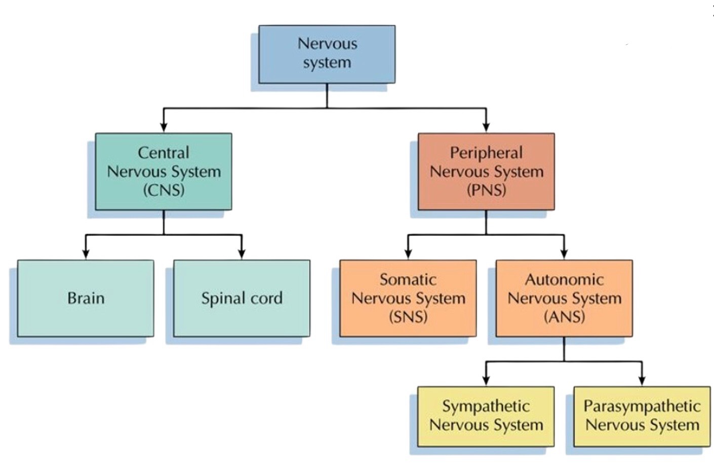
Central nervous system (CNS)
part of the nervous system make up of the brain and spinal chord
Carries sensory information up spinal cod to brain via sensory neurons
Carries motor messages to the PNS via motor neurons
Peripheral Nervous System (PNS)
made up of all the nerves outside of the brain and spinal cord
PNS carries sensory information to the CNS from the body
PNS carries motor messages from the brain to organs and muscles
Key function of the CNS
Input- to receive information
Processing- to integrate/interpret that information based on past experiences
Output- guide actions
Key function of the PNS
made up of out muscles, organs and glands
Consists of all the neurons outside of the CNS
Relay information to the CNS
Carry motor information from the CNS to the muscles and organs via the spinal cord
Parts of the Peripheral Nervous System
Somatic and Autonomic NS
Somatic nervous system
nerves that control voluntary movement through its control of skeletal muscles
Receives motor messages from CNS and transports to skeletal muscles in specific body regions
Autonomic nervous system (ANS)
contains nerves that are connected to the CNS and the involuntary muscles that control activity level of internal organs/glands
By relaying messages between the CNS and the internal systems, ANS controls the body’s internal activities that are essential to survival (heart rate, digestion etc.)
Function and division of the somatic NS
Sensory (function)
transmits sensory information from the body to the CNS via the spinal cord
Motor (function)
sends motor commands from the CNS to skeletal muscles, glands, or organ as for voluntary movement
Divisions of the ANS
Sympathetic and Parasympathetic
Sympathetic nervous system
regular;ages the glands and internal organ function
Physically prepare body during heightened arousal (flight or fight response)
Parasympathetic nervous system
calms the body after ring under control of the SNS
Controls the rest and digest response
Returns body to normal function (homeostasis)
Neurons
neurons are specialised cells located in the CNS and PNS
Cells that receive, transmit, process information
Billions of these cells, each with synaptic connections to more neurons
These connections allow for messages to be passed along throughout the
Diagram of a neuron
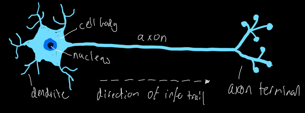
Features of a Neuron
Cell body (soma)
Dendrites
Axon
Axon terminals
Myelin sheath
Cell body (Soma)
contains a nucleus that controls the activities of the neuron
Contains nucleus, and processes information received by dendrites
Dendrites
extensions of cell body that receive neurotransmitters from pre-synaptic neurons and covert them into electrical chemical impulses that are conducted towards cell body
Branch like structures that receive signals from other neurons
Axon
long projection of neuron that conduct electrical nerve impulses and carries away from cell body
Long fibre that transmites electrical signals away from cell body
Axon terminals
enlarged end points of axon branches that store neurotransmitters and release them into the synaptic cleft
end structure that release neurotransmitters to communicate with other neurons
Myelin sheath
fatty covering axon that acts as insulator, protecting from stimuli that could interfere with transmission.
Also assist with speed of electrical impulses
Neurogenesis: The process of growing Neurons
Producing new neurons
the creation of new neurons from neural stem cells
Growing New Branches
development of new dendrite or axon terminal branches
Establishing connections
forming connections between existing neurons to create neural circuits
sensory neaurons
Transmit information from sensory receptors to CNS
Receives sensory information from the sense organs/environment and carry the sensory messages to the spinal cord and brain (CNS)
Motor Neurons
Carry through signals from CNS to muscles and glands
communicates information from the CNS to muscles, allowing movement.
Interneurons
connect and integrate information between other neurons within the CNS
act as the connection between sensory neurons and motor neurons. Activated when sensory neurons receive intense sensory information. Coordinates the reflex arc.
Sensory (differences)
carry nerve impulses from receptor to CNS
Have long dendrites and short axons
Motor (Differences)
Carry nerve impulses from CNS to an effector gland (muscle or gland)
Short dendrites and long axons
Interneurons (Differences)
Found completely in CNS
Provide link within CNS between sensory neurons and neurons
Short dendrites, long or short axons
How can neurons be categorised
Neurons can be categorised by number of processes (sending/receiving( extending from the cell body)
Unipolar, Bịpolar and multipolar neurons
Unipolar
neurons have one axon
Bipolar
neurons have axon and one dendrite, extending from cell body toward opposite poles
Multipolar
neurons have multiple dendrite and a single axon
Neural networks
Neurons form neural networks when axons of one neuron link up with dendrites of another neuron
efficiency of neural transmissions is influenced by
Number of neurons connected and
How often the pathway is used
Neural transmission
Neural transmission is an electro-chemical process
Nerve impulses
Neurons form neural networks when axons form one neuron link with dendrites from another neuron
Nerve impulse is message (electrochemical signal) that travels along fibre nerve
Transmitted very quickly, making possible for body towards respond to change
Described as electrochemical change because involved change in electrochemical signal, within neuron and neurotransmitters are chemical component of signal
Aka action potential, travels in one direction, from dendrites along axon
The electro-chemical signal (4 steps)
1- resting state
2- depolarisation
3- action potential
4- Depolarisation
Resting state
neuron maintains negative charge inside, relative to outside
Depolarisation
stimulus causes sodium channels to open, positive ions flow in
Action potential
If threshold is reaches, electrical signal travels down axon
Depolarisation
Potassium channels open, restoring negative internal charge
Synaptic gap
tiny space between neurons where chemical communication occurs
Neurotransmitters
chemical messengers released by presynaptic neuron to transmit signals
Receptors
Specialised proteins on postynaptic neuron that bind to specific neurotransmitters
What happens at the synapse? Step 1
Axon terminals connect with receptors on neighbouring dendrites
Synapse Step 2
When hit with electrical impulse (action potential), axon terminals of the sending neuron release neurotransmitters
Synapse step 3
Neurotransmitters travel across tiny gap called synapse and attach to receptor sites on the target dendrite of the recieving neuron
Synapse step 4
Attached neurotransmitters generate action potential in recieving neurons Short dendrites (neural impulse has been transmitted)
Synapse step 5
Most of neurotransmitters return to their original axon terminal a “re-uptake” process.
Synapse step 6
Other neurotransmitters are broken down by enzymes and need to be replenished
we replenish neurotransmitters through the food we eat, exercise and sleep
Neurotransmitters and their receptors are affected by drugs, toxins, and emotional states
What happens at synapse simplified
Release: neurotransmitters are released into synaptic gap
Binding: Neurotransmitters bind to receptors
Signal: Postsynaptic neuron is excited or inhibited (depending on the emotion etc)
Removal: excess neurotransmitters are removed
Role of neurotransmitter
Acts as chemical messengers.
Allow neurons to communicate by relaying information between them across synapse
Electrochemical signal
Electrical nerve impulses are ‘electro’ component Neurotransmitters are ‘chemical’ component |
Direction of transmission
One way.
Travels from dendrites down to axon.
Once reached the axon terminals, causes the release of neurotransmitters
role of synapses
Synapse allows neural transmission to occur by converting electrical nerve impulses from one neuron into chemical signal and then back again into electrical |
Acetylcholine: The memory Messenger
Key role
Acetylcholine crucial for memory and learning
Plays role in muscle movement
Deficiency impact
low levels relate to memory problems
Associated with diseases like alzheimers
Boosting levels
diet and certain medications raise acetylcholine
May improve cognitive function
Dopamine: Pleasure, Motivation & more
Reward system: centra to brain’s reward system, reinforces pleasurable behaviours (doesn’t always mean good)
Motivation driver: drives motivation, focus, and productivity. Optimal levels enhance performance
Imbalances: Too little or too much can cause disorders, includes Parkinson’s and schizophrenia.
Serotonin: Mood, Sleep and Digestion
Mood regulation: Serotonin stabilises mood, promotes feelings of well-being and happiness
Sleep cycle: regular;ages sleep patterns, healthy levels promote restful sleep
Digestive health: serotonin influences digestion, imbalances can impact gut function
GABA: The Calmer
GABA inhibits nerve transmission
Reduces anxiety and promotes relaxation
Glutamate” The exciter
Glutamate excites neurons
Vital for learning and memory
GABA & Glutamate Balancing act
proper brain function depends on balance between GABA and Glutamate
Adrenaline & Noradrenaline: Fight or Flight
Adrenaline: surges during stress, boosts energy and awareness
Noradrenaline: sharpens focus, prepares body for action
Survival mechanism: Activate fight-or-flight response, ensures quick reaction to danger
Neurotransmitters in disease: What happens when things go wrong?
Depression: serotonin and dopamine imbalances, causing depression
Parkinson’s: dopamine deficiency leads to motor impairments
Anxiety: GABA imbalances, increase anxiety
Schizophrenia: Excess dopamine triggers schizophrenia symptoms
The Brain
weighs between 1.3-1.5kg
Most water 78%, Fat 10%, Protein 8%
Made up of 100 billion neurons
Each neuron can make up to 100 connections (total 100 trillion connections)
Vital organ and command centre of body
Responsible for all mental processes
Regulates survival processes such as breathing, digestion sleep, blood circulation
Grey matter
brain outer converting
information is stored (Cerebral cortex)
White matter
brain needs neural pathways that connect the different parts of grey matter as well as other inner structures
Forebrain, Midbrain, Hindbrain
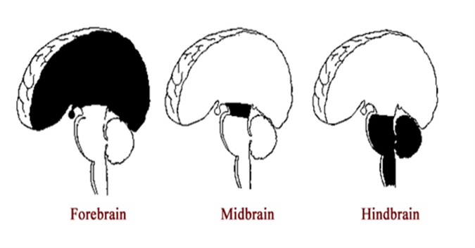
Hindbrain
located at rear base of skull that controls the most basic biological needs for life
Primitive part of the brain
Region of the developing vertebrate brain that is composed of the medulla oblongata, the pons and the cerebellum
Hindbrain- structures
Pons
Medulla oblongata
Cerebellum
Pons: above medulla
regularlates sleep, arousal and respiration
Relays information between cerebellum and cerebrum
Medulla oblongata: middle of the spinal cord
controls involuntary actions such as breathing, heart rate
Also controls reflexes; like swallowing and coughing
Cerebellum
aka little brain
Regulation and coordination of movement- allows us to make accurate and fluid movements
Also involved in learning- storage of lọng term procedural memories
Midbrain
connects hindbrain and forebrain and controls arousl levels, attention and consciousness
Role is to be brains sensory switchboard, involves receiving and processing sensory information (reticular formation), attention and consciousness
Receives messages from our senses, sends these to higher brain regions tha deal with these senses
Also responsible for auditory and visually processing and eye movement
Midbrain- Reticular formation
through centre of midbrain runs network of thick nerves called reticular formation
Job is to screen information so only relevant information gets passed to higher brain, Prevents overload
Reticular activating system (RAS), increases or decreases brain arousal, controlling our level of alertness
forebrain
most highly developed and largest part of the brain.
Affects how we think, feel and
Cerebral cortex consists of the cerebral hemispheres, which account for two this of the brain’s total mass
Part of lambic system (group of structures in your brain that regulated emotions, behaviour, motivation, and memory
Forebrain structures
Thalamus
Hypothalamus
Cerebral cortex
Thalamus
structure beneath cortex, known as body’s communication centre
Processes incoming sensory information and transmits to other parts of brain
Hypothalamus
small structure that controls basic survival actions
Sleep, regulation of body temp., hunger and thirst, and expression of emotions
Cerebral cortex
outer layer
Responsible for receiving information from environment, controlling responses and high order thinking involve in memory, language and emotions
Limbic system
forebrain consists of the cerebrum, thalamus and hypothalamus (part of limbic system)
2 main structures:
Amygdala: controls fear and aggression
Hippocampus: memory formation
CEREBRAL CORTEX
responsible for higher brain function, such as perception, conscious thought, voluntary movement
2 hemispheres (left and right), each with sensory and motor functions in the same place
Each hemisphere is further divided into four lobes
Left (Logic) and Right (Creative) Hemispheres
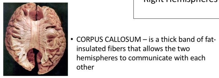
Connected by corpus callous’s
Each hemisphere is dominant in control o specific tasks (Hemispheric specialisation)
Hemispheric specialisation means each hemisphere has greater control over certain functions (but both are involved in almost all functions, just each is Dominant)
Hemispheres have contralateral control of body (ears left hemisphere controls right, right controls left)
Corpus callosum
It’s a set of neural fibres that connect two brain hemispheres
Each hemisphere has central fissure that runs from top of each hemisphere , and sides, separating front of cerebral cortex from rear.
Corpus callosum is part of the mind that allows communication between two hemispheres of the brain. its responsible for transmitting neural messages between both the right and left hemispheres
Lobes of the cerebral cortex
Frontal lobes
Temporal lobes
Occipital lobes
Parietal lobes
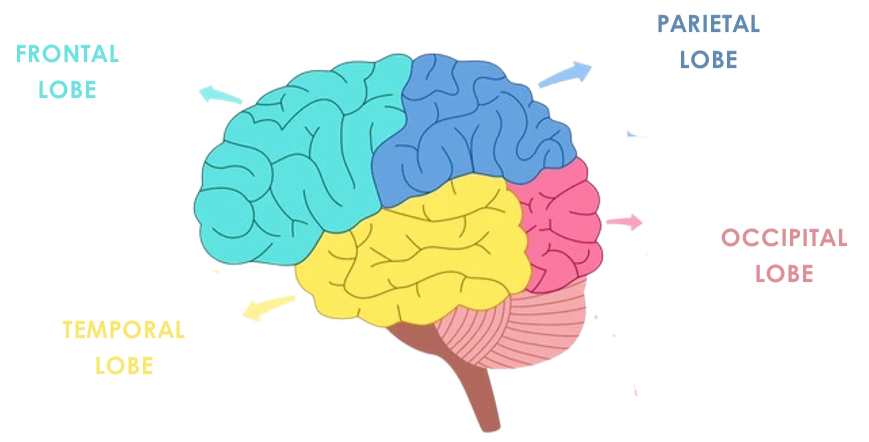
Frontal Lobes
voluntary movement
Planning and decision making
Problem solving
Organise information
Recognition of emotions
Speech production
Impulses control
Temporal lobes
understanding speech
Interprets auditory information
Processing sense of smell
Facial recognition
Partial responsibility for recognition of emotions
Involved in long-term memory formation
Occipital lobes
visual perception and processing
Interpreting visual information
Involved in facial recognition
Depth perception
Parietal Lobes
processing sensory information
Spatial awareness
Proprioception
Integrating sensory information
Localisation of functions
Localisation of function refers to the idea that certain functions have certain localisation or areas within the brain
Supported by brain imaging and case studies (phineas gage)
7 localised areas we focus on
Broca’s area
Wernickes area
Pre-frontal cortex
Primary motor cortex
Primary sensor cortex
Primary auditory cortex
Primary visual cortex
Broca’s area
Location: Adjacent to the primary motor cortex in the left frontal lobe
Function: Controls the fine muscles responsible for clear and articulate speech.
Impairment when damaged: hinders ability to produce articulate speech
Wernickie’s area
Location: Adjacent to the primary auditory cortex in the left temporal lobe
Function: Responsible for the understanding of language and the production of meaningful speech.
Impairment when damaged: hinders ability to understand language and make meaningful speech
Pre-frontal cortex
front layer os the frontal lobe that coordinates executive functions, such as predicting consequences of behaviour, recognise and regulate emotions
Primary motor cortex
strip running through the frontal lobes that control voluntary movement of the body.
Different zones Whithorn cortex correspond to various part of the body, with size of zone representing the importance of body part
Primary sensory cortex (somatosensory)
strip running through parietal lobed that register and processes sensory information
Primary auditory cortex
area within both temporal lobes that register and processes auditory information that is received from the ears
Primary visual cortex
area within the occipital lobes that register and process visual information that is received from the eyes
Pre-frontal cortex (More detail)
part of the brain behind forehead
One of last parts to mature
Area is responsible for skills like planning, prioritising,and making god decisions
Primary motor cortex (more detail)
upper motor neurons in cerebral cortex reside in several adjacent and highly interconnected areas in the frontal lobe
Which all together mediate planning and initiation if complex temporal sequences of voluntary movements
Primary sensory cortex (more detail)
central processing of tactile and nociceptive stimuli take place in primary somatosensory cortex
Fine-touch and proprioception inputs reach these cortical areas through dorsal column-medial lemniscus pathway, and information regarding pain, temperature, and touch reach this cortical areas through the lateral and ventral spinothalamic tracts
Simultaneously received information from other cortical regions and sub cortical areas that modify the output response from this region, similar to operation of primary motor cortex
Primary auditory cortex (more detail)
areas are regions of the cerebral cortex, located bilaterally in the temporal lobes
Primary auditory is positioned in the temporal lobe and associated with auditory areas
Primary auditory area acts as the principal location that receives sounds from peripheral auditory structures and is integral to being the process of complex sound interpretation as well as the conscious perception of noise
Primary visual cortex (more detail)
region of cerebral cortex
Responsible for processing visual data
Situated posterior in occipital lobe
Begins from eyes, travels through lateral geniculate nucleus until reaches visual cortex.