Bold Terms for Aural Surgery ALYSSA
1/76
There's no tags or description
Looks like no tags are added yet.
Name | Mastery | Learn | Test | Matching | Spaced |
|---|
No study sessions yet.
77 Terms
What are the 2 subdivisions of the ear for the purposes of surgery?
pinna and ear canal
The external ear canal is divided into 2 components, what are they?
vertical and horizontal
What is the vertical canal composed of?
auricular cartilage
The canal travels ________ to a point where it makes a ____ degree turn medially to become the horizontal canal
ventromedially, 90
When an animal has a disease of the pinna how will they typically present?
mass, swelling, laceration or bleeding (an obvious anomaly)
Discomfort is a far more common sign when animals have what kind of ear disease?
external ear canal or middle ear disease
What is otic discharge called?
ottorhea
What are some signs of animal pain/discomfort?
whining or biting when head is touched, or reluctance to eat
If an animal has disease extending out of the external ear canal/middle ear into the surrounding tissues what can this cause?
neurologic symptoms related to damaging adjacent nerves
What is an indication of severe external ear canal disease?
Facial nerve palsy (including the inability to blink the ipsilateral eye)
What is Horner's syndrome?
ipsilateral miosis, enophthalmos, drooping upper eyelid, and protruding nicitans

When does Horner's syndrome occur with regards to aural disease?
middle ear disease damaging the vagosympathetic trunk
What nerve is damaged if there is loss of hearing and vestibular disease?
vestibulocochlear nerve
What else can cause hearing loss?
severe external canal inflammation
If there are other cranial nerve deficits or altered mentation what does this mean?
the dog has intracranial extent of their disease
T or F: a generalized PE and an otic exam of the affected ear should be performed on any patient with known or suspected ear disease
false, BILATERAL exam of both ears
What do aural hematomas present as?
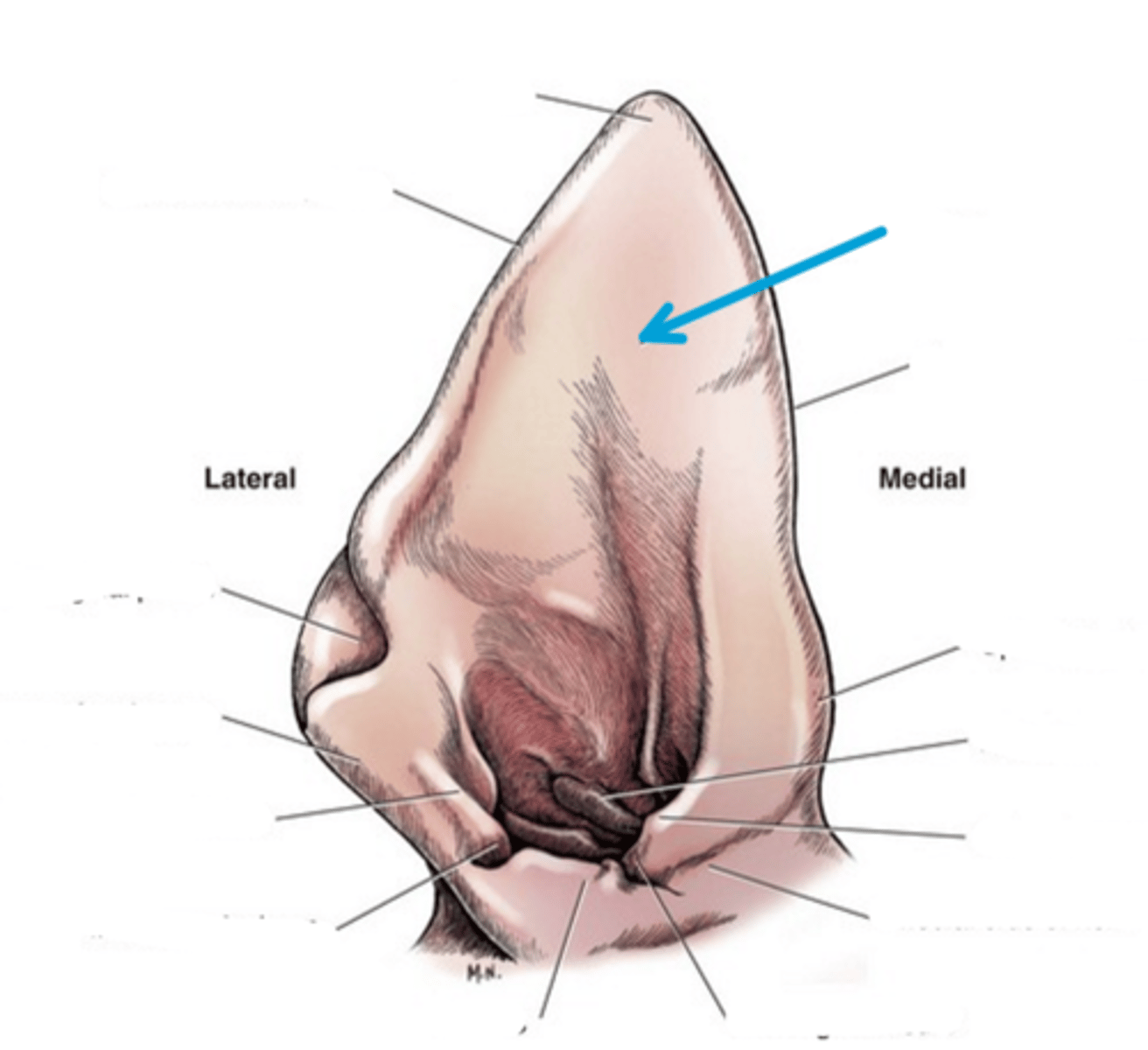
soft, fluctuant swellings on the concave aspect of the scapha (sometimes extending into the ear canal)

What will cytology of a hematoma reveal?
blood and possibly increased fibroblasts (if >3 days old)
What will happen if an aural hematoma is not addressed?
Blood will clot, and eventually replaced by fibrous tissue
If an aural hematoma has fibrosis what will happen then?
chronic disfigurement of ear, abnormal ear carriage, and possibly an exacerbation of underlying disease
What does treatment for an ear hematoma aim for?
removing the blood AND preventing recurrence
How do you prevent aural hematoma reoccurence?
investigation and treatment of underlying cause (often otitis externa)
Conservative treatment of a hematoma:
use a needle and syringe to completely drain the fluid pocket, and a compression wrap holding the ear to its head for at least 2 weeks
T or F: drainage of the hematoma is typically successful
False, drainage alone is unsuccessful as the pocket will rapidly refill
Due to a high rate of recurrence of hematoma with the conservative technique, what is usually combined with this technique to increase the success rate to 90-98%?
anti-inflammatory dose of corticosteroids
(either systemically or locally injected or both)
Surgical management of a hematoma involves what?
Making an incision to open and clean the hematoma pocket, followed by securing the skin and cartilage layers together
T or F: surgical techniques can be used for acute or chronic hematomas, but are the best techniques for chronic hematomas with a large blood clot
true
Where would I incise for the classic surgical approach for a hematoma?
large incision on the concave aspect and on the long axis of the ear, along the entire length of the hematoma
Why is the hematoma incision on the long axis of the ear?
avoid damaging the blood vessels that run from the base towards the apex
After removing the aural hematoma clot, the skin on the (concave/convex) aspect is sutured to the cartilage with simple interrupted suture bites
concave
T or F: it is optional whether you want to suture the skin on the convex aspect of the ear in an aural hematoma surgery
true
The simple interrupted bites should be __ cm long, oriented along the long axis of the ear, and placed in staggered rows so that each suture is __ cm from all other sutures
1
This technique involves making a smaller incision over the hematoma (concave or convex) to remove the clot, which is then sutured completely closed and a drain is placed into the hematoma cavity and exited through the skin of the ear
passive drainage technique
If a pinna has a full thickness wound what should you do?
sutured closed once they are healthy
3 multiple choice options
If the pinna trauma caused severe damage, what may be needed?
partial or total pinnectomy
If a pinna mass is alopecic, discolored, or a crusted lesion, what are some differentials?
- Parasites (demodex, scabies)
- infection (dermatophytes, Malassezia)
- fly strike (common in erect ears)
- immune-mediated or inflammatory disease (pemphigus, allergic dermatitis, vasculitis)
What might be a good idea to do with a alopecic, discolored, crusted mass on the pinna?
-skin scrape
-FNA
- maybe biopsy
What are some benign causes for a pinna mass?
-granuloma
-sebaceous adenoma
-histiocytoma
What are some malignant causes for a pinna mass?
-squamous cell carcinoma
-mast cell tumor
-other cancer types
If neoplasia is known or suspected what may be warranted?
chest radiographs and other staging tests (lymph node aspirates)
What are the margins for a carcinoma?
1 cm
What are the margins for a mast cell tumor?
2 cm
What are the margins for a sarcoma?
3 cm
What is the most common presentation for an ear canal mass?
head shaking, otic discharge, swelling near base of ear, pain on eating/opening mouth or neurologic signs (may be due to visualization of mass but uncommon)
What is ideal to do with an ear canal mass?
FNA or biopsy
T or F: in cats, bilateral aural tumors are common
true
If you find an ear canal mass what do you want to do during surgical removal?
get 1 cm margins
Depending on the ear canal mass location, what are some options for surgical removal?
- lateral wall resection
- vertical canal ablation
- total ear canal ablation
In cats, what is the most common type of polyp origination?
middle ear or auditory tube (nasopharyngeal)
Nasopharyngeal polyps are most common in ____________ cats and are thought to occur secondary to _____________________
young to middle aged
chronic upper respiratory infections
What may middle ear polyps cause?
-head tilt
-head shaking
-Horner's syndrome
If the nasopharyngeal polyp grows into the external ear what may happen?
otic discharge or a visible mass
If the middle ear polyp grows more into the nasopharynx what may happen?
coughing, inspiratory stridor, nasal discharge
T or F: middle ear polyps often grow into both the external ear and nasopharynx
true
What is the recurrence rate if the polyps in the nasopharynx are pulled out with hemostats?
10%
What is the recurrence rate if the polyps in the external ear are pulled out with hemostats?
50%
Recurrent polyps or those confined to the middle ear are best treated with?
ventral bulla osteotomy
What is the initial treatment of otitis media/externa?
sedated cleaning of the ear +/- cutting open the ear drum to lavage the middle ear (myringotomy)
Topical and/or systemic treatment with antibiotics, antifungals and anti-inflammatories for treatment of otitis media/externa should be based on what?
cytology +/- cultures of ears
What otitis media/externa cases are best treated surgically?
-failure of medical management
-severe disease in which medical management is not likely to be successful (stenosis, par-aural abscess)
-animals with neurologic disase
What is the most common surgical treatment for otitis media/externa?
total ear canal ablation
What is a partial pinnectomy?
when masses near the tip of the pinna are resected with a full thickness incision
When is a total pinnectomy necessary?
-large tumors near the middle of the ear
-tumors that involve underlying cartilage
Why do lateral wall resections suck (in Upchurch's opinion)?
leaves behind most of the external ear epithelium, which is grossly diseased in most animal with otitis externa
When might a lateral wall resection be useful?
benign mass of the lateral aspect of the vertical canal or those with no disease of the horizontal canal
Why does the vertical ear canal ablation also suck?
leaves behind the entire horizontal canal, which is grossly diseased in most animals with otitis externa
T or F: vertical ear canal ablation can be useful in many situations
false, Uppie hates them
When may a vertical ear canal ablation be useful?
rare animal with a tumor confined to the vertical canal or those with no disease of the horizontal canal
what is the treatment of choice for most cases of external ear canal neoplasia or otitis externa?
total ear canal ablation and lateral bulla osteotomy
What all must be removed in a TECA?
-entire external canal
-lining of the middle ear (secretory)
T or F: there is an external opening left open to let the middle ear drain after a TECA is complete
false
TECA's have higher likelihood or ______ or ______ damage and is best performed by a specialist
nerve or vascular
What are some post operative complications of a TECA?
facial nerve paralysis, Horner's syndrome, vestibular disease, deafness, incisional infection, or development of an abscess under the surgical site
What procedure gives the best exposure to the middle ear but does not approach the external ear? (only used for middle ear disease)
ventral bulla osteotomy
What is a ventral bulla osteotomy most commonly used for?
removal of nasopharyngeal polyps in the middle ear
Complications of Ventral bulla osteotomy?
facial nerve paralysis, Horner's syndrome, vestibular disease, incisional infection
What is the risk of doing a bilateral ventral bulla osteotomy?
respiratory compromise due to incisional swelling as the pharynx lies directly between both bulla