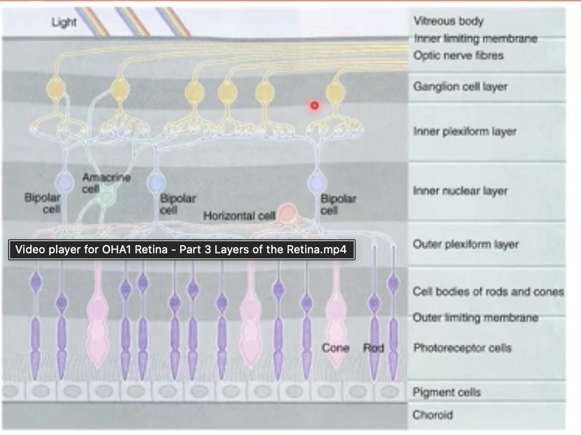
retina revision (layers of the retina)
1/16
There's no tags or description
Looks like no tags are added yet.
Name | Mastery | Learn | Test | Matching | Spaced | Call with Kai |
|---|
No analytics yet
Send a link to your students to track their progress
17 Terms
Retinal pigment epithelium
outermost layer of retina
lies between outer segments of photoreceptors and brutes membrane of choroid
RPE functions
absorption of scattered light by melanin granules
physical support of photoreceptors outer segments
transport of nutrients from choriocapillaris
breakdown of photoreceptor discs and recycling vitamin A
Photoreceptor layer
= sight of light absorption
remember its not the only layer with photoreceptors as they extend through 4 layers
outer and inner segments in photoreceptor layer
external limiting membrane - thin layer
nucleus lies in outer nuclear layer
axons and terminal endings in the outer plexiform layer
external limiting membrane
region of attachment between photoreceptors and muller cells
selective barrier for nutrients ( tight adherens junctions)
stabilise uppor portion of photoreceptors
muller cells
they are a type of glial cell
provides insulation between rods and cones
extend all the way through retina for structural support
largest cells in retina
outer nuclear layer
rod and cone nuclei in this layer
location of rod ad cone nuclei differ
rod- several rows about external limiting membrane
but cone nuclei close to ELM
thickest at the fovea
outer plexiform layer
contains axons and terminal endings of photoreceptors
contains dendrites of bipolar cells
terminals referred to as outer synaptic layer
axial portion referred to as Henles fibre layer
where communication occurs between photoreceptors and bipolar cells and modified by horizontal cells
Inner nuclear layer
information passed on from photoreceptors in outer plexiform layer
contains cell bodies of horizontal, bipolar, amacrine and interplexiform neurons- they communicate with eachother
muller cells for structural insulation
inner plexiform layer
laminated
On responses(signals about increases in light intensity) go to the inner part of the layer- bipolar cells terminating closer to ganglion cells
OFF responses ( signals about decrease in light intensity) go to outer part of inner plexiform layer- synapse is closer to bipolar cells
dendrites of ganglion cells receive input from bipolar and amacrine cells
ganglion cell layer
contains cell bodies of retinal ganglion cells and some amacrine cells
ganglion cell axons make up optic nerve but is located in next layer
dendrite of ganglion cells are in inner plexiform layer(receive input from Bipolar and amacrine cell)
nerve fibre layer
made up of axons of ganglion cells(nerve fibres)
nerve fibres run perpendicular to photoreceptors
thickest at nasal edge of disc
no more editing information by retina by this point
only transmission of information to brain for processing
inner limiting membrane
innermost layer of retina
outermost layer of vitreous(hyaloid membrane)
composed of :
collagen fibrils
HCL
basement membrane of muller cells
plasma membrane of muller cells
layers f the retina
what happens to layers at the fovea
all layers above photoreceptor layer are moved to the Sid or thinner to all all light to meet cones in photoreceptor layer
causes a dip shape
how are the photoreceptors arranged at the fovea
arranged in a cone mosaic
photoreceptors arrangment
cones are much larger than rods
at fovea - only cones in mosaic
moving further out rods and. cornea are equal in number
even further out- rods outnumber cones about 4:1
into the further periphery - rods become larger and reduce in number
blood vessels in retina
fewer further out into periphery then transitions to ora serrata