Lab 02: Anatomy of the Peripheral Nervous System
1/68
There's no tags or description
Looks like no tags are added yet.
Name | Mastery | Learn | Test | Matching | Spaced | Call with Kai |
|---|
No analytics yet
Send a link to your students to track their progress
69 Terms

1
Suprascapular Nerve (C6)
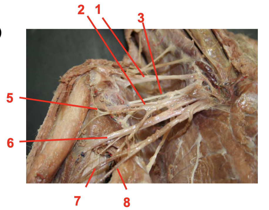
2
Axillary Nerve (C7)

3
1st Subscapular Nerve

4
2nd Subscapular Nerve

5
Musculocutaneous Nerve

6
Radial Nerve (C8)

7
Median Nerve

8
Ulnar Nerve

9
1st Thoracic Nerve

10
Ventral Thoracic Nerve

11
Long Thoracic Nerve

12
3rd Subscapular Nerve
13
Phrenic Nerve

14
Vagus Nerve (C10)

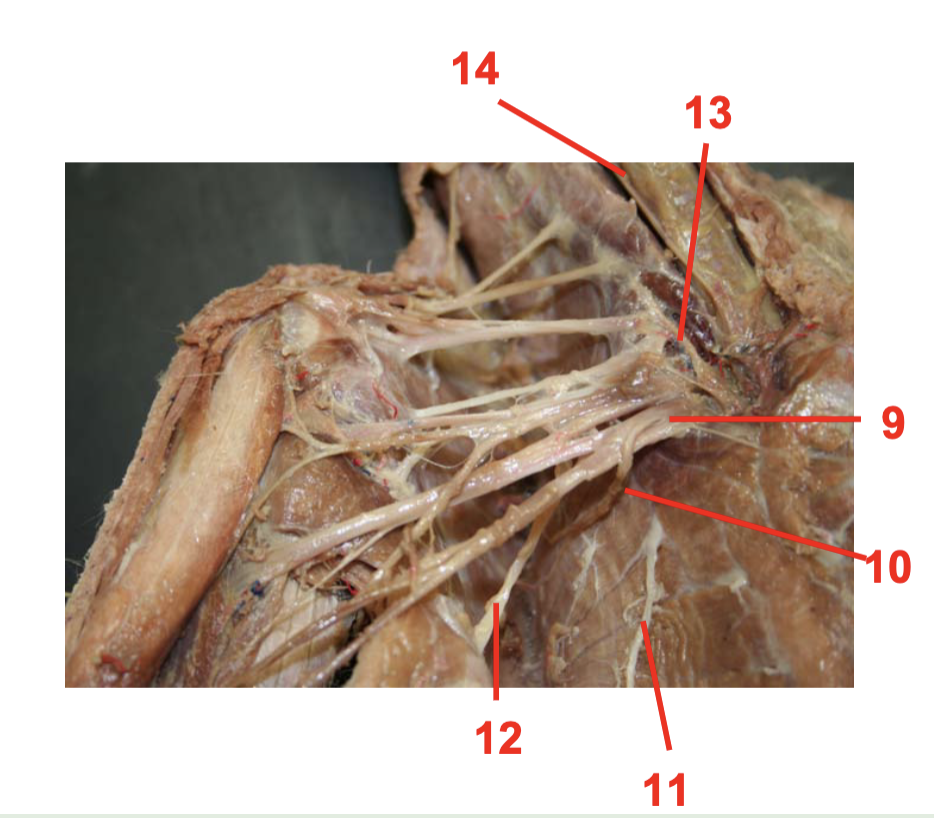
Suprascapular Nerve (C6)
1

Cervical Nerve 5
2

Cervical Nerve 4
3

1st Subscapuar Nerve
4

2nd Subscapular Nerve
5

Axillary Nerve (C7)
6

Musculocutaneous Nerve
7

Radial Nerve (C8)
8
Medial Nerve
9

Ulnar Nerve
10

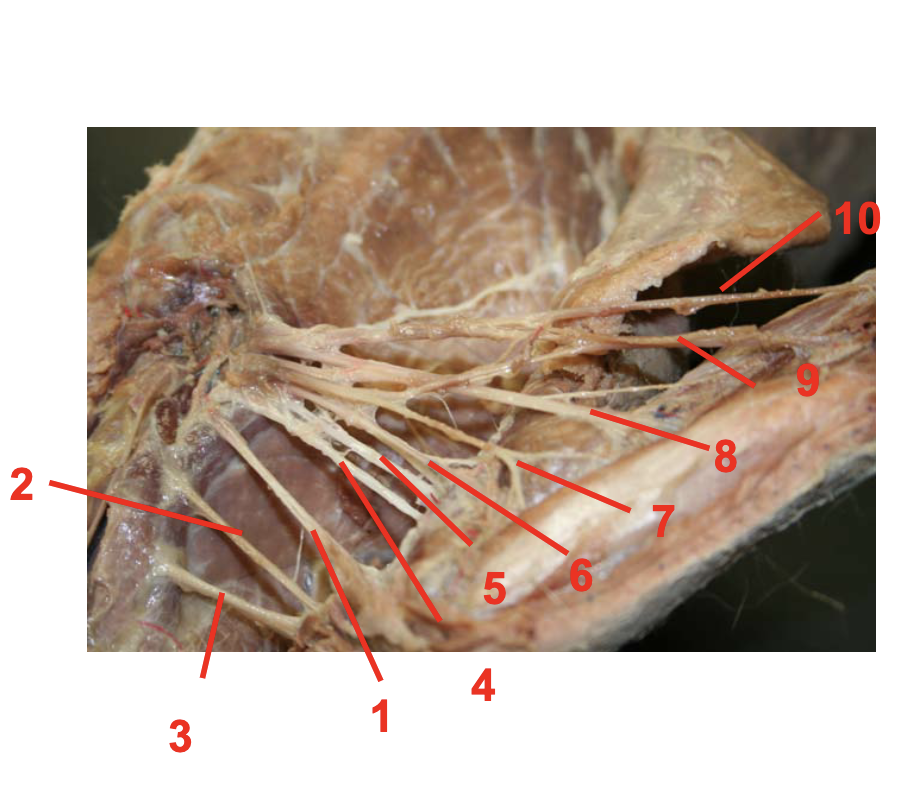
1
Cervical Nerve 1
2
Cervical Nerve 2

3
Cervical Nerve 3

4
Cervical Nerve 4

5
Cervical Nerve 5

6
Cervical Nerve 6 (Subscapular Nerve)

7
Vagus Nerve (10)

8
Spinal Accessory Nerve
Peripheral Nervous System (PNS)
consists of a sensory & motor division; components are the receptors, nerves, ganglia, & plexuses; responsible for detecting stimuli in & around the body, conducting action potentials to the Central Nervous System for interpretation
Sensory Division
transmits electrical signals from specialized receptor in the body towards the CNS (aka afferent division)
Motor Division
transmits electrical signals from the CNS to the effector organs (aka efferent division)
Somatic
Voluntary
Autonomic
Involuntary
Dendrites
recieve information from other neurons or from sensory receptors
Neuron cell body
contains nucleus & other organelles, processes information
Axon
central processes of neuron that conducts action potential away from the cell body
Schwann Cell
form myelin sheath
Nodes of Ranvier
gaps in myelin sheath that aid in signal conduction
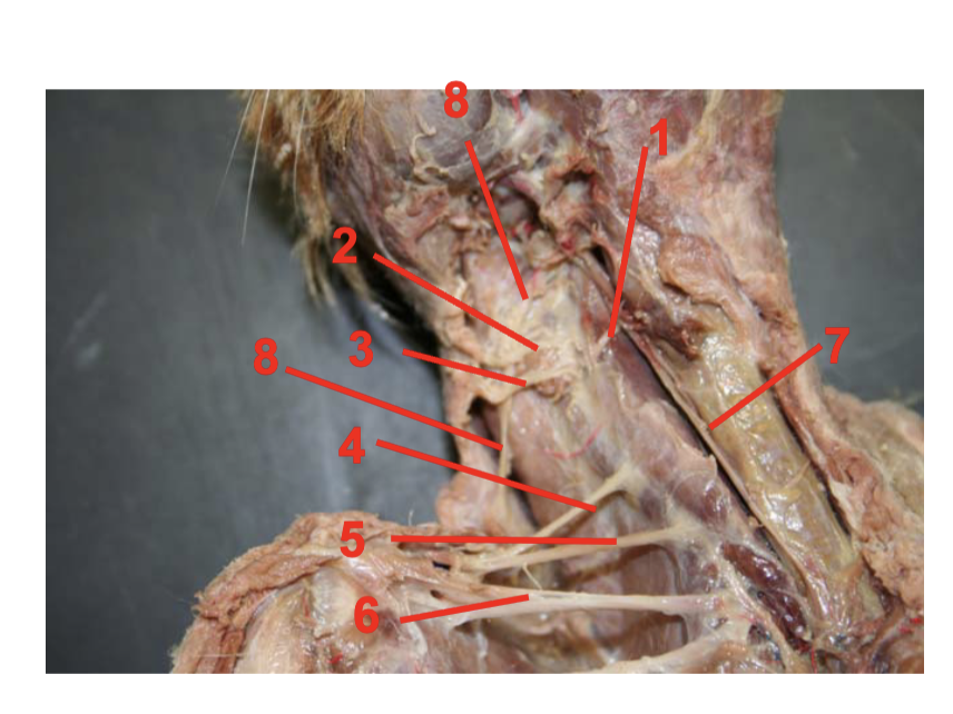
Spinal Accessory n. XI (motor)
sternocleidomastoid & trapezius muscles
Hypoglossal n. XII (motor)
muscles of the tongue
Vagus n. X (both motor & sensory)
both motor & sensory fibers to visceral body organs
Infraorbital n.
lower eyelid & upper lip; infraorbital canal
Phrenic n.
diaphragm muscles to control breathing; both nerves run from C3-C5 along the anterior scalene muscles before dividing into the thorax to pass between the lungs & heart
Suprascapuar n.
supraspinatus & infraspinatus muscles; suprascapular notch of the scapula
Subscapular n.
subscapularis, teres major, & latissimus dorsi muscles
Axillary n.
detoid & teres minor muscles
Musculocutaneous n.
coracobrachialis, biceps brachii, & brachialis muscles
Radial n.
triceps brachii & all muscles of the posterior forearm, sensation to lateral posterior surface of the hand; radial groove of the humerus bone
Median n.
sensation to the later anterior hand
Ulnar n.
sensation to the anterior & posterior medial hand; behind the medial epicondyle of the humerus
Long thoracic n.
Serratus anterior muscle
Sciatic n.
biceps femoris, semitendinosus & semimembranosus muscles; greater sciatic notch of the pelvis
Tibial n.
gastrocnemius, soleus & plantaris muscles
Common fibular nerve
anterior muscles of the leg (not the thigh)
Femoral n.
rectus femoris, vastus medialis, vastus lateralis, & vastus intermedius
Lateral cutaneous n.
sensation to the lateral thigh
Obturator n.
sensation to the medial thigh; obturator foramen of the hip
Saphenous n.
sensation to the medial leg & medial ankle/foot
Sympathetic trunk & ganglia
distributes fibers through the ramus communicans for sympathetic influence into spinal nerves
Vagus nerve pathology
clinical malfunctions may include dysphagia, vocal cord weakness & alterations of the parasympathetic tone of the thorax & abdomen
radial nerve pathology
when damaged, an individual cannot draw their wrist up & thus this condition is referred to as wrist drop
Median nerve pathology
carpal tunnel syndrome is generally associated with compression of this nerve
ulnar nerve pathology
superficial & thus often exposed contact stimulation & pain in the funny bone
long thoracic nerve pathology
when this nerve is damaged, the scapula wings out as it can no longer be held against the body wall
common fibular nerve pathology
when damaged, muscles in the posterior leg are no longer antagonized by contracting muscles from the anterior leg resulting in an unsynchronized foot drop with each step