Biopsychology
1/53
There's no tags or description
Looks like no tags are added yet.
Name | Mastery | Learn | Test | Matching | Spaced | Call with Kai |
|---|
No analytics yet
Send a link to your students to track their progress
54 Terms
structure of the nervous system
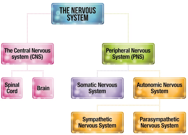
central nervous system
brain
spinal cord
coordinator of everything
receives information from the environment
processes, makes decisions and makes these actually happen
brain: decision maker
spinal cord: major connection to the peripheral. does reflex actions
peripheral nervous system
millions of neurons transmit messages to and from the CNS
then subdivided into the autonomic and the somatic
somatic nervous system
voluntary, conscious, deliberate actions
muscle coordination and info from sensory receptors
autonomic nervous system
involuntary, unconscious actions
e.g. breathing, HR, digestion
sympathetic and parasympathetic (antagonistic pair)
sympathetic
gut: slows digestion
salivary glands: inhibits production
heart: increases rate
eye: dilates pupil
lungs: dilates bronchi
parasympathetic
increases digestion
increases saliva
decreases HR
constricts pupil
constrict bronchi
flight or fight response
the hypothalamus identifies a threat and instructs the sympathetic system to act.
stress hormone adrenaline is released from the adrenal glands into the blood stream
adrenaline prompts a number of physical changes in the body to prepare for fight or flight
following the fight or flight response the parasympathetic nervous system is activated to return the body back to a normal resting rate e.g. slows HR, breathing rate, reduces blood pressure
evaluating the fight or flight response
useful in evolutionary terms. when we need energy to deal with the situation it prepares us to have the energy to run away or stay and fight.
→ not useful for stressers that do not require physical activity e.g. modern ones like debt. these bodily changes can be unhelpful and lead to illness.
research in this area of flight and fight is gender biased (alpha- androcentric). the research is only done on male Ps then generalised to females as well. females are more likely to show tend and befriend response. produces less adrenaline so gather together and support more.
the endocrine system (glands and their use)
pituitary
thyroid
adrenal
ovaries
works alongside the nervous system to control glands which release hormones into the bloodstream.
→ pituitary (base of brain under hypothalamus)
stimulated other endocrine glands e.g. controls growth, blood pressure, water levels
oxytocin (childbirth and breastfeeding), vasopressin (balances salt and water)
- thyroid (front of neck)
controls speed of metabolism,breathing, temperature, brain development
thyroxine
- adrenal (top of kidneys)
regulates metabolism, blood pressure, development of sexual characteristics
cortisol: controls body’s use of fats, proteins, carbs and the blood pressure
adrenaline: flight or fight- increases HR etc
- ovaries (either side of uterus)
produces and stores eggs
oestrogen- breasts, wide hips, thickens uterine lining
progesterone- thickens uterine linings, breasts to produce milk
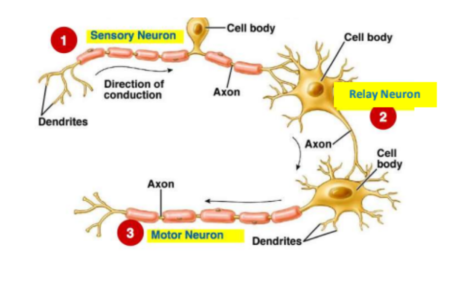
types of neurons
sensory: carries messages from PNS to CNS. long dendrites and short axons.
relay: connect sensory to motor (or other relay). short dendrites and short axons
motor: connect CNS to affects like muscles and glands. short dendrites and long axons
structure of a neuron
cell body with nucleus that contains genetic material
dendrites → axon → terminal buttons
myelin sheath protects and speeds up impulse. has nodes of ranvier to force it to ‘jump’
reflex arc
uses relay neurons in spinal cord to produce automatic response to environmental issues which need a quick response e.g. moving hand from heat
detected by sense organs in PNS which convey message along sensory neuron
message researches CNS → relay neuron → motor neuron
then carries messages to effectors such as a muscle
electrical transmission (done within neurons)
in a resting state the inside of the neuron is negatively charged compared to the outside
when activated by a stimulus, the inside becomes positively charged for a split second causing an action potential to occur creating an electrical impulse
this travels down the axon towards the end of the neuron (and triggers the release of neurotransmitters)
chemical transmission (done between neurons)
each neuron is separated from the next by a tiny gap called a synapse
signals between neurons are transmitted chemically
neurotransmitters are released from tiny sacs called synaptic vesicles.
they mostly diffuse across the synapse to the next neuron
(or are broken down by enzymes or reabsorbed into vesicles)
once a neurotransmitter crosses the gap it is taken up by the receptor sites and converted back into electrical impulses.
each neurotransmitter has its own specific molecular structure that fits to receptor site (lock and key)
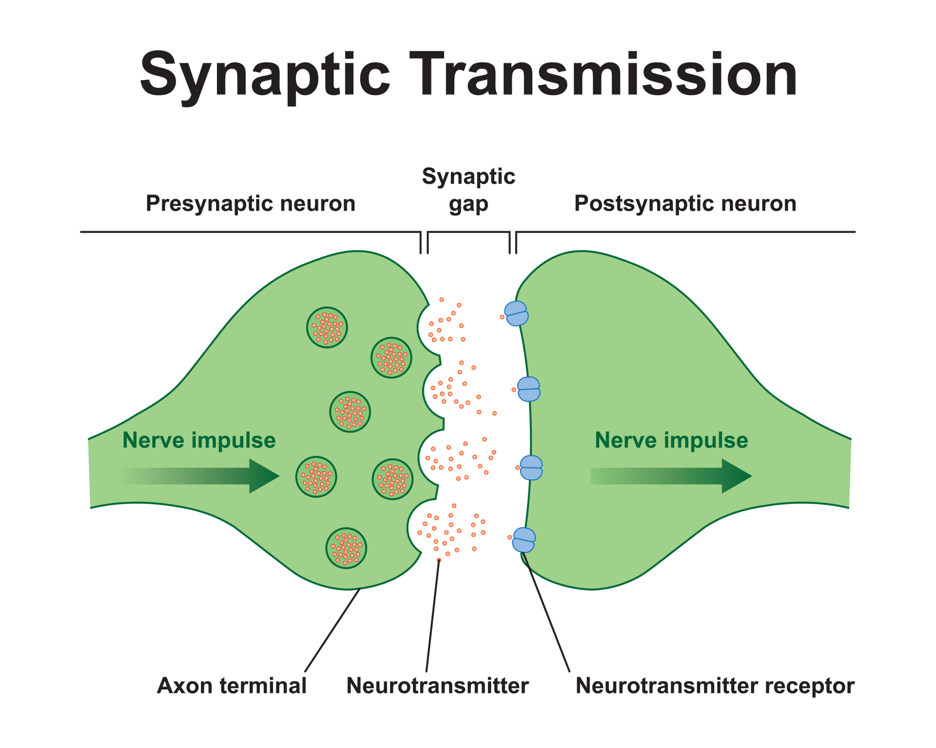
excitation
neurotransmitter increases the positive charge of the post synaptic neuron which increases the likelihood that the post synaptic neuron will pass on the electrical impulse e.g. adrenaline
inhibition
neurotransmitter increases the negative charge of the post synaptic neuron which decreases the likelihood that the post synaptic neuron will pass on the electrical impulse e.g. serotonin
summation
whether a postsynaptic neuron fires
sum of excitatory and inhibitory influences
net effect either excitatory or inhibitory
Localisation
specific areas of the brain are associated with particular physical and psychological functions (rather than holistic- the brain working as a whole)
Lateralisation
dominance of one hemisphere of the brain for particular physical and psychological functions e.g. language areas are only found on the left. (mostly 2 hemispheres are quite similar)
3 concentric layers
central core
Central core/ brain stem
regulated most primitive and involuntary behaviours e.g. breathing, sleeping, sneezing
includes structures like the hypothalamus in the mid brain
regulates eating, drinking and the endocrine system to maintain homeostasis (how the body keeps a constant physiological state)
3 concentric layers
limbic system
controls our emotions
interconnected with the hypothalamus
contains structures like hippocampus which is associated with memory
3 concentric layers
cerebrum
regulates our higher intellectual processes
outermost layer, cerebral cortex: appears grey due to location of cell bodies ‘ grey matter’
each sensory system sends messages to and from cerebral cortex
middle of left and right hemispheres connected by corpus callosum: can convey messages to both left and right
each hemisphere then further divided into four lobes

temporal lobe
auditory
location for auditory ability and memory acquisition
auditory centres
cochlea: sound waves converted to nerve impulses
auditory nerve
brain stem- decodes duration and intensity of sound
thalamus- further processing
auditory cortex- recognises and generates an appropriate response

Occipital lobe
visual
location for vision
light enters and strikes photoreceptors (rods and cones) in retina
nerve impulses to optic nerve
nerve impulses to thalamus
some to other areas to coordinate circadian rhythms
visual cortex- contains areas that process different types of visual information such as colour, shape and movement.
right receives input from left hand side of the visual field and vice versa

Parietal lobe
somatosensory
location for sensory information and coordination
somatosensory cortex: detects sensory events from different regions in the body. uses sensory information from the skin to produce sensations such as touch, pressure and pain.
cortex on one side of the brain receives information from opposite side of the body
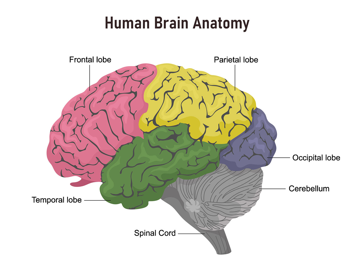
Frontal lobe
Motor
awareness of interactions with environment, consciousness and motor cortex
motor cortex: responsible for voluntary motor movements. in the right hemisphere the cortex controls muscles on left and vice versa. different parts control different parts of the body, and is arrange logically (part that controls foot near part that control leg)
damage to this area results in impaired movements
Language centres
Broca’s
Paul Broca, French Neurosurgeon
treated a patient who was only able to say ‘tan’ but did understand language
also studied 8 other patients who h\d similar language deficits along with lesions in their left frontal hemisphere (people with damage on right did not have the same issues)
identified language centres in back portion on left frontal lobe that is critical for speech production
near where the mouth, tongue and vocal cords are controlled
Neuroscientists found when people do cognitive tasks (unrelated to language) their Broca’s area is active
Federonka: 1 part involved in language and 2 responds to demanding cognitive tasks like maths problems
Language centres
Wernicke’s
German neurologist
back left temporal lobe
patient with legion’s on Wernicke’s area could speak but could not understand language
sensory area- responsible for auditory and visual input processing
neural loop runs between Wernicke’s and Broca’s
Evaluating localisation of the brain
evidence from neurosurgery- damage to areas of brain linked to mental disorders
neurosurgery treats mental disorders by targeting specific areas of the brain that may be involved.
DOUGHERTY: 44 people who had had cingulotomy (isolate cingulate gyrus involved in OCD) after 32 weeks: 30% had successful response, 14% partial response
behaviours in mental disorders are localised.
evidence from brain scans.
PETERSON: used brain scans to demonstrate how Wernicke’s area was active during listening task and Broca’s during a reading task.
BRACKNER + PETERSON: semantic and episodic memories reside in different parts of the prefrontal cortex.
confirms localised areas for everyday behaviours.
→ LASHLEY: removed areas of cortex (10%-50%) in rats learning route through maze. no area was proven to be more important than any other area in terms of rat’s ability to learn the route. more about the process of learning and needing more of the cortex. more holistic as emphasises involvement of the whole brain.
When the brain has been damaged and a function has been impacted the rest of the brain helps to recover it. Law of equipotentiality- surviving brain circuits chip in so the same function can be achieved e.g. stroke victim’s recovery- learning relies on the whole brain.
Language not just localised to Broca’s and Wernicke’s area
only 2% of researchers believe it is completely controlled by these areas.
advances in brain imaging e.g, fMRI means neural processes can be studied with more clarity.
language function distributed more holistically in brain e.g. language streams across cortex- brain regions in right hemisphere, thalamus.
holistic so contradicts localisation
lateralisation
and how stimuli is processed
the 2 hemispheres of the brain are functionally different and certain mental processes and behaviours are mainly controlled by 1 hemisphere rather than the other
→ most stimuli is processed contralaterally meaning if stimuli enters on the left it is processed in the right hemisphere and vice versa.
analyser and synthesiser for language
analyser: left is dominant in language e.g. broca’s and wernicke’s
synthesiser: right hemisphere can only produce basic words and phrases but provides emotional context.
commissurotomy
the corpus callosum is severed
often for people with severe epilepsy
the main communication line between each hemisphere is removed
so the information can not be conveyed from 1 hemisphere to the other. (cannot generate whole picture)
split brain research, sperry
image usually projected on Ps right visual field is processed by the left hemisphere and then vice versa.
for people with a commissurotomy the information cannot be conveyed between each hemisphere.
therefore sperry could see extent to which 2 hemisphere were specialised for certain functions and whether the hemispheres performed certain tasks individually.
→ investigated hemispheric lateralisation with 11 split brain patients
Sperry’s research methods
describe what you see
recognition by touch
describe what you see
picture presented to the left visual field (processed by right): patient COULD NOT describe what was shown or said nothing was present.
picture presented to the right visual field (processed by left); patient COULD describe what they saw.
recognition by touch
object placed in the left hand (processed by right): COULD NOT describe what they felt but could identify by selecting similar appropriate object from a group of objects.
object placed in right hand (processed by left): COULD describe what they felt and could also identify object by selecting similar appropriate objects from a group of objects.
evaluating sperry’s research
even in connected brains the 2 hemispheres process information differently
FINK: used PET scans to identify which brain areas were active in visual processing task
Ps were asked to look at big elements of an image (e.g. whole forest) and right hemisphere was much more active.
when focusing on finer details (e.g. individual trees), specific areas of the left hemisphere were more dominant
→ for visual processing, there is lateralisation
scientific methodology due to high control: presented visual stimulus for short amount of time to ensure only one hemisphere was receiving at a time high internal validity.
lacking generalisability as Ps were compared to neurological control group where none had epilepsy. this is a major confounding variable as differences/ cognitive abilities may be due to epilepsy not split brain
there is an exaggerated difference between hemispheres, when a situation requires it e.g. damage from illness or trauma, function can be effectively performed by another hemisphere (plasticity)
What is functional recovery?
SR
3 ways
following physical injury or trauma areas of the brain are often able to adapt and compensate for areas that are damaged through neural plasticity
spontaneous recovery: can happen quickly
brain is able to rewire and organise itself by forming new synaptic connections e.g. unmasking secondary neural pathways
axonal sprouting: growth of new nerve endings which connect with other undamaged nerve cells to form new neural pathways.
denervation supersensitivity: axons are aroused to do function to a higher level
recruitment of homologous areas (in the opposite side of the brain): specific tasks can still be performed e.g. if Broca’s area was damaged on the left, the right sided equivalent could carry out functions.
What is brain plasticity?
SP
brain’s tendency to change an adapt through life due to experience and learning
synaptic pruning: at age 3 we have around 15,000 synaptic connections but this has halved by adulthood as rarely used connections are deleted.
Research for brain plasticity
maguire (taxi)
draganski (medicine)
Maguire: followed a group of 79 trainee taxi drivers and 31 controls using MRIs and their performance on memory tasks over time.
volume of grey matter in hippocampus significantly higher for those who had passed and also were better at recalling london landmarks.
supports plasticity as taxi drivers learnt knowledge which altered their brain structure.
Draganski: imaged brains of medical students 3 months before and after their final exams. changes in posterior hippocampus and parietal cortex were induced by learning.
Evaluating plasticity and functional recovery of the brain after trauma
meditation and neuroplasticity:
MRI scans showed experienced meditators had thicker cortex than non meditators particularly in areas associated with attention and sensory processing.
meditators also had a higher volume of grey mater in brain areas associated with emotional regulation and response control.
compared to control group, Ps in 8 week mindfulness based stress reduction programme had increased grey matter in hippocampus.
→ people may be drawn to meditations as brains were different rather than the difference being a result of meditation.
human echolocation is a learned ability used by some blind people to navigate their environment and sense surroundings through echoes. (click with tongue and hear the impact on environment)
studies using fMRI have shown that parts of the brain associated with visual processing are adapted for this new skill of echolocation e.g. echoes processed by brain regions usually devoted to vision.
→ human evidence is restricted to small scale studies which have individual differences.
research support from animal studies.
TAJIRI: provided evidence for the role of stem cells in recovery from brain injury. randomly assigned rats with traumatic brain injury to 1 of 2 groups: one received transplants of stem cells intro geion affected by traumatic brain injury, control group had a saline solution infused into the brain. brains of stem cell rats had development of neuron like cells in area of injury showing research led to new treatments to aid recovery.
→ animal studies limited as have ethical concerns and are hard to generalise.
BANERJEE: 5 stroke victims showed complete recovery using stem cell treatment compared to typical 4% recovery. → only 5Ps.
level of education SCHNEIDER
more time people with a brain injury spent in education, the greater their chances of recovery without disabilities.
college education, 7x more likely to recover from severe brain injury than people who didn’t finish high school.
concluded cognitive reserve from greater educational attainment could be factor in neural adaptation to recover from brain injury.
→ have high level of education due to being richer so can afford better level of education.
fMRI
evaluation
functional magnetic resonance imaging
detects the change in blood oxygenation and flow which happens due to neural activity in different parts of the brain.
when a brain area is more active it consumes more oxygen so therefore has more blood flow (haemodynamic response).
produces 3D images showing which parts of the brain are involved in particular mental processes.
does not rely on use of radiation so is virtually risk free as non invasive.
high spatial resolution creating clear picture on how the brain is localised.
expensive
poor temporal resolution as 5s time lag between image on screen and initial firing of neuronal activity so does not represent moment to moment activity.
postmortem examinations
evaluation
analysis of a person’s brain following their death
they are likely to have had rare disorder or unusual deficits in cognitive processes or behaviour in lifetime.
understand cause of affliction by comparing with a neurotypical brain to ascertain extent of difference.
vital in providing foundation for early understanding of key processes in brain e,g, Broca, Wernicke, HM
causation- observed damage may not be linked to deficits but unrelated trauma or decay.
raises ethical issues of consent as cannot provide informed consent.
EEG
electroencephalogram
measures electrical activity within the brain via electrodes that are fixed to an individual’s scalp using a skull cap.
it records brainwave patterns that are generated from the action of thousands of neurons creating overall account of brain activity.
useful in diagnosing as can see unusual patterns of activity which may indicate abnormalities e.g epilepsy, tumours
high temporal resolution so detects brain activity within a millisecond.
generalises information so not useful pinpointing exact source of neural activity and therefore exact location of it.
event related potentials (ERP)
uses statistical averaging technique from the EEG to filter only the responses to specific stimuli or performance of a task.
therefore sees brain waves triggered by particular events.
more specificity than the EEG. high temporal resolution e.g. can measure cognitive functions and deficits like working memory.
lack of standardisation between different research studies making it difficult to confirm finding.
need to completely eliminate background noise and extraneous material which is hard to achieve.
biological rhythms: circadian
sleep wake cycle
rhythms that last around 24 hours e.g. core body temperature, sleep wake cycle
sleep wake cycle
daylight (exogenous zeitgeber) effects sleep/wake cycle e.g. drowsy at night, awake in day
suprachiasmatic nucleus (SCN) endogenous pacemaker- governs cycle. located above the optic chiasm and provides information about light.
research on circadian cycle
siffre’s cave study
wever bunker
follkard: clock
Siffre: spent several extended periods underground to study effects on his own biological rhythms. deprived of exposure to natural light.
developed free running biological rhythm of ~ 25 hours.
Wever: group of Ps spent 4 weeks in a bunker deprived of natural light and almost all had rhythm of 24-25 hours.
natural sleep/wake cycle slightly longer than 24 hours but entrained by exogenous zeitgebers in our 24 hour day such as daylight hours.
→ exogenous zeitgebers cannot override our circadian rhythm, they have a limited influence.
Folkard: studied group of 12 people who agreed to live in a dark cave for 3 weeks.
clock 11:45 = sleep, 7:45= wake
sped up clock so 24 hour day pushed into 22 hours and only 1 comfortably adjusted.
evaluating circadian rhythms
shift work: provides understanding of consequences when circadian rhythms are disrupted (desynchronisation)
night workers have reduced concentration at 6am meaning mistakes and accidents are more likely.
relationship between shift work and poor health.
workers 3x more likely to develop heart disease than those working typical work patterns.
→ real world economic implications to best manage worker productivity.
been used to improve medical treatments
circadian rhythms coordinate basic processes such as heart rate, digestion, hormone levels.
led to chronotherapeutics: medical treatments administered in way that corresponds with biological rhythms.
e.g. treatment for heart attack most helpful when taken last thing at night.
→ led to increase in effectiveness of drug treatments.
research limited as generalisation difficult to make due to such small samples of participants
sleep/wake cycles vary to each person
Duffy: people have natural preference being larks or owls.
confounding variables: e.g. artificial light
Biological rhythms: Infradian
menstrual
take longer than 24 hours to complete.
menstrual cycle: governed by monthly changes in hormone levels which regulate ovulation (28 days).
oestrogen: ovary develops egg and releases it (ovulation)
progesterone: womb lining gets thicker. when there is no pregnancy it leaves body (period)
although endogenous system, evidence suggests it is influenced by exogenous factors such as the cycles of other women.
STERN +McCLINTOCK: studied 29 women with a history of irregular periods. pheromones collected from 9 women at different stages. these pads were then treated with alcohol and then frozen and rubbed on upper lip of other Ps.
68% women experienced changes to cycle bringing them closer to cycle of odour donor.
Infradian; seasonal affective disorder
SAD
depressive disorder with seasonal patterns of onset: persistent low mood, lack of activity and interest in life winter blues
hormone melatonin: pineal gland secretes it at night until dawn when there is an increase in light. in winter this secretion process continues for longer which impacts serotonin levels.
→ linked to circadian rhythms as disruption of sleep/wake cycle due to more darkness.
evaluating infradian
menstrual synchrony research explained by natural selection
synchronisation has evolutionary values because it would be advantageous to menstruate together and become pregnant at the same time.
allows babies who have lost their mothers in childbirth to still have access to breast milk.
most effective treatment for SAD is light therapy which uses a box with very strong light to reset body’s internal clock.
reduces effects of SAD in about 80% of people.
→ led to headaches and eyestrain, CBT reduces relapse rate much more.
research has methodological limitations: many factors may affect changes to the menstrual cycle e.g. stress, diet, exercise
may act as confounding variable so pattern of synchronisation could have been chance.
other studies have not replicated these findings.
small samples.
Biological rhythms: ultradian
sleep cycle
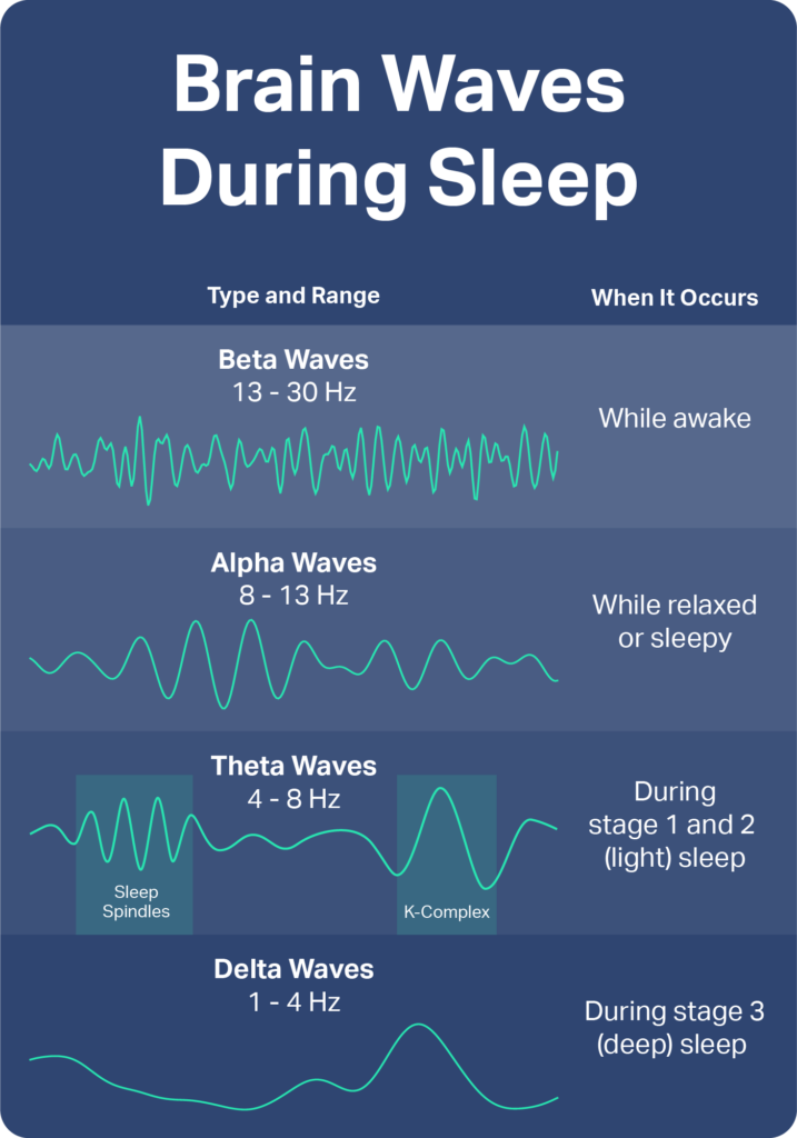
less than 24 hours to complete e.g stages of sleep. each characterised by different brainwave activity. (90 mins)
light sleep: high frequency and short amplitude. (alpha waves)
alpha waves but occasional random changes (sleep spindles)
+ 4. deep sleep: delta waves with low frequency and high amplitude.
REM: body paralysed but brain activity closely resembles awake brain.
rapid eye movement
most likely time of dreams
theta waves
evaluating ultradian rhythms:
improved understanding of age-related changes in sleep
slow wave sleep (deep sleep) reduces with age.
deep sleep is when the growth hormone is produced so this sleep deficit can lead to practical issues such as reduced alertness.
in order to increase deep sleep, use relaxation and meditation → practical value.
conducting studies in a lab means there is a control of extraneous variables so a researcher can exclude variables like noise and temperature.
→ being hooked up to complicated machines does not represent ordinary sleep habits.
research limited because there is significant differences between Ps in terms of duration of each sleep stage especially 3+4. TULVER believes these differences are biologically determined.
endogenous pacemakers
SCN
pineal gland
+ 1 ao3 point
suprachiasmatic nucleus: bundle of nerve cells located in the hypothalamus in each hemisphere of the brain.
lies just above the optic chiasm which are linked to eyes so receive info about light
passes information on the light in receives to pineal gland.
pineal gland: produces melatonin during night which induces sleep and so is inhibited during the day.
darkness increases → less light penetrates eyelids → optic nerve sends signal to SCN → sends signal to pineal gland → releases melatonin → sleep
endogenous pacemakers hard to study in isolation e.g. Siffre’s cave study: not just natural sleep/wake cycle as artificial light would have had impact. (lowers validity and makes little sense attempting to separate these)
animal studies on SCN
Decoursey: destroyed SCN connections in the brains of 30 chipmunks who were then returned to natural habitat and observed for 80 days.
sleep wake cycle disappeared → supports role of SCN
significant proportion had been killed by predators as vulnerable (ethical issues)
Ralph: bred mutant hamsters with 20 hour sleep/wake cycle. then transplanted cells from SCN into brains of normal hamsters who then defaulted to 20 hours.
exogenous zeitgebers
light
social cues
+ao3
reset biological clocks through entrainment
brought into like by environmental cues e.g. light, social cues.
LIGHT: Campbell and Murray: demonstrated that light will be detected by skin receptors on body, not just necessarily by the eyes.
15Ps woken at random times and light shone on backs of knee
could alter their sleep/wake cycle by up to 3 hours
shows light is a powerful exogenous zeitgebers.
lack control as replication found no difference and confounding variables e.g. light reaching eyes
SOCIAL CUES:
babies learn to adjust cycles by schedules decided by parents
research on jet lag shows entraining and adapting to cues of local times helps to entrain circadian rhythms.
exogenous zeitgebers do not have the same effect in all environments. experience of people (e.g. in artic circle) who live in places with very little light in winter (the opposite in summer) have similar sleep patterns all year round despite this. suggests cycle primarily controlled by endogenous pacemakers that can override changes in light.