Breast Cancer: Nottingham Grading System and Tumour Differentiation
1/15
There's no tags or description
Looks like no tags are added yet.
Name | Mastery | Learn | Test | Matching | Spaced | Call with Kai |
|---|
No analytics yet
Send a link to your students to track their progress
16 Terms
What is the fundamental difference between tumour Grade and Stage?
Grade refers to how abnormal the cells look (degree of differentiation), while Stage refers to how far the cancer has spread (tumour size, lymph nodes, metastasis).

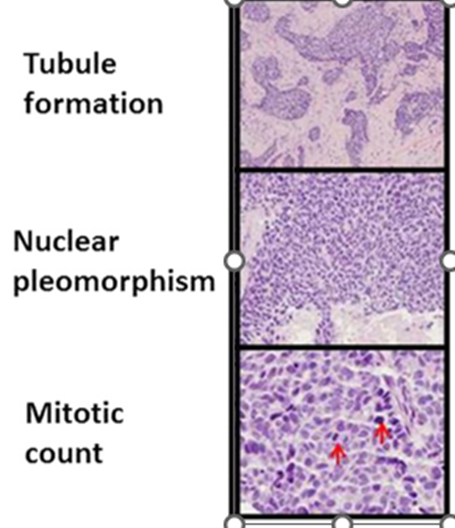
What are the three criteria scored in the Nottingham grading system?
1. Tubule formation 2. Nuclear pleomorphism 3. Mitotic count
How is tubule formation scored in the Nottingham grading system?
Score 1: >75% tubules; Score 2: 10-75% tubules; Score 3: <10% tubules.
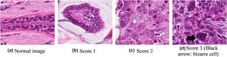
How is nuclear pleomorphism scored in the Nottingham grading system?
Score 1 (mild pleomorphism)
Nuclei are small, uniform, and regular, with minimal variation in size and shape.Score 2 (moderate pleomorphism)
Nuclei show moderate variation in size and shape, with visible irregularity.Score 3 (marked pleomorphism)
Nuclei are large, irregular, and highly variable, often hyperchromatic, with prominent nucleoli.
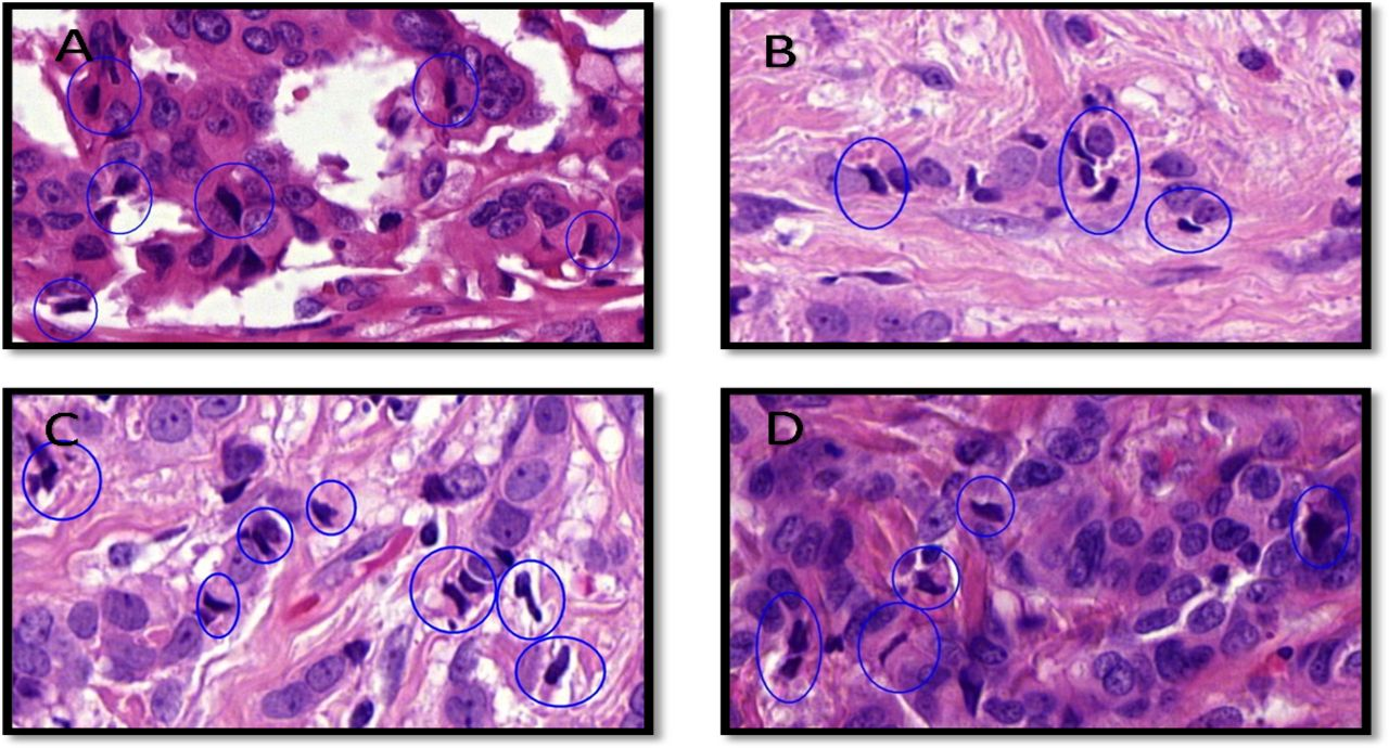
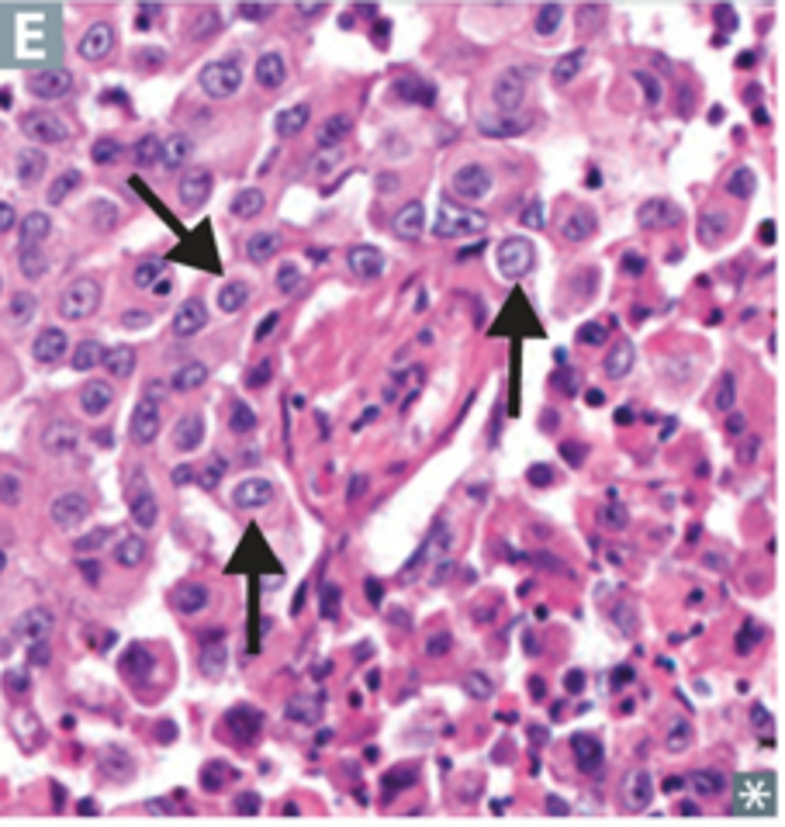
How is mitotic count assessed in the Nottingham grading system?
For mitotic count per 10 high-power fields (HPFs) (as used in systems like the Nottingham histologic grade), the usual range and scoring is:
Score 1 (low): 0–5 mitoses per 10 HPFs
→ Few mitotic figuresScore 2 (intermediate): 6–10 mitoses per 10 HPFs
Score 3 (high): ≥11 mitoses per 10 HPFs
How do Nottingham scores translate to grade?
3-5 → Grade I (well differentiated)
6-7 → Grade II (moderately differentiated)
8-9 → Grade III (poorly differentiated).
What does 'poor differentiation' mean histologically?
Loss of normal tissue architecture, pleomorphic nuclei, high mitotic activity, and little or no tubule formation.

What is the nuclear-to-cytoplasmic (N:C) ratio and why does it matter?
Malignant cells have a high N:C ratio, indicating large dark nuclei and minimal cytoplasm, which is a sign of aggressive behaviour.
Why is histology preferred over cytology for tumour grading?
Histology preserves tissue architecture, which is essential for assessing tubule formation and invasion.

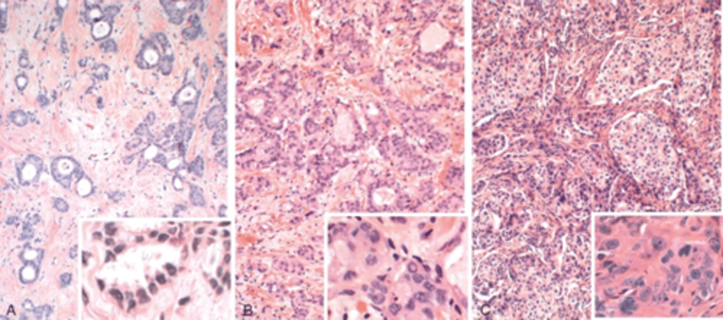
What does a well-differentiated (Grade I) breast tumour look like?
This is a well-differentiated (Nottingham Grade 1) breast carcinoma with a combined score of 3 (tubule formation = 1, nuclear pleomorphism = 1, mitotic count = 1). It shows abundant well-formed glands, uniform nuclei with minimal pleomorphism, and low mitotic activity, closely resembling normal breast tissue.
What does a poorly differentiated (Grade III) breast tumour look like?
This is a poorly differentiated (Nottingham Grade 3) breast carcinoma with a combined score of 9 (tubule formation = 3, nuclear pleomorphism = 3, mitotic count = 3)

What is an anaplastic tumour?
A very high-grade tumour with complete loss of differentiation, making the tissue of origin difficult to identify.
What is the basement membrane, and why is it critical?
It is the boundary between epithelium and stroma; crossing it marks the transition from in situ disease to invasive carcinoma.
Why does tumour grade predict prognosis?
Higher-grade tumours grow faster, invade earlier, and have a greater risk of metastasis.
How does Nottingham grade influence treatment decisions?
High-grade tumours are more likely to receive chemotherapy, despite having a worse overall prognosis.
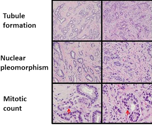
What does 'moderate differentiation' (Grade II) look like?
This is a moderately differentiated (Nottingham Grade 2) breast carcinoma with a combined score of 6 (tubule formation = 2, nuclear pleomorphism = 2, mitotic count = 2).