Lower Limb Bones and Features
1/31
There's no tags or description
Looks like no tags are added yet.
Name | Mastery | Learn | Test | Matching | Spaced | Call with Kai |
|---|
No analytics yet
Send a link to your students to track their progress
32 Terms
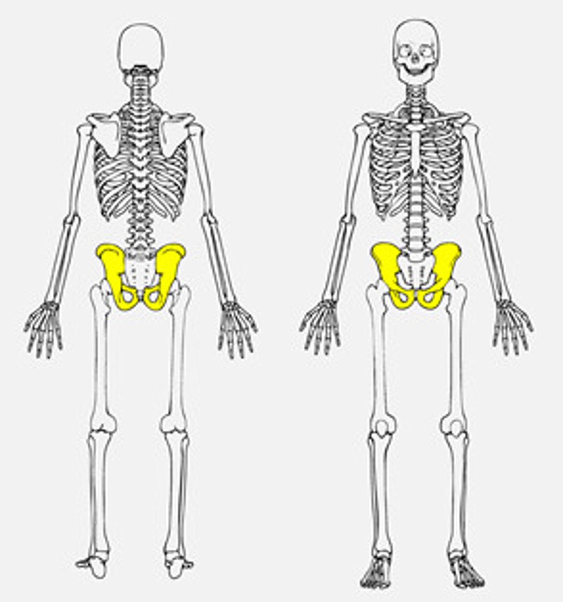
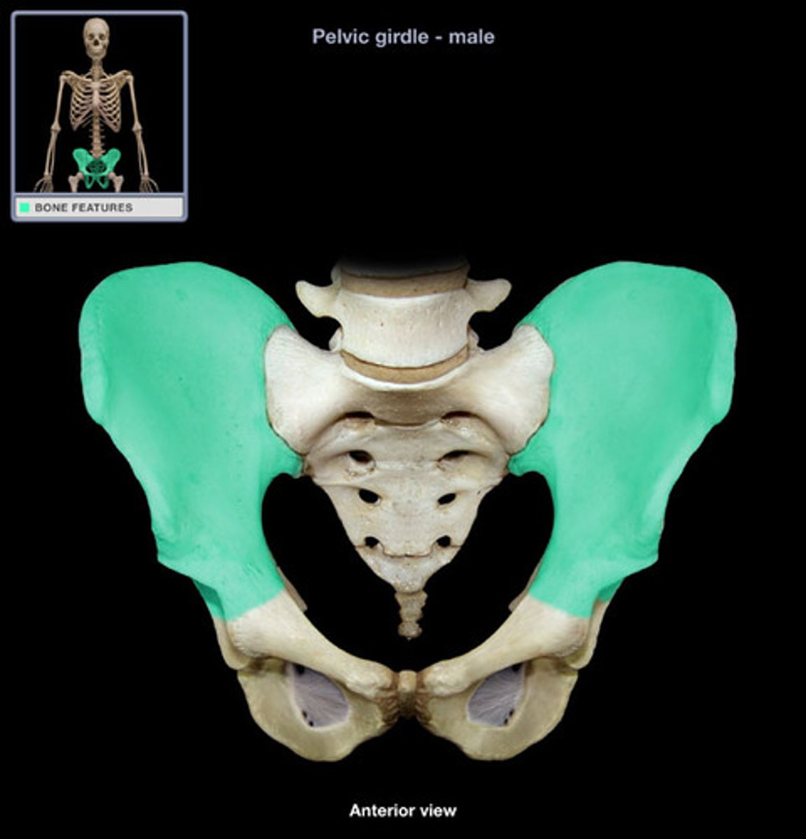
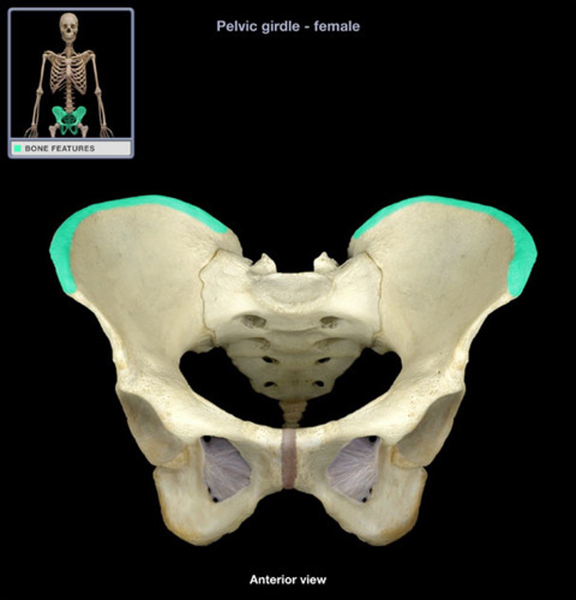
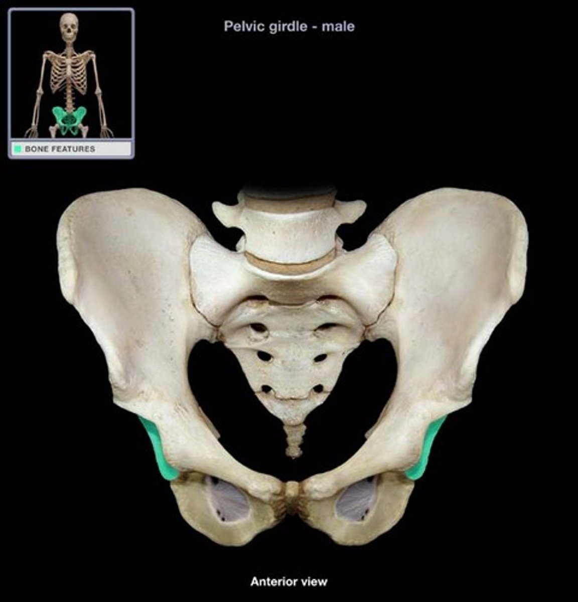
pelvic girdle
enclosing structure formed by the pelvis, providing attachment for the hind limbs
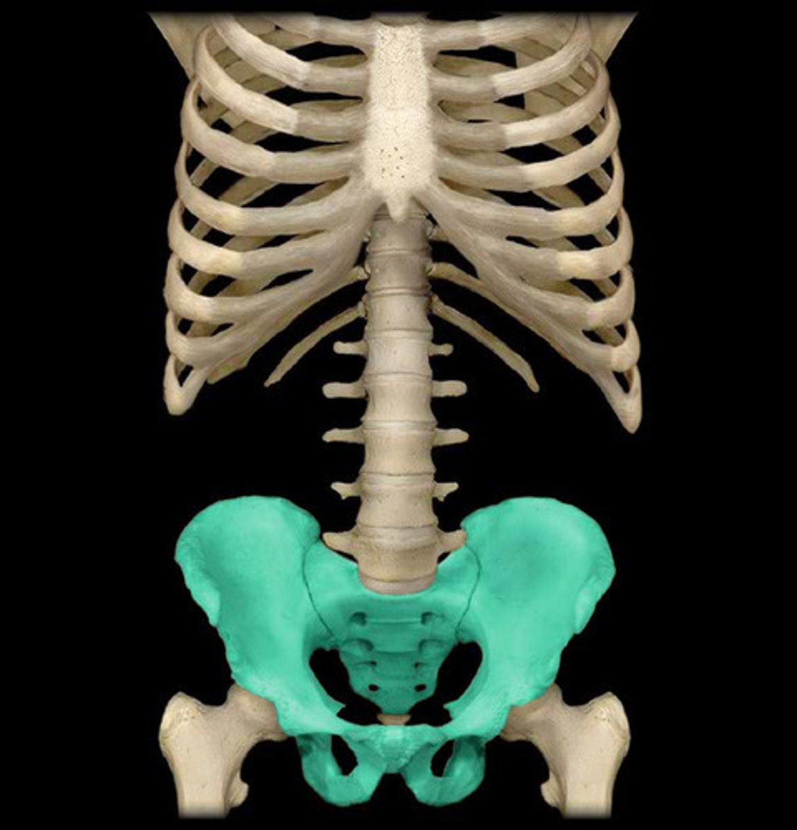
pelvis
large bony structure near the base of the spine to which the hind limbs or legs are attached
coxal bones / os coxae
a large flat bone, consists of ilium, ischium, and pubis
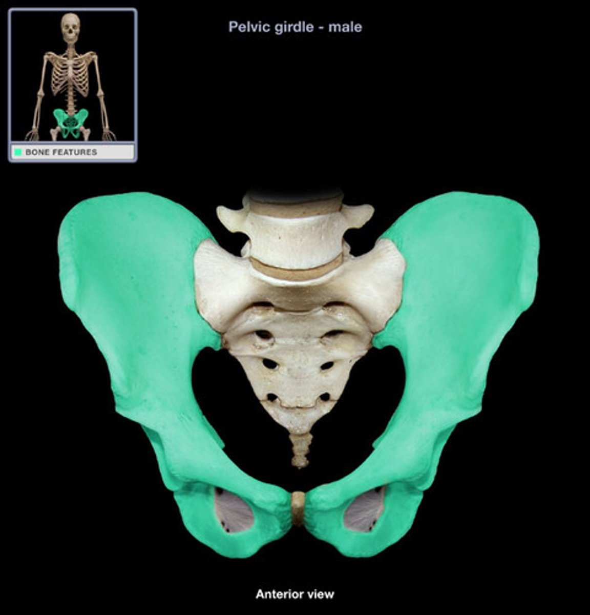
ilium
large broad bone forming the upper part of each half of the pelvis
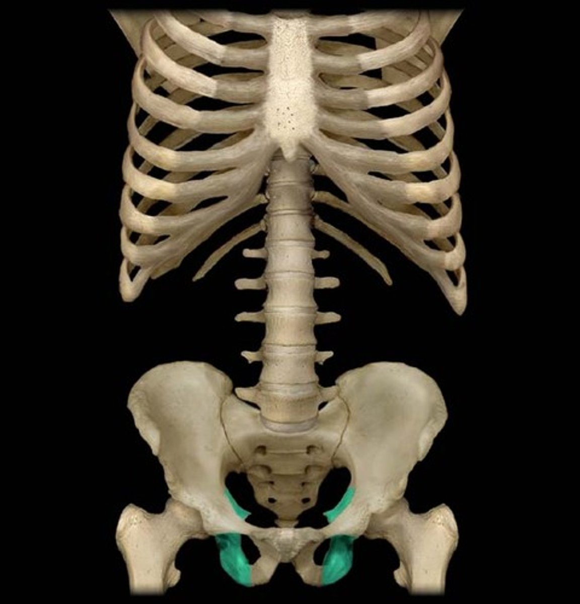
ischium
curved bone forming the base of each half of the pelvis
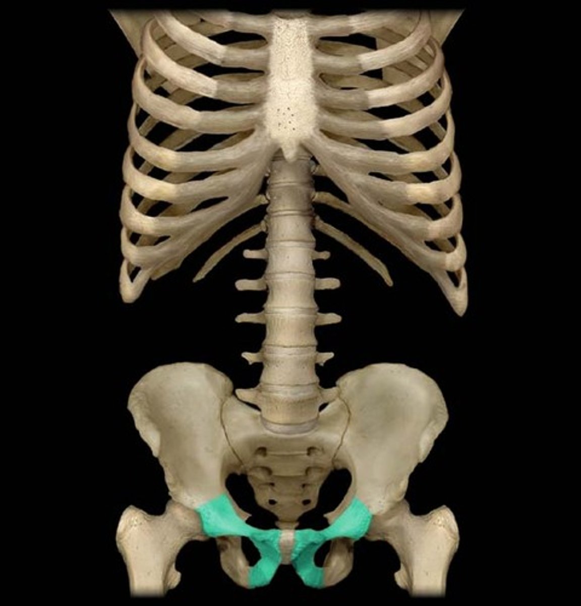
pubis
pair of bones forming the two sides of the pelvis
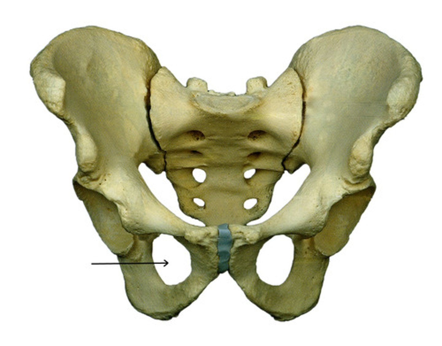
obturator foramen
large opening in the hipbone between the pubis and the ischium
iliac crest
thick curved upper border of the ilium, the most prominent bone on the pelvis
acetabulum
socket of the hipbone, into which the head of the femur fits
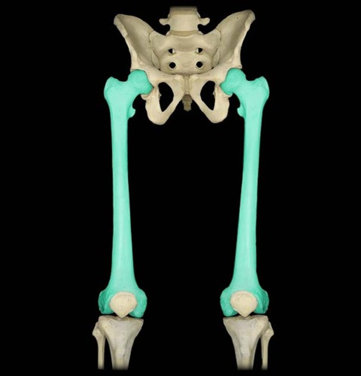
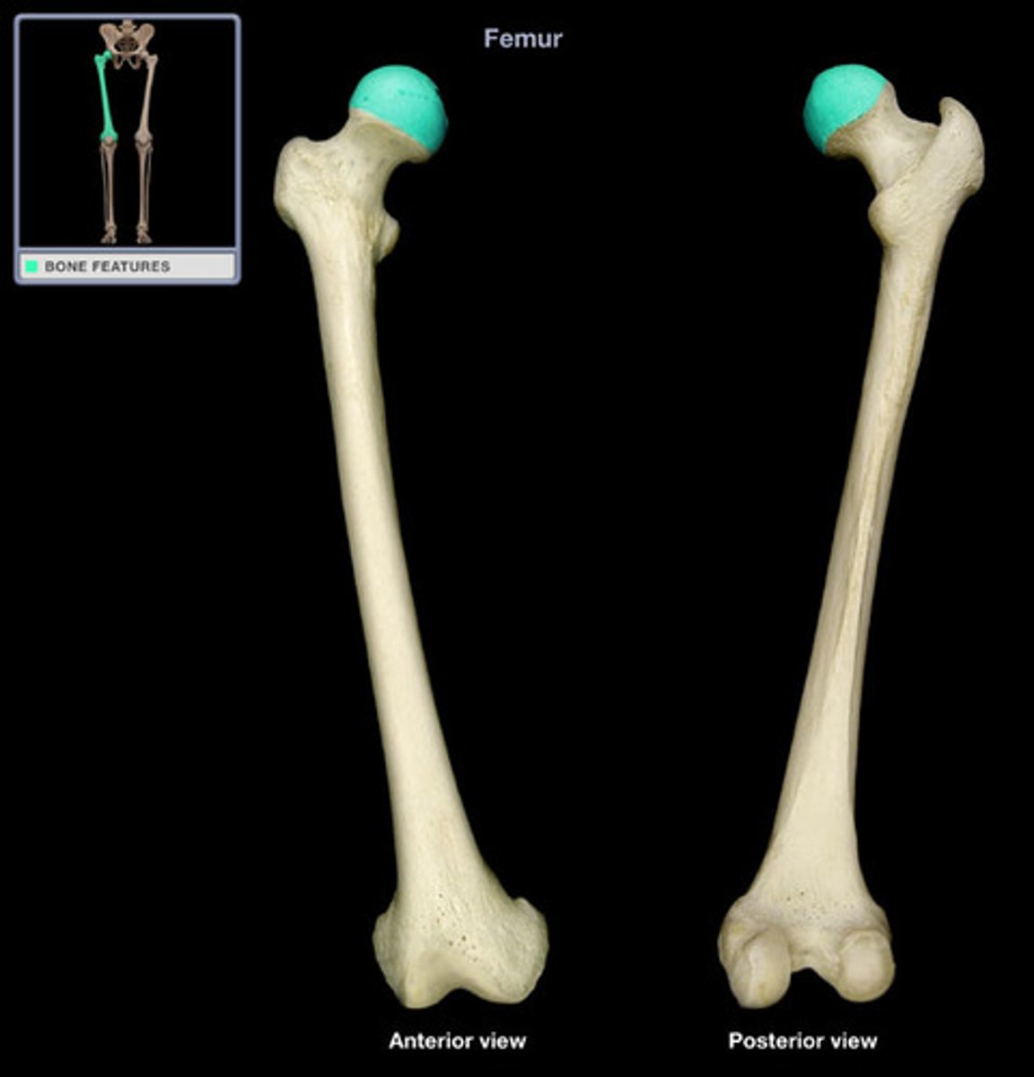
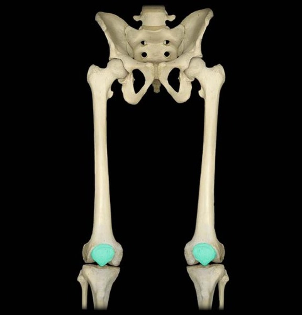
femur
bone of the thigh or upper hind limb, articulating at the hip and the knee
neck of femur
flattened pyramidal process of bone, connecting the femoral head with the femoral shaft

head of femur
the highest part of the thigh bone, supported by femoral neck, articulates with acetabulum in pelvic bone to form hip joint
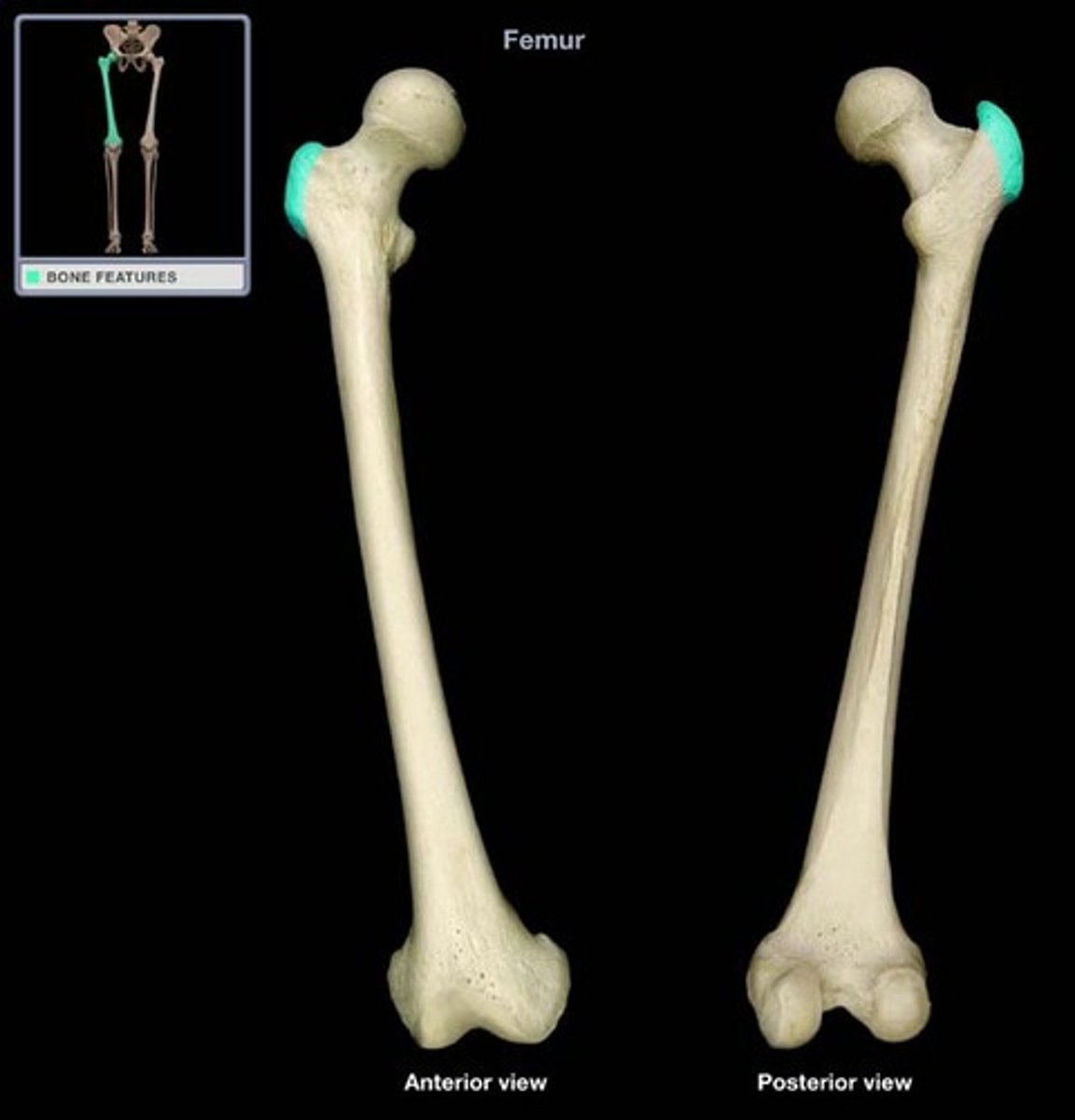
greater trochanter
large, irregular, quadrilateral eminence, slightly posterior, attachment for muscles
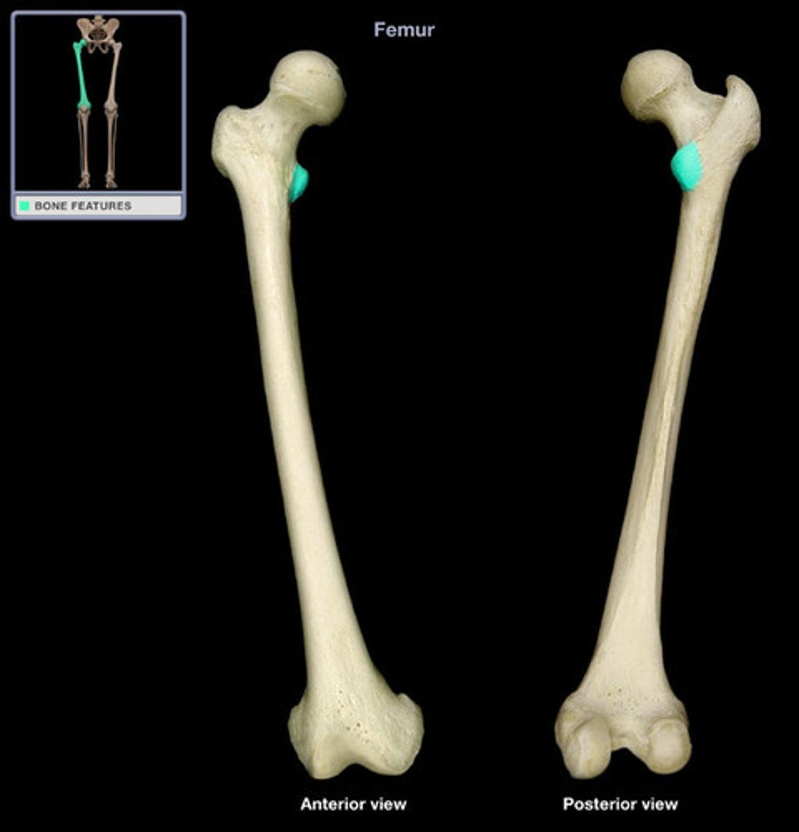
lesser trochanter
small protuberance of bone that projects from the posterior aspect of the femur at the base of the femoral neck
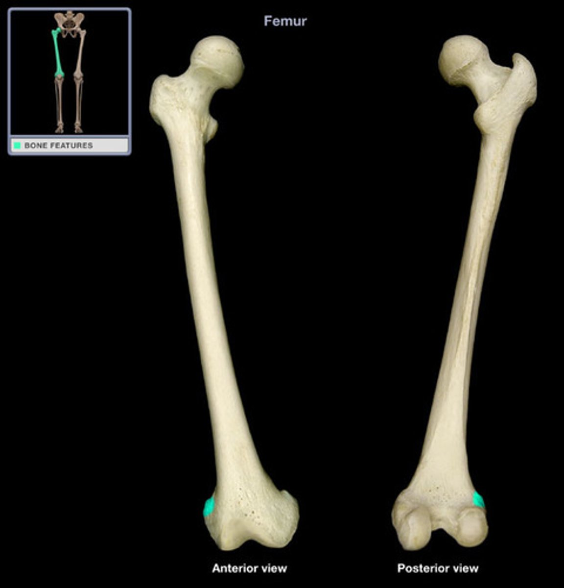
medial epicondyle of femur
bony protrusion located on the medial side of the bone's distal end, it bears an elevation, the adductor tubercle
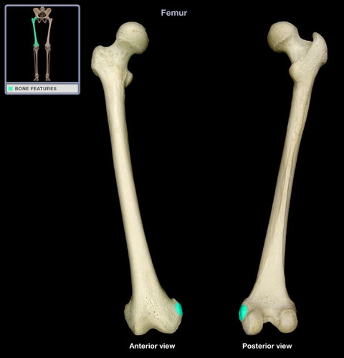
lateral epicondyle of femur
smaller and less prominent than the medial epicondyle, gives attachment to the fibular collateral ligament of the knee-joint
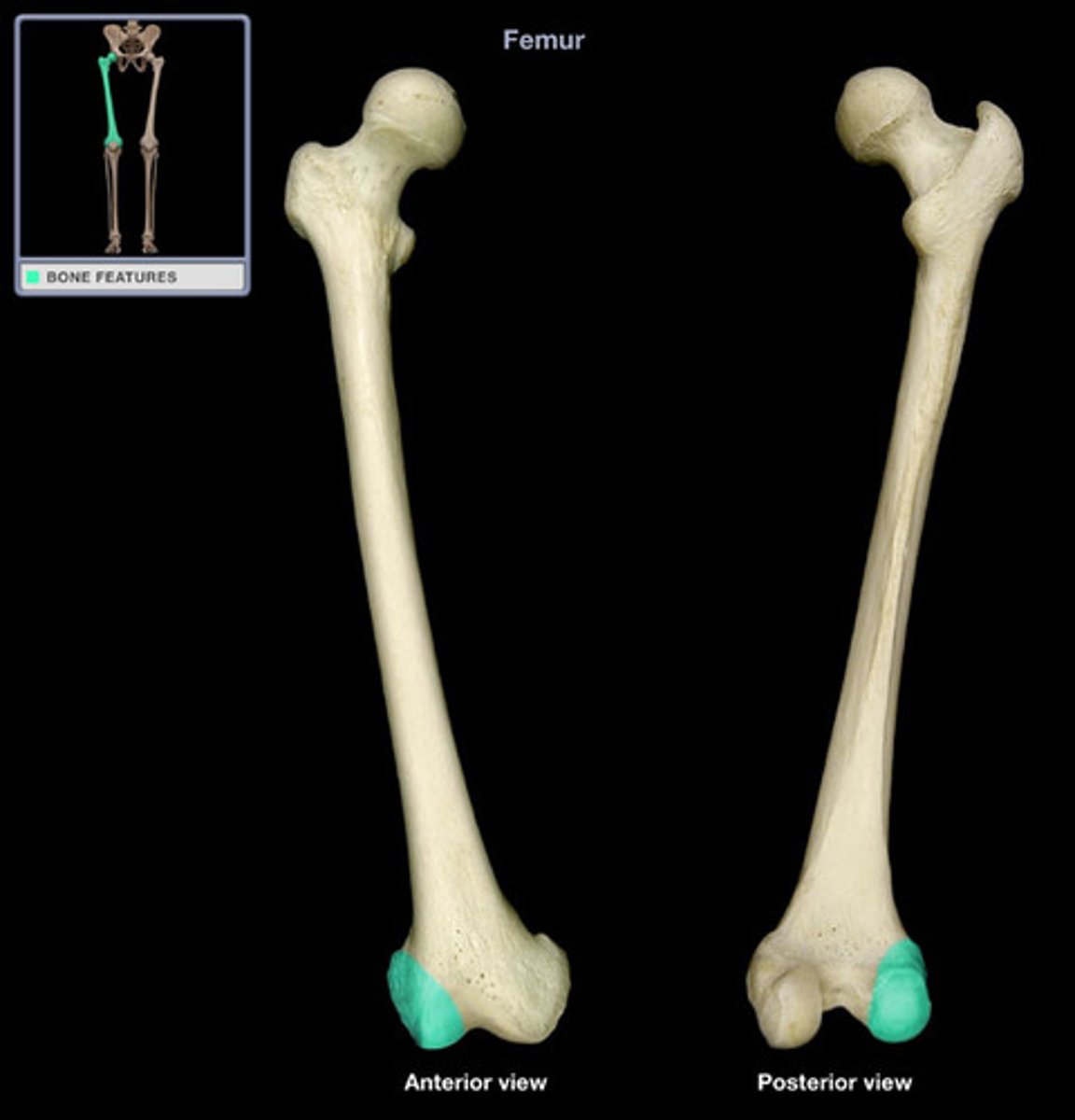
medial condyle of femur
medial projection on lower part of femur, help make up knee joint, articulates with meniscus above medial tibia condyle
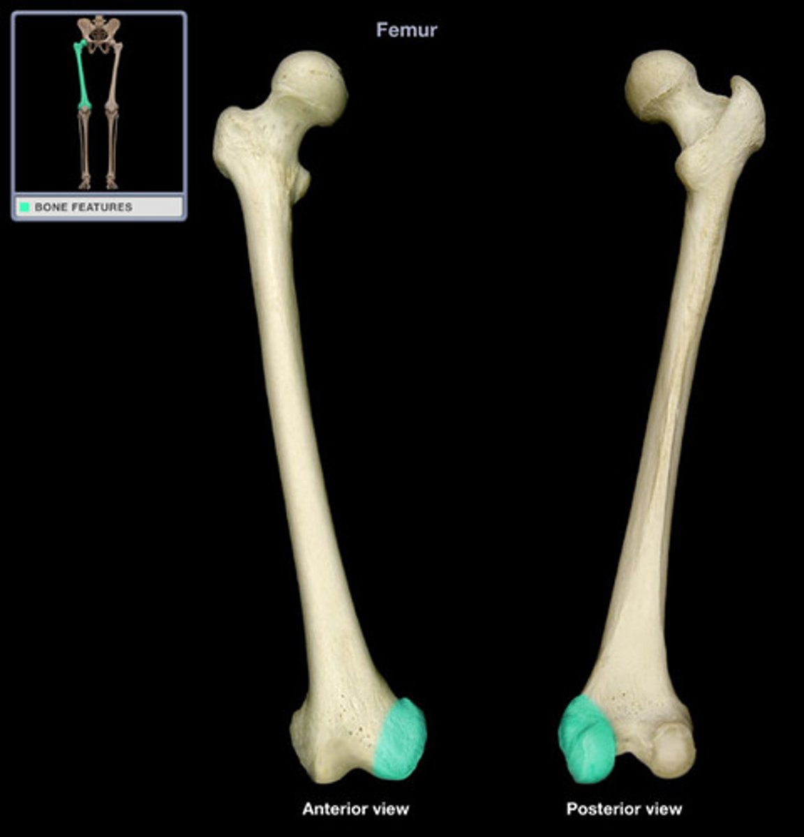
lateral condyle of femur
lateral projection on lower part of femur, help make up knee joint, articulates with meniscus above lateral tibia condyle
patella
kneecap
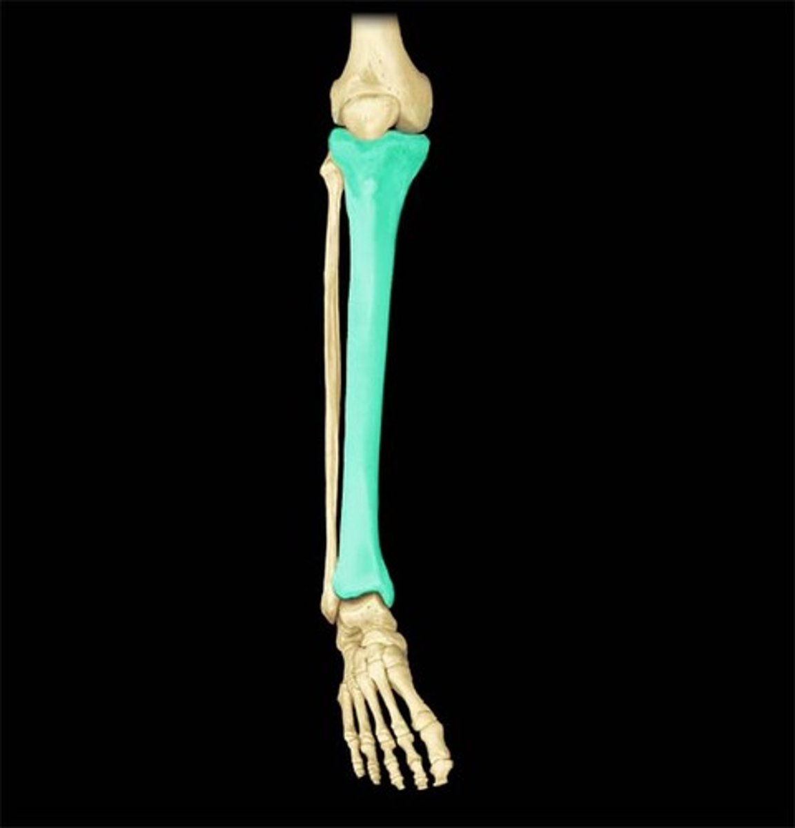
tibia
inner and larger of the two bones between the knee and the ankle
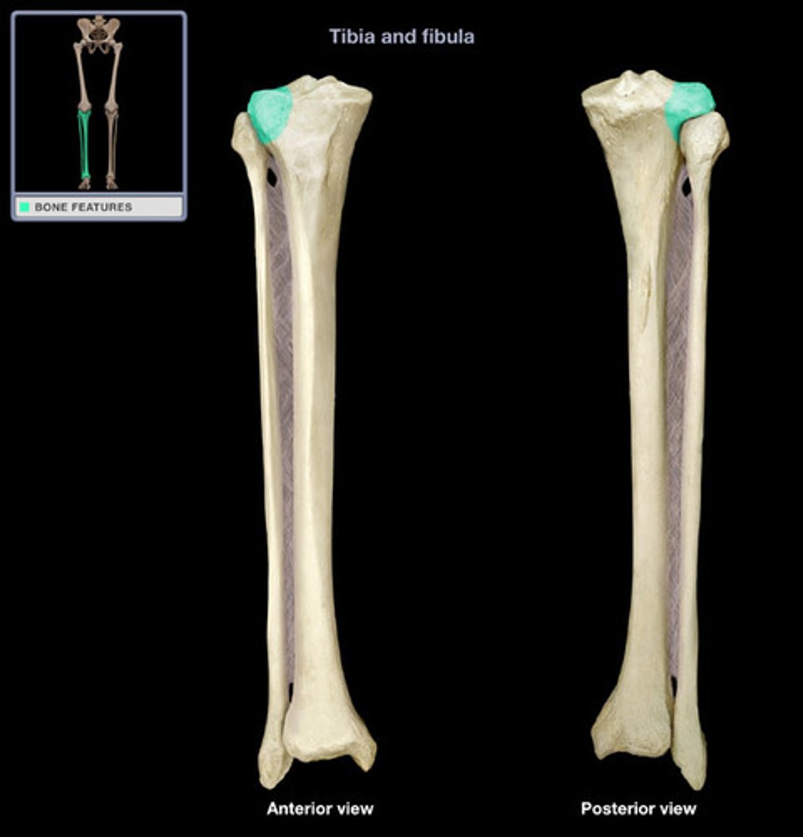
lateral condyle of tibia
lateral portion of the upper extremity of tibia, it serves as the insertion for the Biceps femoris muscle
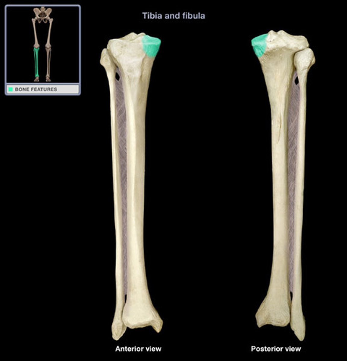
medial condyle of tibia
medial portion of the upper extremity of tibia, site of insertion for muscle
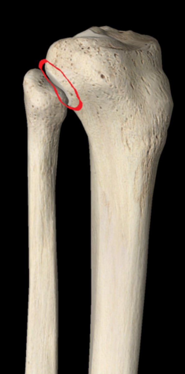
fibular articular facet
flat circular articular facet on the inferior and lateral aspect of the lateral condyle of the tibia for articulation with the head of the fibula
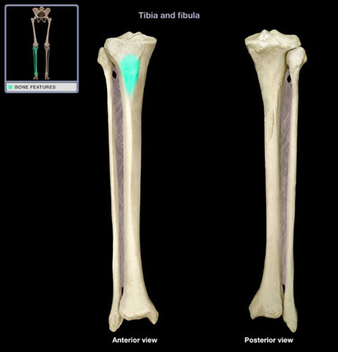
tibial tuberosity
oval elevation on the anterior surface of the tibia, giving attachment to patellar ligament
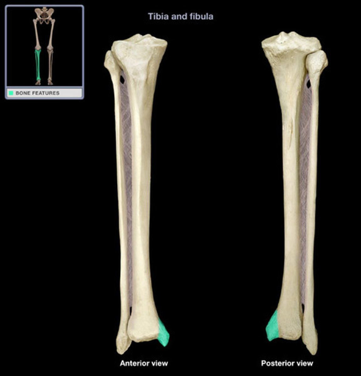
medial malleolus of tibia
prominence on the inner side of the ankle, formed by the lower end of the tibia
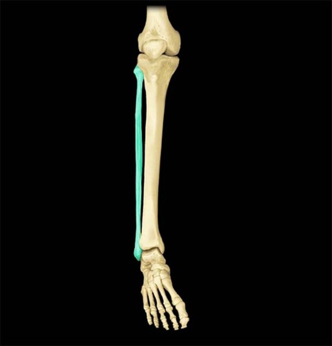
fibula
lateral and smaller of the two bones of the leg; does not bear weight and articulates with the tibia
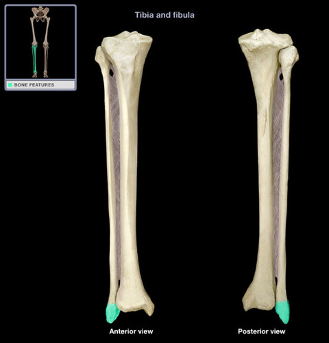
lateral malleolus of fibula
prominence on the outer side of ankle, formed by the lower end of the fibula
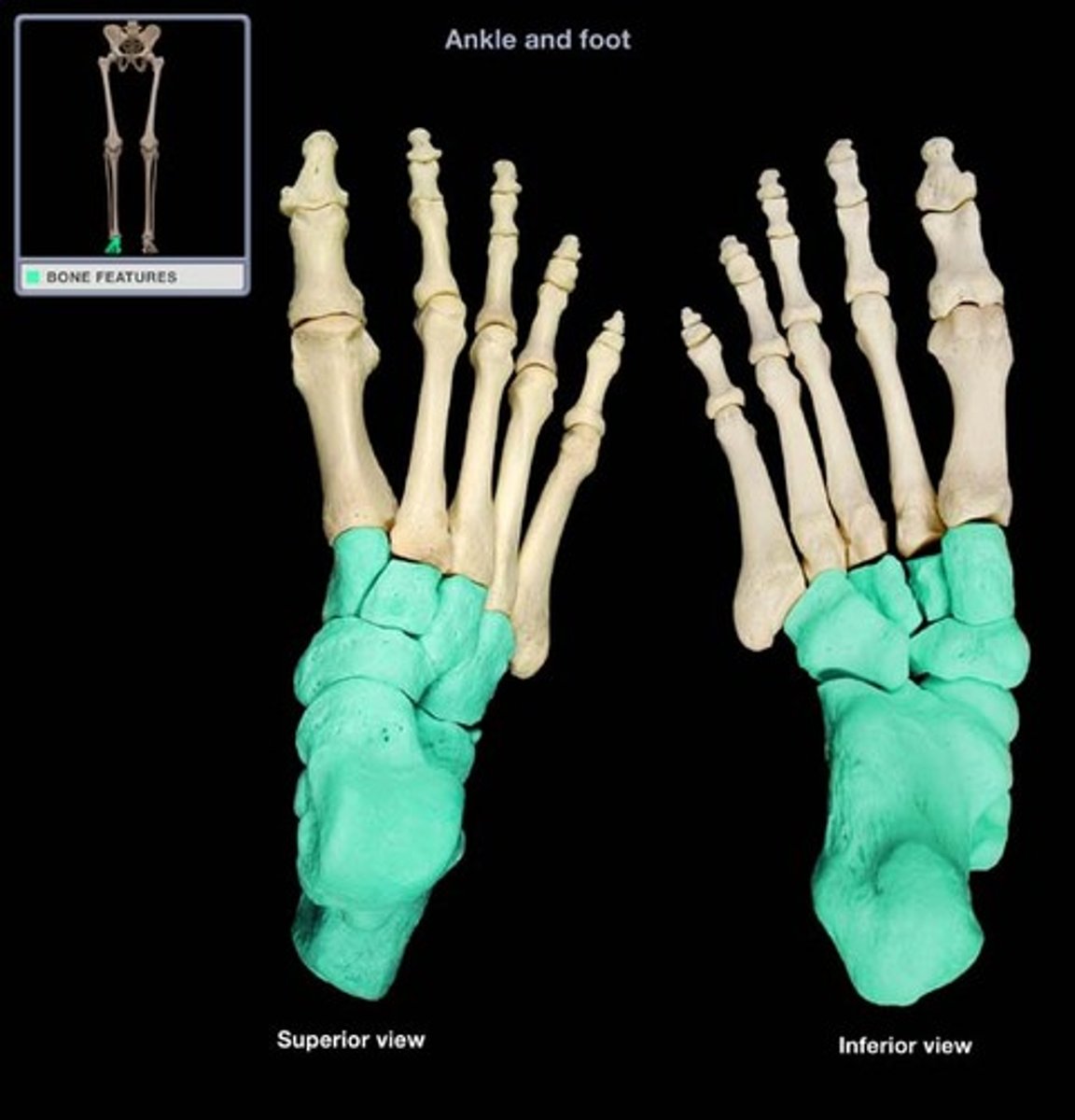
tarsal bones
seven bones between lower end of tibia/fibula and metatarsals
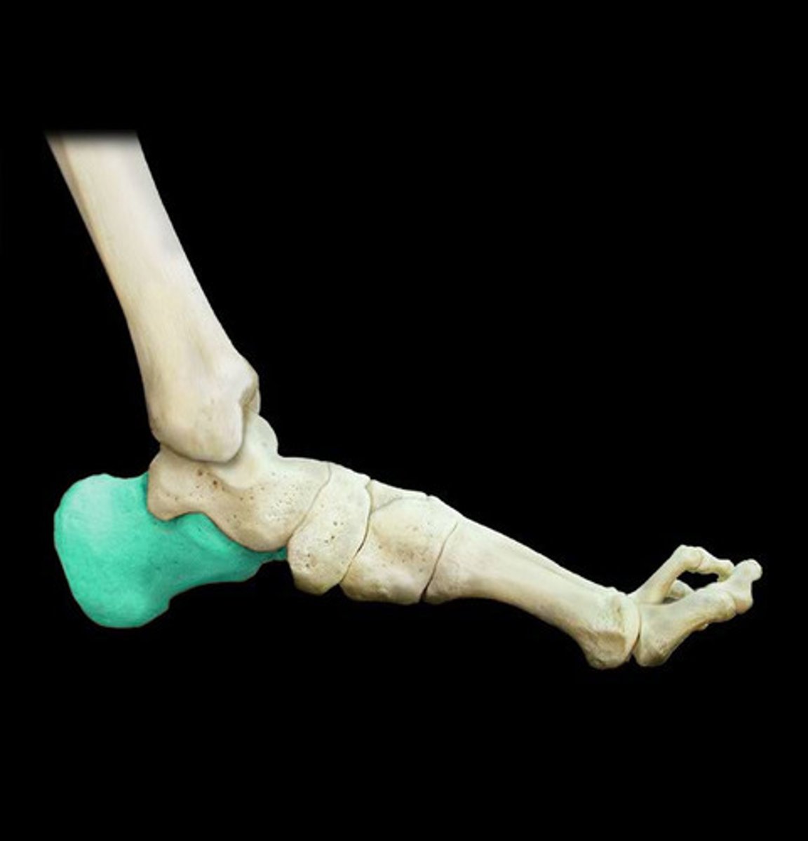
calcaneus
largest of the tarsal bones; it forms the heel and articulates with the cuboid anteriorly and the talus superiorly
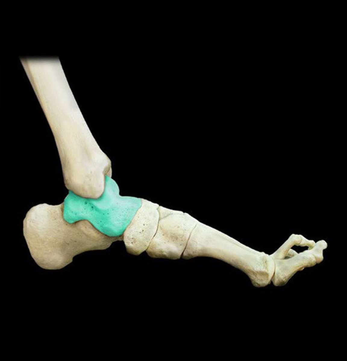
talus
bone of the foot that articulates superiorly with the tibia and fibula to form the ankle joint inferiorly with the calcaneus to form the subtalar joint
metatarsals
five long bones numbered I-V beginning with the bone on the medial side forming the skeleton of the anterior portion of the foot
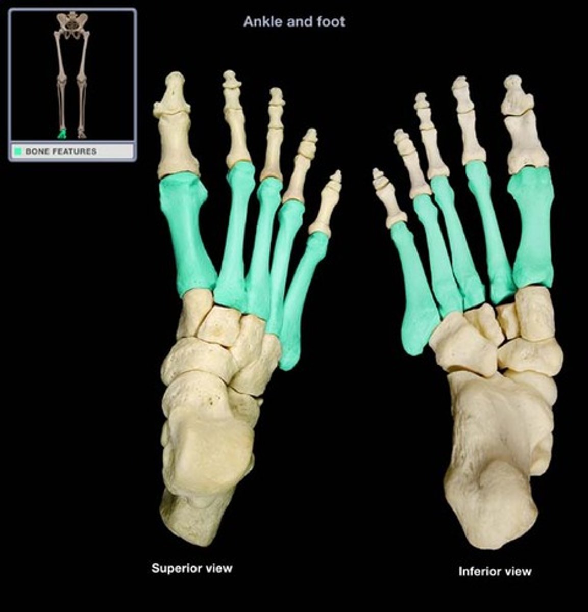
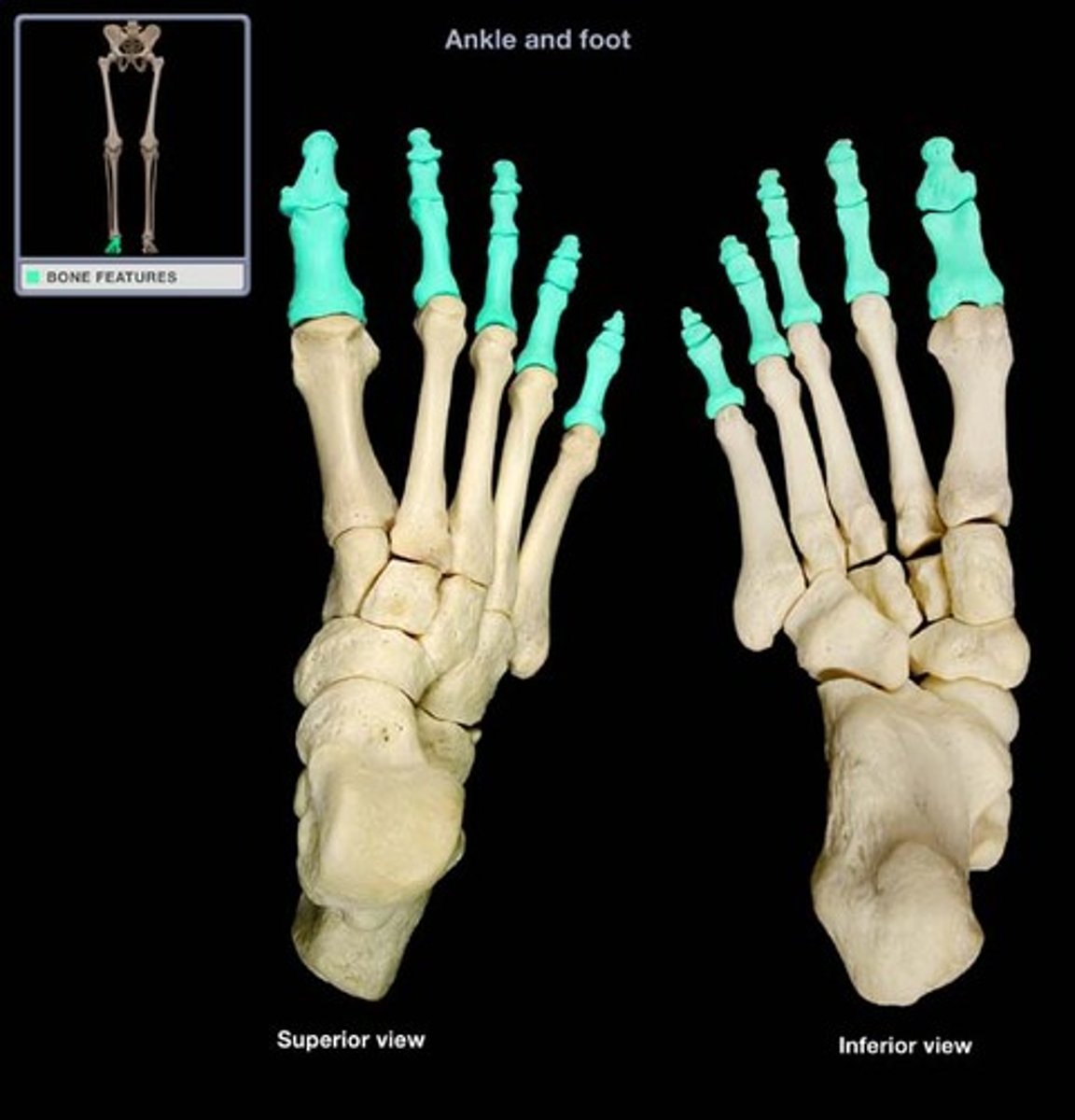
phalanges
five bones of the toes