cell signaling and nervous system
1/129
There's no tags or description
Looks like no tags are added yet.
Name | Mastery | Learn | Test | Matching | Spaced | Call with Kai |
|---|
No analytics yet
Send a link to your students to track their progress
130 Terms
three types of cell signalling
paracrine signaling
synaptic signaling
endocrine signaling
paracrine signaling
cells within an organ
cell targets a nearby cell
how can cells adjacent to each other communicate
through gap junctions
synaptic signaling
only between NEURONS and a target cell
neuron releases neurotransmitters
endocrine singaling
cells from endocrine glands release hormones into the blood
second messengers in cell signaling
RELAYS the signal from the extracellular environment to cytoplasm
how are second messengers in cell signaling detected
by receptor proteins at cell surface, or within the cell (if non polar)
how are second messengers produced
after first messenger triggers the system, they’re indirectly produced to spread the message
how is signal relayed from receptor to enzyme (after second messenger has triggered the process)
via G proteins
what are the G proteins coupled to
GPCRs (G protein-coupled receptors)
three subunits of G proteins
alpha, beta, gamma
beta and gamma are always together
GPCR when it’s inactive
G-proteins are coupled with the GPCRs, with alpha beta gamma together.
GDP is attached to alpha.
GPCR when it’s triggered by a ligand
alpha subunit releases GDP to the cell and binds with GTP instead.
then, alpha with GTP splits from beta and gamma
the two teams cause the effectors to work
how does GPCR cycle end
alpha units will hydrolysis GTP → GDP
GPCR goes inactive again
ligand leaves
depolarization
positive charges entering neuron cell
hyperpolarization
negative charges entering neuron cell
repolarization
neuron cell returning to the resting membrane potential
depolarization: excitatory vs inhibitory
excitatory (depolarization = going up)
hyperpolarization: excitatory vs inhibitory
inhibitory (hyperpolarization = going down)
voltage gated channels
channel only opens after a change of the resting membrane potential occurs
how are sodium channels gated in general?
by voltage
how are potassium channels gated?
by voltage or simple diffusion (leakage channels)
example of sodium channel blocker
tetrodotoxin (pufferfish)
what happens if depolarization doesn’t reach a certain threshold
goes back to the resting membrane potential, the stable equilibrium. either threshold is reached or not reached.
happens through leakage of charges through the axon
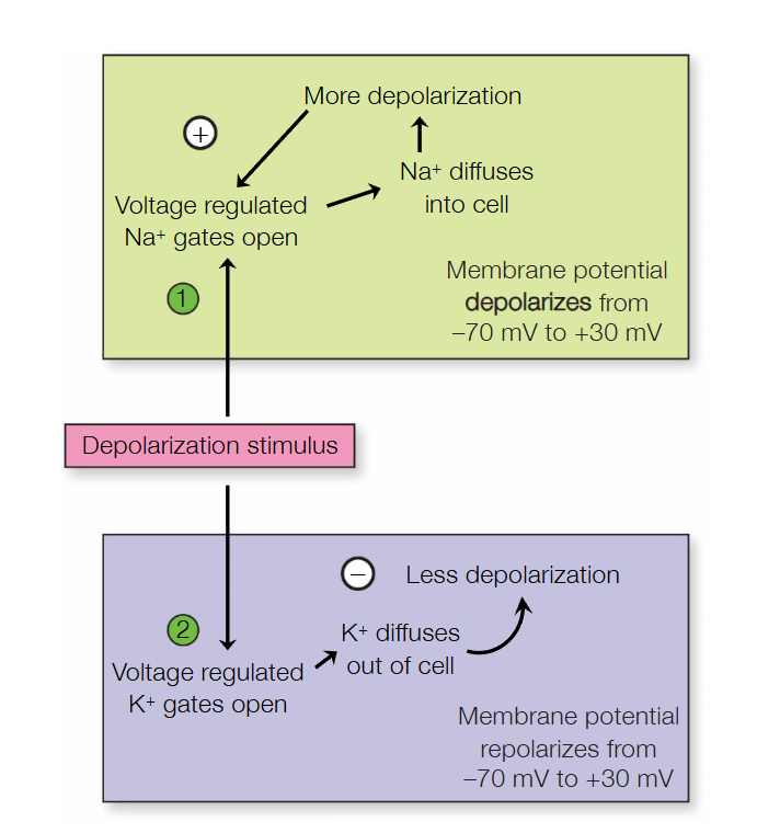
action potential process
depolarization reaches a threshold
sudden rapid change in membrane potential spikes up
voltage-gated sodium channels open, and sodium rushes INTO cell
membrane potential hits peak
Na+ channels close
voltage-gated K+ channels open
K+ rushes OUT of cell
resting membrane potential in neurons = -70mV. At this point, we’re at + mV.
as K+ goes out, membrane potential spikes down under -70mV.
hyperpolarization
goes back to equilibrium at -70 mV
what kinds of loops does the Na+/K+ channels opening cause?
Na+ positive feedback loop
K+ negative feedback loop
does stronger stimulus = greater action potential?
no. all action potentials of a specific neuron has the same amplitude, shape, and duration.
does stronger stimulus = more action potentials?
yes, frequency depends on strength of stimuli
what happens if a neuron is stimulated during its action potential?
its incapable to respond to further stimulation
its REFRACTORY to further stimulation.
refractory period
when neuron cannot generate another action potential even while its stimulated
absolute refractory period
completely incapable of responding to further stimulation
due to inactivation of voltage-gated Na+ cannels.
(inhibitory domain blocks the sodium channel)
relative refractory period
during repolarization of membrane.
VERY STRONG depolarization will trigger a new action potential.
action potential conduction
stimulus starts near the head of the neuron
as it is depolarized, the nerve impulse travels down the axon.
why is the action potential unidirectional (one direction)
because as soon as one part of the axon is fired up, it inactivates the Na+ channel and cannot reopen. (refractory period)
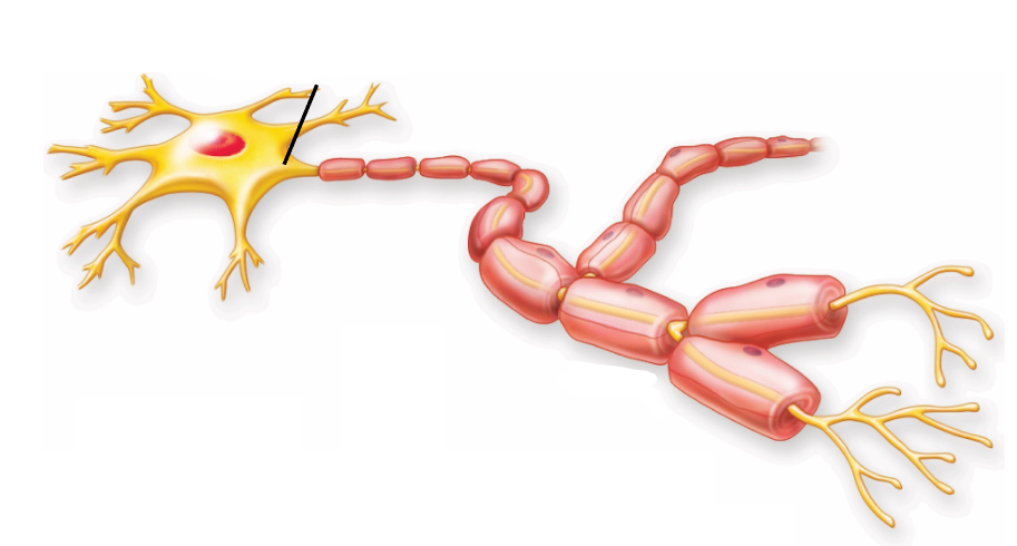
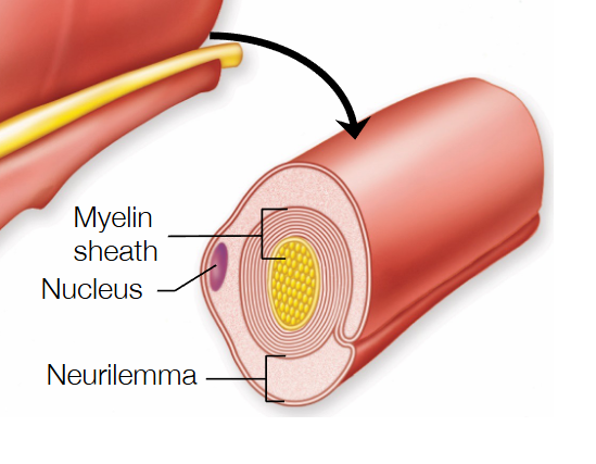
what is myelin
insulating layer of phospholipids and proteins wrapped around axons
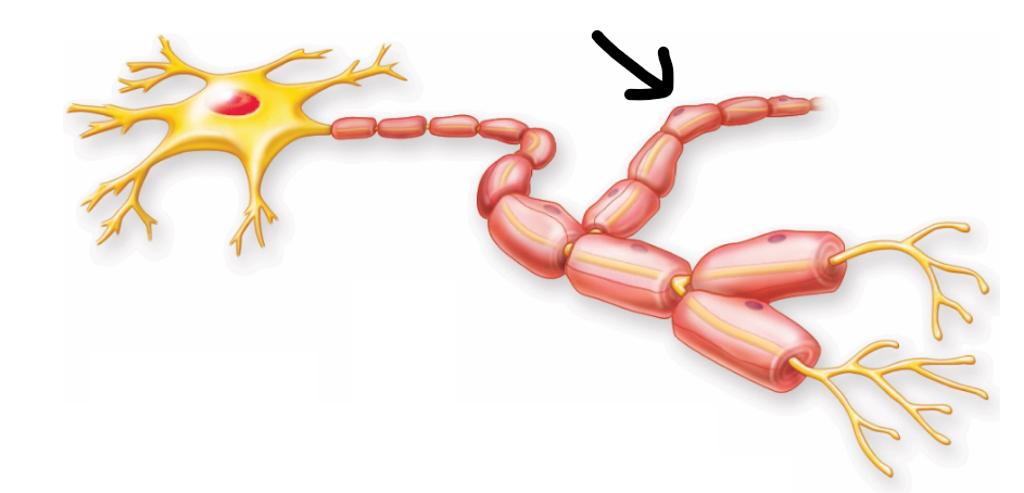
nodes of ranvier
parts that don’t have myelin on the axon. The only spots where action potential can occur, since sodium can’t escape through myelin
myelin function
prevents movements of ions, speeds up the process of neurotransmission
saltatory conduction
the rapid “jumping” of nerve impulses thanks to myelin. speeds up action potential.
what is action potential used for
to relay signals through the body
central nervous system
composed of brain + spinal cord
peripheral nervous system
made of nerves outside the CNS (basically everything else)
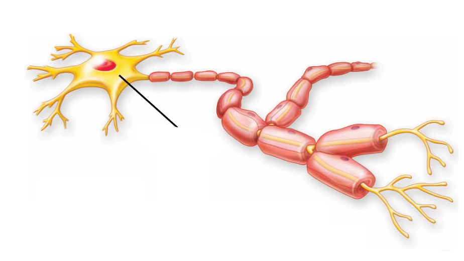
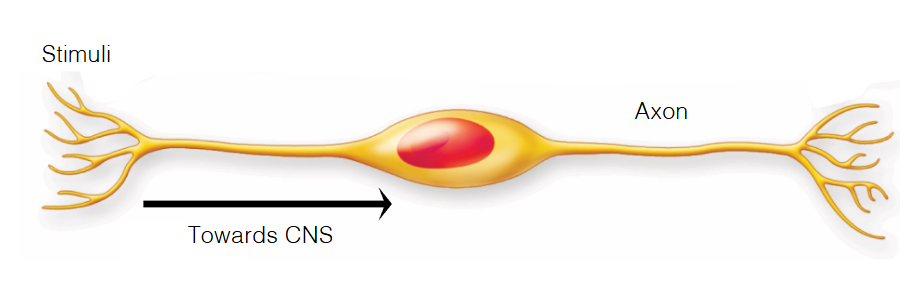
three principal regions of neurons
cell body (has nucleus of neuron)
dendrites
axon
cluster of cell bodies in CNS
nucleus
cluster of cell bodies in PNS
ganglion
contains nucleus of the neuron
cell body
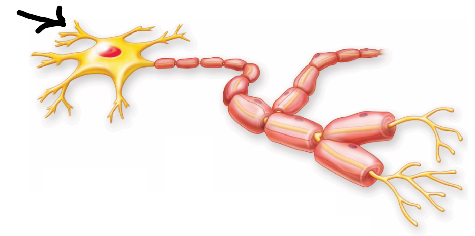
cytoplasmic extensions that provide a receptive area for electrochemical stimulation
dendrites
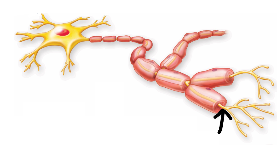
long cytoplasmic projection that conduct action potential
axon
where axon originates from
axon hillock
branched out part of the neuron
collateral axon
motor neurons
carry out the impulse from CNS to effector organs
messenger
motor neurons aka
efferent neurons
somatic motor neuron
stretches from CNS to skeletal muscles
autonomic motor neuron
connects the end of a CNS neuron signal to the smooth and cardiac muscle or glands
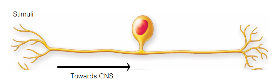
sensory neurons
conduct (pass) impulses from sensory receptors into the CNS
stretches from PNS to CNS
lets you feel things (woah sensory)
sensory neurons aka
afferent neurons
interneurons
relays the signals between two other neurons, only located in CNS
multipolar neurons
several dendrites and one axon
most common
pseudounipolar neurons
one branch receive stimuli
other branch relay signal to CNS
branched off cell body in the middle
(most sensory neurons are pseudounipolar)
bipolar neurons
neurons with two extensions, one either side of cell body
cell body in the middle, part of axon
neuron type?
multipolar
neuron type?
pseudounipolar
neuron type?
bipolar neuron
multiple axons form together in PNS
nerve
multipole axons form together in CNS
tract
mixed nerve
has both sensory and motor neuron
neuroglia
nervous system cells, five times more abundant than neurons
brings protection and support to the nervous system
can’t produce impulse
neuroglia is composed of
schwann cells
neuroglia function
produces myelin sheaths around axons and surround all PNS axons to form a neurilemmal sheath
neurilemmal sheath
outermost layer of schwann cell. Made up of Schwann cells cytoplasm and nucleus
schwann cell function
wraps around the axon to form protective layer (myelin sheath)
do schwann cells exist next to unmyelinated axons
yes, they’re just not wrapped around the axons
neuroglia (PNS) is made of
satellite cells
satellite cells
cover the surface of neuron cell bodies
provide support
might control microenvironment
4 types of neuroglia in CNS
astrocytes (structural support)
oligodendrocytes (myelin formation)
microglia (immune defense)
ependymal cells (cerebrospinal fluid circulation).
oligodendrocytes
produce myelin sheaths around axons
white matter
area of CNS where myelin sheaths are prominent
because myelin sheaths give a white color
grey matter
cell bodies and dendrites of neurons in the CNS
neuroregeneration in PNS
when axon is cut, the severed portion degenerates and is phagocytosed by schwann cells
schwann cells then form a regeneration tube, acts as guidance track for regenerating axon
neurotrophins
secreted by schwann cells to promote axon regeneration
neuroregeneration in CNS
neurons die upon injury or via apoptosis
regeneration prevented by inhibitory proteins (e.g. Nogo)
forms a glial scar
further prevents axon repair
microglia
main form of immune defense in CNS
able to detect sites of infection or damage
also involved in synaptic pruning
synaptic pruning
eliminating unused or weaker neural connections (synapses) to create more efficient, specialized neural circuits
microglia origin + classification
distinct embryonic origin from other types of neuroglia
considered myeloid cells
microglial activation
cells become ameboid and become phagocytic cells
ameboid cells
able to change shape due to low consistency of cytoplasm
what can cause microglial activation
altered state of the extracellular environment
how do microglia find damaged cells
detect ATP released from damaged cells
how do microglia end inflammatory responses
releasing anti-inflammatory chemicals, contributes to neuroprotection
astrocytes
help regulate external environment of neurons.
what do astrocytes look like
numerous cytoplasmic projections rating outwards
(thus astro- aster: star in greek)
most abundant neuroglia in CNS
astrocytes
how do astrocytes work
they encircle the endothelial cells of blood capillaries with projections named end feet
in other words, the end feet attach themselves to different blood vessels and axons
blood brain barrier is formed
what things can astrocytes do
K+ uptake (kidnap K+ that’s exiting the neuron during action potential)
glucose uptake from the blood, release lactate for neurons
how do astrocytes help synapse
maintenance, formation, and maturation
what could astrocytic dysfunction be a sign of
neurodevelopmental disorders
alexander’s disease
caused by a gain of function point mutation in an astrocyte-specific protein
gliotransmitters
messengers released by astrocytes to regulate neuron function
fenestration
new opening is formed
endothelial cell
surrounds the blood cell
sometimes leaves gaps (through fenestration) to allow some filtering to go through