108 Hand, Forearm, Humerus A&P
1/165
There's no tags or description
Looks like no tags are added yet.
Name | Mastery | Learn | Test | Matching | Spaced |
|---|
No study sessions yet.
166 Terms
What should be done with a patient's sling or splints before imaging?
Do not remove them without permission from the provider.
What is the recommended Source-to-Image Distance (SID) for forearm and elbow imaging?
40 inches.
What is the positioning requirement for the patient's wrist, elbow, and humerus during imaging?
They should be on the same plane.
What is the central ray (CR) entry point for an AP forearm projection?
At the midpoint of the forearm.
What structures should be included in an AP forearm projection?
Entire forearm, including wrist, distal humerus, and proximal row of carpal bones.
What is the patient position for a lateral forearm projection?
Elbow centered to the middle of the IR with humeral epicondyles parallel to the IR.
What should be the CR orientation for a lateral forearm projection?
Mid forearm
What is the evaluation criteria for an AP elbow projection?
Radial head, neck, and tuberosity slightly superimposed over the proximal ulna and the elbow joint open.
What is the purpose of the partial flexion position for an AP elbow?
To image the distal humerus and proximal forearm separately when the patient cannot fully extend the elbow.
What is the CR orientation for the AP elbow in the partial flexion position?
Perpendicular to the humerus, passing through the elbow joint.
What structures are shown in the AP elbow—distal humerus partial flexion position?
Distal humerus without rotation, proximal radius superimposed over the ulna, and a closed elbow joint.
What is the CR angle for the axiolateral (Coyle) projection for the radial head?
Angled 45 degrees toward the shoulder.
What is the positioning for the AP humerus projection?
Epicondyles perpendicular to the IR with the top border of the IR approximately 1½ inches above the humeral head.
What structures should be visible in a lateral humerus projection?
Both elbow and shoulder joints, with superimposed humeral epicondyles.
What is the CR orientation for the transthoracic lateral projection of the humerus?
Perpendicular to the midportion of the humerus, entering at the surgical neck.
What is the respiration instruction for the transthoracic lateral projection?
Exposure made on inspiration or using a breathing technique with slow, deep breathing.
What is the evaluation criteria for the trauma horizontal beam lateral projection?
Both proximal and distal humerus joints should be included.
Fossa
Shallow depression in a bone
Styloid
Pointed projection
Condyle
Rounded articular surface
Epicondyle
Prominence above a condyle
Tubercle, Tuberosity
Roughened bump for muscle attachment
Head
Rounded proximal end of a bone
radiocarpal joint
Wrist joint formed by articulation between the distal radius and the proximal row of carpal bones
intercarpals
the space between the carpals
distal radioulnar joint
The articulation between the distal ends of the radius and ulna that allows for pronation and supination of the forearm
MCP
Metacarpophalangeal
DIP
Distal interphalngeal
PIP
Proximal interphalangeal
Humerus, elbow, forearm positioning
Must be in same plane. Long bones: include both joints. Always do AP & Lateral.
SID
40 inches.
Collimation
Collimate ~1 inch beyond skin line.
Grid usage
Used for proximal humerus/transthoracic.
Forearm - AP Positioning
Patient seated, arm extended, palm up, epicondyles parallel to IR. CR → mid-forearm. Shows entire radius & ulna, wrist + elbow joints.
Forearm - Lateral Positioning
Elbow flexed 90°, thumb up, wrist & elbow same plane. CR → mid-forearm. Shows radius/ulna superimposed distally, elbow flexed.
Elbow - AP Positioning
Arm extended, palm up, epicondyles parallel to IR. CR → mid-elbow. Shows distal humerus, proximal radius & ulna.
Elbow - AP Partial Flexion
Used if elbow cannot fully extend. CR perpendicular to elbow joint, angled through humerus or forearm.
Elbow - Lateral Positioning
Elbow flexed 90°, thumb up. CR → mid-elbow. Shows olecranon, fat pads, superimposed epicondyles.
Elbow - AP Medial Oblique
Hand pronated, elbow extended. CR → mid-elbow. Shows coronoid process free of superimposition.
Elbow - AP Lateral Oblique
Hand supinated, elbow externally rotated. CR → mid-elbow. Shows radial head & neck free of superimposition.
Elbow - Coyle Method
Elbow flexed 90°, CR angled 45° toward shoulder. Shows radial head & capitulum.
Humerus - AP Positioning
Arm extended, palm up, epicondyles parallel to IR. CR → mid-humerus. Shows entire humerus, shoulder + elbow joints.
Humerus - Lateral Positioning
Elbow flexed 90°, palm on hip or abdomen. Epicondyles perpendicular to IR. CR → mid-humerus. Shows lateral humerus, superimposed epicondyles.
Humerus - Transthoracic (fractures)
Affected arm against IR, unaffected arm raised. CR → surgical neck. Breathing technique or full inspiration. Shows lateral humerus through thorax.
Forearm Bones
Ulna = medial (pinky side). Radius = lateral (thumb side).
Radius - Proximal Structures
Head, neck, radial tuberosity.
Radius - Distal Structures
Base with styloid process, ulnar notch.
Ulna - Proximal Structures
Olecranon process, coronoid process, trochlear notch, radial notch.
Ulna - Distal Structures
Head, styloid process.
Humerus - Proximal Structures
Head (articulates with scapula), anatomic neck, surgical neck (fracture site), greater tubercle, lesser tubercle.
Humerus - Distal Structures
Medial/lateral epicondyles, trochlea (ulna), capitulum (radius), coronoid fossa (anterior), radial fossa (anterior), olecranon fossa (posterior).
Elbow Joint - Components
Humeroulnar, humeroradial, proximal radioulnar joints, all within one capsule.
Elbow Joint - Movements
Flexion/extension (hinge) and supination/pronation (pivot).
Fat Pads of the Elbow
Anterior fat pad, posterior fat pad (hidden unless pathology), fat pad sign = displaced triangular shadows → occult radial head fracture.
Bony Surface Landmarks
Fossa = depression. Styloid = pointed projection. Condyle = rounded articular surface. Epicondyle = process above condyle. Tubercle/tuberosity = roughened bump. Head = rounded proximal end of bone.
AP forearm part position
Elbow extended with hand supinated ; humeral epicondyles parallel to IR
AP forearm CR placement
midpoint of forearm
Lateral forearm part position
Elbow is flexed 90 degrees, thumb side up ; ulnar and radial styloid processes superimposed (stacked)
forearm, elbow, and humerus patient positioning
wrist, elbow, and humerus on the same plane. Patient is seated with legs not under table
Lateral forearm central ray
midpoint of forearm
AP elbow part positioning
elbow extended and hand supinated, elbow centered to middle of IR
AP elbow central ray centering
perpendicular to elbow joint
Lateral elbow part positioning
Elbow flexed 90 degrees, thumb side up, humeral epicondyles perpendicular to IR
Lateral elbow CR centering
perpendicular to elbow joint
AP elbow partial flexion (distal humerus) part positioning
Supinate hand and center IR to condyles of humerus with elevated forearm
AP elbow partial flexion (distal humerus) CR centering
Perpendicular to elbow joint, passing through elbow joint
AP elbow partial flexion (proximal forearm) part positioning
Leave elbow flexed, patient is standing, supinate hand and center IR to condyles of humerus
AP elbow partial flexion (proximal forearm) CR centering
Perpendicular to elbow joint and long axis of forearm
Elbow AP medial oblique part positioning
Elbow centered to IR, rotate the arm 45 degrees medially
Elbow AP medial oblique CR centering
perpendicular to elbow joint, collimatation is 3 inches above and below elbow crease
Elbow AP lateral oblique part positioning
Laterally rotate arm 45 degrees, first and second digit will touch table
Elbow AP lateral oblique CR centering
Perpendicular to elbow joint, collimation 3 inches above and below elbow joint
When to use the coyle method for radial head
When patients cannot fully extend the elbow for medial and lateral oblique projections
Axiolateral (Coyle method) positioning
the CR is at a cephalic angle of 45 degrees towards shoulder, the patients hand is pronated with the elbow flexed at a 90 degree angle
Axiolateral (Coyle method) CR centering
entering elbow joint at mid elbow
AP humerus part positioning
Patient is standing with back to board, arm out with supinated hand
AP humerus CR centering
Perpendicular to midportion of humerus, collimated to include the elbow joint and 1 ½ inches above humeral head with 1 inch on the sides
Lateral humerus part positioning
Patients body facing board, top of IR 1 ½ inches above humeral head, elbow flexed with hand over stomach, shoulder and elbow touching board
Lateral humerus CR centering
at midpoint of humerus
Transthoracic lateral part positoining
Patients left side is toward the board, with non injured arm raised with hand on back of head
Transthoracic lateral CR centering
Centered at surgical neck of humerus to IR
Structures shown in Lateral Forearm
Entire forearm with wrist and distal humerus
Structures shown in AP elbow
Medial and lateral epicondyle, Trochlea, proximal ulna, capitulum, radial head, radial neck, radial tuberosity
Structures shown in Lateral elbow
Humeral epicondyles, radial head, coronoid process, Olecranon process
Structures shown in AP medial oblique elbow
Coronoid, coronoid process, ulna, radial head, radial neck, olecranon process, olecranon fossa
Structures shown in AP lateral oblique elbow
Radial head, Radial neck, Tuberosity, ulna, capitulum
Structures shown in Axiolateral (Coyle) radial head
Open joint space between radial head and capitulum, humeral epicondyles
Structures shown in AP Humerus
Elbow and shoulder joints, Humeral epicondyles, Humeral head, greater tubercle
Structures shown in Lateral Humerus
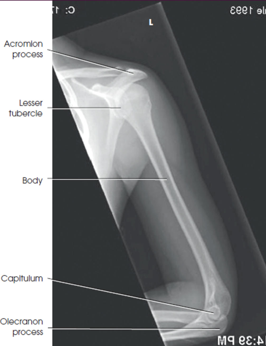
Elbow and shoulder joints, superimposed humeral epicondyles, Lesser tubercle, Greater tubercle superimposed over humeral head
Structures shown in Transthoracic Lateral
Unaffected humerus above shoulder, proximal half of humerus projected through thorax
Where is the radial tuberosity?
On the radius, below the radial head
What is the number one thing to remember when doing forearm, elbow, and humerus
Always keep each of them on the same plane
technique for wrist
55 kvp at 1.5 to 2 MA
KvP for forearm
60-65
Technique for humerus
65-70 kvp at 2.5 - 3 MAs
Instruction for upper extremities
Don’t breathe, don’t move
technique for transthoracic lateral
80 kvp at 40 ma
How can you tell if it is a AP humerus just by looking at the humerus
The greater tubercle is shown on the lateral side of the humerus
Breathing technique
Have the patient breathe normally
Condyle over the ulna
Trochlea