Cell Injury, Cell Death, and Autopsy- PRELIMS L2
1/54
There's no tags or description
Looks like no tags are added yet.
Name | Mastery | Learn | Test | Matching | Spaced | Call with Kai |
|---|
No analytics yet
Send a link to your students to track their progress
55 Terms
CELLULAR INJURY
Results when cells are stressed so severely that they are no longer able to adapt.
When cells are exposed to inherently damaging agents or suffer from intrinsic abnormalities.
CAUSES OF CELLULAR INJURY
Oxygen deprivation
Reduced blood flow
Physical agent
Chemical agents and drugs
Infectious agent
Immunologic reaction
Genetic derangement
Lack of nutrition
REVERSIBLE CELL INJURY
Cell may return to its normal and functional state
CELLULAR DEATH
(irreversible) – apoptosis or necrosis
REVERSIBLE INJURY
CELLULAR SWELLING/ HYDROPIC CHANGE/ VACOULAR DEGENERATION
FATTY CHANGE
CELLULAR SWELLING/ HYDROPIC CHANGE/ VACOULAR DEGENERATION
Cells are incapable of maintaining ionic and fluid homoeostasis failure of energy-dependent ion pumps which results in swelling of the cell
FATTY CHANGE
Occurs in hypoxic injury
Manifested as lipid vacuoles in the cytoplasm
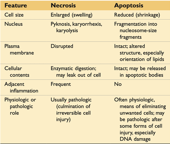
TYPES OF CELL DEATH
Apoptosis
Necrosis
APOPTOSIS
Is a form of cell death that is generally triggered by normal, healthy processes in the body.
Apoptosis, which can also occur as a defense mechanism during healing processes, is almost always normal and beneficial to an organism.
NECROSIS
Is cell death that is triggered by external factors or disease, such as trauma or infection.
Necrosis is always abnormal and harmful.
It is considered an "unprogrammed" (unnatural) cell death process at this time
TYPE OF NECROSIS
“CoLiG C. FaG F“
Coagulation Necrosis
Liquefaction Necrosis
Gangrenous Necrosis
Caseous Necrosis
Fat Necrosis
Gaseous Necrosis
Fibrinoid Necrosis
COAGULATION NECROSIS
The architecture of the dead tissues is preserved
Denatures not only the proteins but also the intracellular enzymes
This type is seen most often in the heart after an infarction, as well as in kidneys and spleen
LIQUEFACTION NECROSIS
Transformation of the tissues into liquid viscous mass
Seen in focal bacteria/ fungal infection
Accumulation of leukocytes and liberation of enzymes- pus
GANGRENOUS NECROSIS
Refers to massive death of the tissue caused by a combination of ischemia and superimposed by bacterial infection
Applied to a limb:Coagulative→ arterial occlusion
Liquefactive→ venous occlusion
CASEOUS NECROSIS
Conversion of destroyed cells into granular, friable mass made up of a mixture of coagulated protein and fats.
Fragmented or lysed cells enclosed in an inflammatory border- granuloma
FAT NECROSIS
Focal area of fat destruction
Release of pancreatic lipase into the pancreas and peritoneal cavity
Acute Pancreatitis
GASEOUS NECROSIS
This combination of liquefactive and coagulative necrosis is caused by dead cells that are not completely digested by macrophages; they leave a granular residue that impedes circulation.
FIBRINOID NECROSIS
Fibrin is the main reason for this type of necrosis to happen.

BIOPSY
Removal of cells or tissues for examination from a living subject to determine the presence or extent of a disease
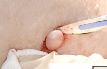
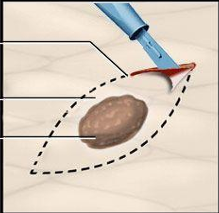
EXCISIONAL BIOPSY
When an entire lump or suspicious area is removed
INCISIONAL/ CORE BIOPSY
When only a sample of tissue is removed with preservation of the histological architecture of the tissue’s cell
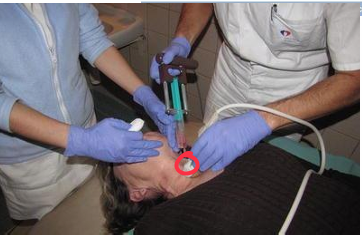
FINE NEEDLE ASPIRATION BIOPSY
When a sample of tissue or fluid is removed with a needle.
OTHER TYPES OF BIOPSIES
Punch Biopsy
Shave Biopsy
Curettage Biopsy
PUNCH BIOPSY
A punch biopsy is done with a circular blade ranging in size from 1 mm to 8 mm. The blade, which is attached to a pencil-like handle, is rotated down through the epidermis and dermis, and into the subcutaneous fat, producing a cylindrical core of tissue.
SHAVE BIOPSY
A shave biopsy is done with either a small scalpel blade or a curved razor blade.
CURETTAGE BIOPSY
A curettage biopsy can be done on the surface of tumors or on small epidermal lesions with minimal to no topical anaesthetic using a round curette blade.
CURETTE
A surgical instrument that has a scoop, ring, or loop at the tip.
NECROPSY; AUTOPSY
After death examination of the body and dissection of its internal organs to confirm or determine the cause of death.
Autopsy can uncover the existence of disease not detected during life, determine the extent of injuries may have contributed to a person’s death.
PRELIMINARIES FOR PME
Written consent from the next of kin-abide by the extent or restrictions allowed
Death certificate (blue form)
Medical abstract / Clinical data
Medico Legal Clearance
PME IS PERMITTED W/O CONSENT IN THE FOLLOWING CIRCUMSTANCES
When it is ordered by the police or coroner
When it is necessary to complete the death certificate
When the deceased himself has given consent before he died
Deceased military personnel who dies in active/training duty or military service
THE CORONER HAS AUTHORITY IN THE FOLLOWING CASES
All natural deaths occurring in the hospital within 24 hrs. of admission, unless the case was attended by a private physician within 36 hours of death
Newborns in the first 24 hours of life
All injury cases, old or recent
All deaths due to unknown cases
All deaths due to suspicious cases
All abortion cases, whether self-induced or otherwise
All violent deaths
All accidental deaths
All sudden deaths
All cases without medical attendance within 36 hours prior to the hour of death
All deaths due to drowning, hanging or strangulation
All deaths due to shooting, stab wounds, burns, electricity, lightning, tetanus, etc.
All homicide
All suicide
All cases in which there is suspicion of poisoning
Stillborns
Prematures
POSTMORTEM CHANGES
Algor Mortis
Rigor Mortis
Livor Mortis or Postmortem Hypostasis
Postmortem clotting of blood
Discoloration of tissues
Putrefaction- decaying/ rotting
ALGOR MORTIS
At room temp.: 2-2.5°F/hr – 1st hour
As a rule, the body cools at 1.5°F/hr (50% of cases)
Not reliable indicator
12 hours: 1.5-2°F/hr
12-18 hours: 1°F/hr
RIGOR MORTIS
Rigidity of the body due to hardening of the skeletal muscles caused by a series of physicochemical events after death
Lacks ATP regeneration
Sets within 2 hrs. after death (head & neck)
Complete with 12 hrs.
3-4 days longevity
LIVOR MORTIS OR POSTMORTEM HYPOSTASIS
Becomes evident as early as 20 min. after death
Blood supply will gravitate to the skin which become toneless and dilate
FOUR MAIN TYPES OF AUTOPSIES
Medico-Legal Autopsy or Forensic or Coroner's autopsy
Clinical or Pathological autopsy
Anatomical or Academic Autopsy
Virtual or Medical Imaging Autopsies or Virtopsy
MEDICO- LEGAL AUTOPSY OR FORENSIC OR CORONER’S AUTOPSY
Death is classified into one of five headings: (HANSU)
a. natural
b. accident
c. homicide
d. suicide
e. undetermined
cause and manner of death
CLINICAL OF PATHOLOGICAL AUTOPSY
Is performed to diagnose a particular disease
It aims to determine, clarify, or confirm medical diagnoses that remained unknown/ unclear prior to the patient's death
ANATOMICAL OF ACADEMIC AUTOPSY
Is performed by students of anatomy for study only.
VIRTUAL OR MEDICAL IMAGING AUTOPSIES OR VIRTOPSY
Is performed utilizing imaging technology only, primarily magnetic resonance imaging (MRI) and Computed Tomography (CT)
EXTERNAL EXAMINATION
Photograph
Note the clothing, position
Evidence-residues- uses UV Light
Samples of hair, nails.
After external evidence is collected-
Clean the body, weighed, measured
General description of the body as regards ethnicity, sex, age, hair color and length, eye color and other distinguishing features
Handheld voice recorder
INTERNAL EXAMINATION
Y shaped incision can be made starting at the top of each shoulder and running down the front of the chest, meeting at the lower point of the sternum.
T-Shaped Incision made from the tips of both shoulders in a horizontal line across the region of the collar bones to meet at the sternum in the middle
Single Vertical Cut is made from the middle of the neck
KINDS OF AUTOPSIES
Autopsy of Virchow
Autopsy of Letulle
Autopsy of Rokitansky
Autopsy of Ghon
AUTOPSY OF VIRCHOW
Where each organ is taken out one by one
GOOD FOR: Demonstrating pathological change in individual organs
ADVANTAGE OF VIRCHOW
Simple to perform
Systematic examination
DISADVANTAGE OF VIRCHOW
Destroys anatomic relationship
AUTOPSY OF LETULLE
Where organs are taken out En bloc
(En bloc- all together in a united group, En masse- in a mass; all together; as a group)
ADVANTAGE OF LETULLE
Preserving vascular supply
AUTOPSY OF ROKITANSKY
Where organs are examined in- situ (in placed)
ADVANTAGE OF ROKITANSKY
ADVANTAGE:
Practical for single examiner
Good preservation of anatomic structures in-situ.
Good preservation of pathologic anatomic relationships
Subsequent autopsies can be easily performed on the same body
DISADVANTAGE OF ROKITANSKY
Requires a certain degree of expertise and skill
Thorough examination may not be possible
If cause of death is determined, less attention is paid to other
Pathologies
AUTOPSY OF GHON
There organs are taken out in three separate blocks
A. Cervical region
B. Abdominal region
C. Urogenital region
ADVANTAGES OF GHON
Anatomic relationships are preserved without a bulky mass of organs
Systems examined within their structural integrity
Good observation of pathologic lesions
ADVANTAGES OF GHON
Not ideal for multiple organ involvement
Requires skill and time