Test 2 - Visual Pathways (4)
1/29
There's no tags or description
Looks like no tags are added yet.
Name | Mastery | Learn | Test | Matching | Spaced | Call with Kai |
|---|
No analytics yet
Send a link to your students to track their progress
30 Terms
what is a special feature of the fovea?
It takes up .01% of the retina BUT 8-10% of the cortical map’s area. (over-represenation of fovea) (CORTICAL MAGNIFICATION)
why does the special feature of foveas exist?
no neural convergence (cones < bipolar cells < ganglion cells), needs more neurons to support lack of neural convergence
what are the three parts that make up a hypercolumn?
location columns
orientation columns
ocular dominance columns
what is a retinotopic map?
an electron map of the retina on the cortex
what is positron emission tomography (PET)?
injection of a radioactive tracer monitors changes in blood flow which represent changes in brain activity
what is functional magnetic resonance imaging (fMRI)?
can determine areas of activity in the brain by looking at changes in the magnetic response of hemoglobin. this is cause hemoglobin has oxygen which carries a magnetic molecule.
does cortical over-representation exist in our other senses?
Touch - over-representation of cortical neurons in hands and face because more sensory information is coming in
what is a location column?
receptive fields at the same location on the retina are within a column
what is an orientation column
exists within location columns. all neurons within same column fire to the same orientation. all following columns will have slightly different orientation preferences but they are different in a consistent pattern. 1 mm across cortex represents the entire range of orientation.
what is an ocular dominance column?
neurons in the cortex respond preferentially to one eye
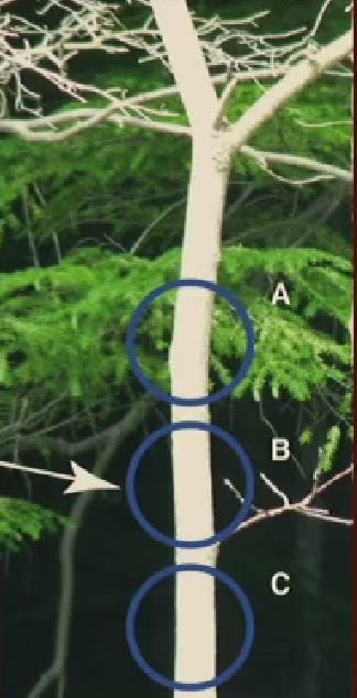
What type of activity would you expect to see in the hyper columns for points A, B, and C?
Neurons in the 90 degree orientation columns would light up for A, B, and C. It is worth noting that A, B, C are different areas meaning they are different receptive fields meaning there are gonna be three separate orientation columns within three separate location columns.
For the tree question, what type of coding would that be?
sparse coding - minimal neurons responding to photo NOT ONE & NOT A SHIT TON

describe what going on in your brain when you attempt to perceive this image
For each of those yellow circles, they represent separate receptive fields. For each of those receptive fields, there exists a separate location column and for each of those location columns, appropriate orientation columns would fire in response to the picture depending on the angle of the bars of light.
how do feature detectors respond to a scene?
tiling - columns working together to cover the entire visual field
where does the dorsal pathway lead? what type of information does that lobe process.
parietal lobe, processes ‘where/how’ information
where does the ventral pathway lead? what type of information does that lobe process?
temporal lobe, processes the ‘what’ information
how did we find out about the functional specialization of certain lobes?
through lesioning and ablation experiments. we’d cut off parts of animal brains and see what works and what doesnt.
what is the object discrimination problem? what does that study reveal?
Object discrimination is telling the different between two separate objects. Monkeys were measured on their ability to tell where the food was at. Experimenters taught monkeys by hiding food under specific types of objects. Revealed that temporal lobe contained ‘what’ information. (what object has food hur dur?)
what is landmark discrimination? what does that study reveal?
Monkeys were trained to find food based on closeness to landmark object. Revealed that parietal lobe contained the ‘where/how’ information. (where the food at? how the food always here? because its close to a landmark.)
how did neuropsychology prove localization of function for the parietal lobe and the temporal lobe?
a double dissociation using two people who had brain damage for the temporal lobe and parietal lobe.
what was the findings of the double dissociation study?
alice (temporal lobe damaged patient) could determine object location but could not name it. bert (parietal lobe damaged patient) could name the object but not determine the location.
who was patient D.F?
had damage to ventral pathway. Was not able to match orientation of card with slot.
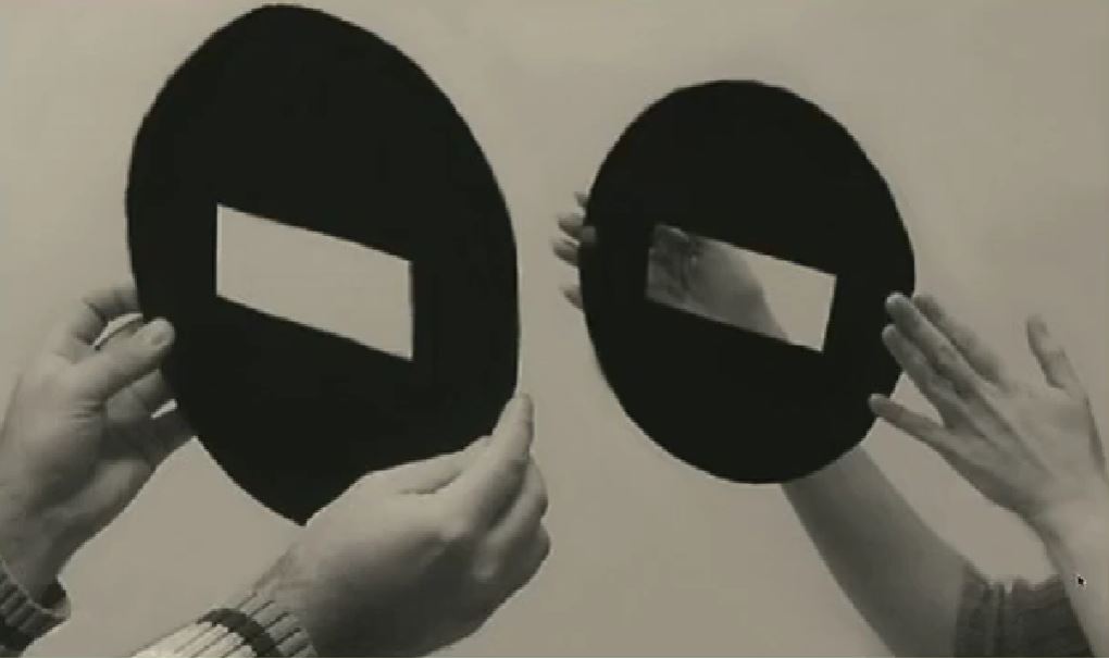
how did they help patient D.F?
rather than matching the slot, researchers changed the conditions of the task and had her insert a card into a slot. ‘what’ based task turned into ‘where’ based task
whats a module?
a brain structure that processes information about specific stimuli
what does the inferotemporal (IT) cortex best respond to? (monkey)
faces
which area in humans best responds to faces? where is it located?
fusiform face area (FFA), in temporal lobe
what happens when fusiform face area damaged?
This causes face blindness. (prosopagnosia)
what does the parahippocampal place area (PPA) respond best to?
responds best to places/spatial layout
what does the extrastriate body area best respond to?
responds best to pictures of full bodies and body parts
which area of temporal lobe is important for memory?
medial temporal lobe (hippocampus)