Intro to Sonography Chapter 3 and 4
1/104
Earn XP
Description and Tags
PPT notes
Name | Mastery | Learn | Test | Matching | Spaced |
|---|
No study sessions yet.
105 Terms
The Study of Sound - Acoustics
Sound is a form of energy that is produced when a vibrating source causes molecules within a medium to move back and forth.
The back-and-forth motion allows waves of sound energy to travel.
The human body is made of different mediums that allow sound to propagate.
The scientific study of sound is referred to as acoustics.
Boethius identified the pebble theory, which visualizes sound waves traveling like the waves created by a pebble dropped in water.
da Vinci also assumed the sound traveled in waves.
Robert Boyle recognized there must be a medium through which sound can travel in order for it to propagate.
His research laid the groundwork for the use of coupling gel.
The Study of Sound- Doppler effect
Abbe Lazzaro Spallanzani, the “father of ultrasound,” studied how bats use sound waves to detect their victims and to guide their flight.
By recalling this, we can recognize how the ultrasound transmitter utilizes the pulse-echo technique.
Christian Johann Doppler discovered that the pitch of a sound wave varies if the source of the sound was moving (the Doppler effect).
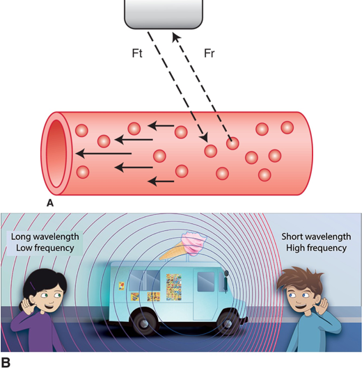
The Study of Sound- Doppler sound beam
A. The transmitted Doppler sound beam (Ft) encounters red blood cells (RBCs) moving toward the transducer within the blood vessel. The RBC motion causes an increase in frequency of the returning echo (Fr) due to the Doppler effect. The ultrasound instruments detect and measure the frequency of the returning Doppler signal, confirming the presence of blood flow and the direction in which the blood is traveling. B. Illustration of the Doppler effect where the frequency of sound increases when object moves toward the boy and decreases when object recedes away from the girl.
The Study of Sound- piezoelectric effect
The Currie brothers recognized the piezoelectric effect.
This is the process whereby a material, such as a crystal or element within an ultrasound transducer, generates electricity and changes shape with application of pressure.
The crystals in the transducer produce ultrasound waves.

Ultrasound in Medicine- Reflectoscope
During World War I, ultrasound was used to detect submarines.
This led to the development of sonar technology, which used sound that was sent through the water, bounced off an object, and then returned to the source.
Floyd Firestone used this to develop and use the reflectoscope, which used ultrasound to detect flaws in metal.
- This was the technique first used in medicine.
The first application of ultrasound in medical diagnosis was in 1941.
Karl Dussik used it to image the lateral ventricles in the brain.
As research progressed, scientists realized the ultrasound waves returned to the transducer and may be able to form an image.
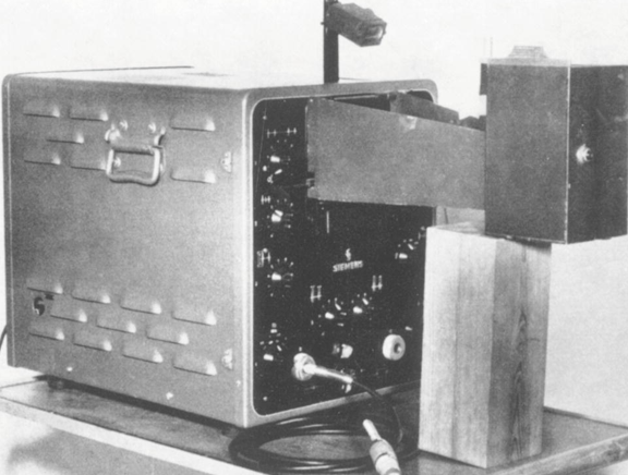
Ultrasound in Medicine- Early Echocardiograms
Edler and Hertz used an ultrasonoscope, seen here, to record their early echocardiograms
Ultrasound in Medicine- pulse-echo technique
The pulse-echo technique sought to exploit reflected sound back from within the body to create an image.
- Sound must be pulsed or allowed to be alternated rapidly on and off so the transducer can listen for the echo.
Upon hearing the echo, the machine calculates distance and presents the reflector’s location on the monitor.
- Efforts to use the technique were first made in the late 1940s and early 1950s.
One early attempt demonstrated reflections from a gallstone.
In another, a Swedish cardiologist borrowed a sonar device from a shipyard and recorded echoes from his own heart.
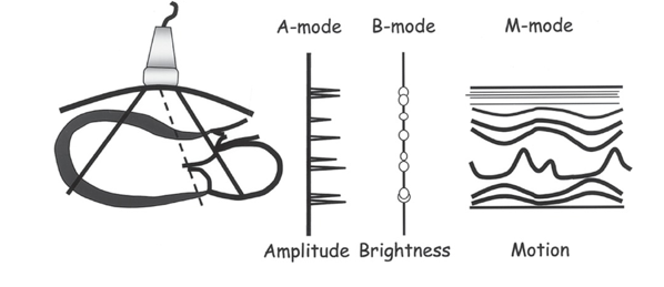
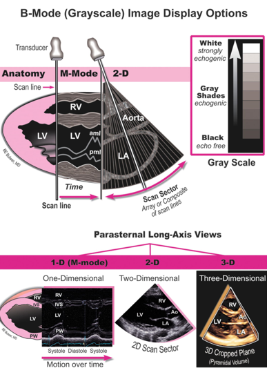
Imaging Modes and Doppler- A-mode and B-mode
Display Options
- A-mode (amplitude mode) represents the depth of the returning echo on the x-axis and the strength (amplitude) of the reflector on the y-axis.
A pulse of sound was sent out to create one scan line of information interpreted to represent depth and amplitude.
This is used in echocardiography and ophthalmic ultrasound.
B-mode (brightness mode) displays the returning ultrasound signal as a dot on the monitor.
The dot has varying degrees of brightness, based on the strength of the returning echo.
The stronger the returning echo, the brighter the dot.
This is also referred to as grayscale sonography.
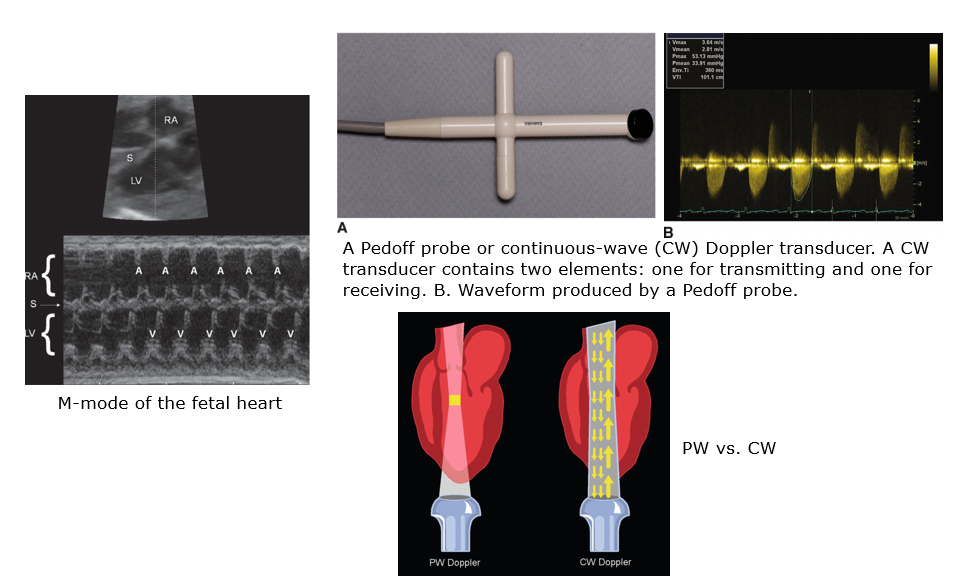
Imaging Modes and Doppler - M-mode
Display Options (cont.)
- M-mode (motion mode) documents the movement of structures in the body along a single scan line.
The y-axis shows depth; the x-axis shows time.
M-mode is used to demonstrate fetal heart rate and in echocardiography as a critical part of standard protocols.
- The original ultrasound machines provided static images; now we are able to use real-time scanners.
Imaging Modes and Doppler- Doppler Technology
Doppler Technology
- Robert Rushmer and his colleagues established the varying uses of continuous-wave (CW) Doppler and spectral analysis in 1963.
CW transducers combine an element continuously sending waves with one that continuously listens for the return signal.
- In the 1970s, there were several advancements, including:
Pulsed-wave Doppler
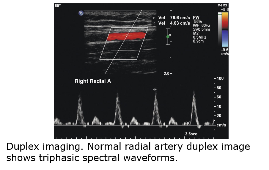
Duplex imaging, a handheld duplex pulsed system
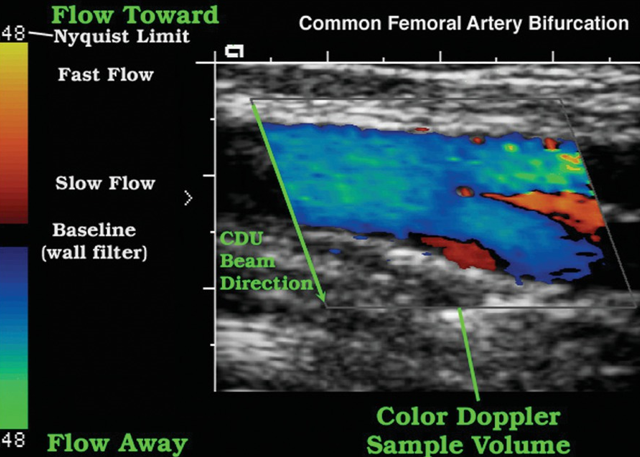
Advancements in color Doppler imaging and instrumentation
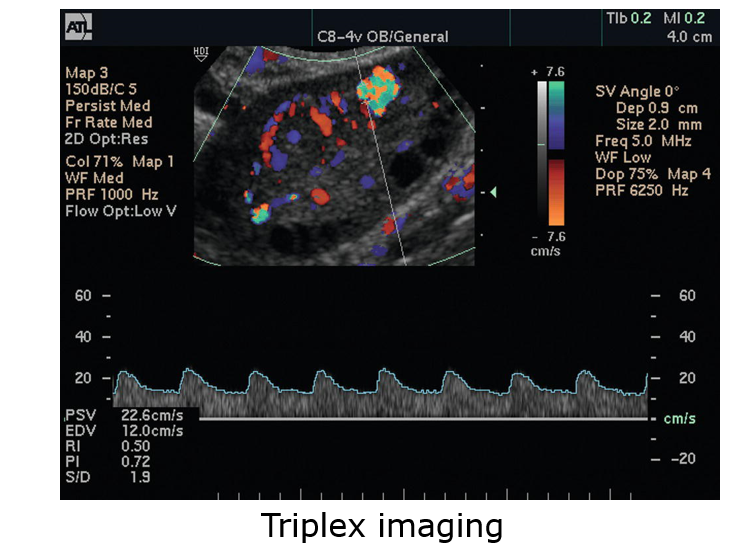
The combination of B-mode, spectral, and color Doppler is called triplex imaging.
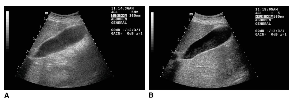
Imaging Modes and Doppler- B-Mode (Gray-scale) Image
…
Imaging Modes and Doppler- M-mode, PW, CW etc examples
…
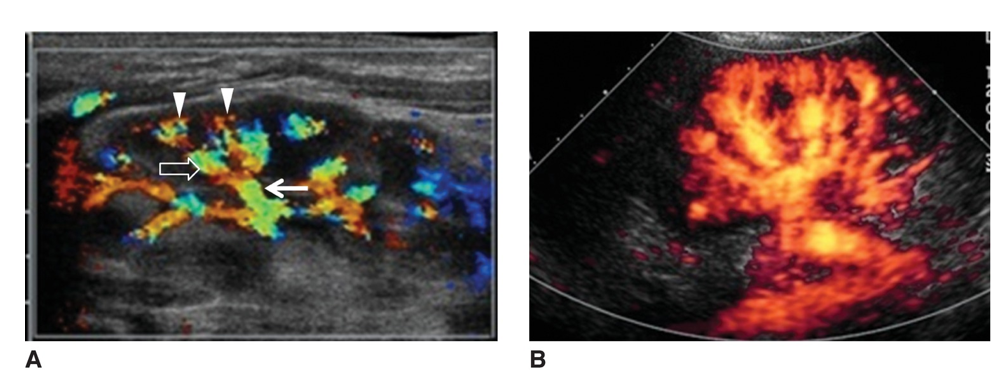
Imaging Modes and Doppler- Color Doppler
Beam direction going downwards.
Imaging Modes and Doppler- Duplex imaging
…
Imaging Modes and Doppler- Triplex imaging
…
Imaging Modes and Doppler- Color Doppler and Power Doppler
…
Tissue Harmonic Imaging
Harmonics are additional frequencies, other than the transmitted frequency sent into the body, that are generated by differing human body tissues.
These are collected by the transducer and used to create a crisper, higher-resolution image.
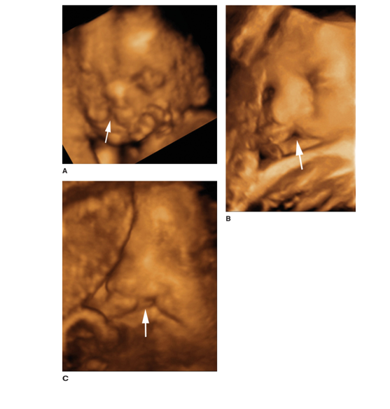
3D and 4D Technology - 3D allows one to see the width, height, and depth of Images
3D allows one to see the width, height, and depth of images.
- It is useful in obstetrics for clear visualization of the form of the fetal face.
It is also used in breast, vascular, gynecologic, and abdominal sonographic imaging.
The images are made of two 2D images placed next to each other and reconstructed by a computer into a 3D format.
A 3D image can be created through several processes:
- Manual movement of the transducer across a specific path
- Use of a mechanical 3D transducer
- Use of a 2D transducer
3D and 4D Technology- Volumetric imaging
Volumetric imaging helps address the concern of depending on the skill of the sonographer to acquire diagnostic imaging.
- An offline work station is used to do digital postprocessing of images.
- The sonographer finds a good window and completes just one sweep from lateral to medial.
- A computer recreates 3D images.
There are some limitations to 3D technology:
- Optimal imaging of the fetal face depends on enough amniotic fluid and favorable fetal positioning.
- For some applications, a 2D image provides enough information for diagnosis; a 3D image simply confirms.
3D and 4D Technology- 3D images of a fetus
…
3D and 4D Technology- Keepsake imaging
4D ultrasound offers real-time images in 3D.
- The fourth dimension is time.
The use of the technology is still evolving.
Keepsake imaging centers exploit 3D and 4D images for economic and entertainment purposes.
- The American Institute of Ultrasound in Medicine’s official statement calls for certified professionals and licenses physicians to maintain appropriate patient care.
Specialties in Sonography - Abdominal Sonography
Abdominal Sonography
Abdominal sonographers must appreciate relevant normal abdominal anatomy and pathology of each organ and system within the abdomen and small parts.
Transducer frequency ranges 2 to 5 MHz for general imaging.
CW and PW Doppler are often utilized to assess vascular structures and provide evidence of blood flow within abdominal masses and organs.
Abdominal sonographic imaging includes a wide variety of abdominal structures and can be ordered for numerous reasons.
Abdominal sonographers may assist the physician in invasive procedures or evaluate for renal artery stenosis or assist during endoscopic ultrasound.
Patient prep is typically nothing by mouth for 6+ hours prior.
Specialities in Sonography- Small Parts Sonography
Abdominal Sonography: Small Parts Sonography
- Small parts include the thyroid, scrotum, and prostate gland.
- Abdominal sonographers may also:
Perform breast sonography
Evaluate the penis, chest, specific painful joints or tendons, bowels, the abdominal wall for hernias, and palpable masses to confirm evidence of foreign bodies.
Scan any external body part to which both acoustic gel and the transducer can be applied.
- The majority of small parts require the use of a linear transducer and, sometimes, the use of an acoustic stand-off device.
- Challenges can arise from large patient body habits, bowel gas, surgical bandages, patient preparation, lack of compliance, or intolerance.
Specialities in Sonography- Breast Sonography in Conjunction with Mammography and Physical Examination
Breast Sonography
- This is used in conjunction with mammography and physical examination.
- Sonography is the initial modality of choice for patients under 30 and those who are pregnant or lactating.
- Sonography can differentiate cystic versus solid masses; mammography often cannot.
- Other uses include possible breast implant rupture, during needle placement for biopsy, cyst drainage, and radio-frequency ablation.
- Breast sonography should be performed with a high-resolution, real-time linear array transducer with a frequency of at least 10 MHz.
- A supine-oblique position with the ipsilateral arm raised is often used.
Specialties in Sonography- Breast Sonography like a Face of a Clock
Breast Sonography (cont.)
- The breast is visualized like the face of a clock.
- Imaging is performed in transverse and longitudinal planes or radial and antiradial planes.
- Breast sonographers should have a thorough appreciation of mammography techniques and breast pathology noted on a mammogram.
- Interpretation often integrates Breast Imaging Reporting and Data System (BI-RADS).
- One of the main concerns is that sonography is highly operator dependent.
- Automated whole breast scanners allow for better reproducibility and correlation with other modalities.
Specialties in Sonography- Neurosonography and Pediatric Sonography
Neurosonography includes neonatal brain imaging, newborn infant spine imaging, and intraoperative sonography.
Sonography is a portable, high-resolution, economic alternative to other imaging studies.
Images are obtained routinely through the anterior fontanelle.
The infant spine is imaged with a high-frequency linear array transducer with frequency ranges 7 to 10 MHz.
The patient is placed prone.
Infants may be scanned as the result of suspicious intrauterine findings during an obstetric sonogram.
Neurosonography was once a distinct certification, but it has been replaced with certification in pediatric sonography.
Pediatric images follow protocols similar to adult imaging.
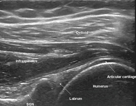
Specialties in Sonography- Musculoskeletal Sonography
This includes evaluation of the shoulder, wrist, knee, and any other joints, tendons, and muscles of the extremities.
The search for foreign bodies may also be a requirement.
Specialties in Sonography- Gynecologic Sonography - Filling the Urinary Bladder
Patient preparation includes filling the urinary bladder.
This provides an acoustic window for visualizing the uterus, ovaries, and other structures in the adnexa.
Transabdominal sonography employs a transducer 3.5+ MHz.
Transvaginal sonography employs a transducer 5+ MHz.
Transvaginal sonography has several advantages:
Better resolution of organs and structures in large patients
Does not require a distended bladder
Saline infusion sonohysterography allows clear visualization of the endometrial lining and uterine cavity.
Sterile saline is injected with a catheter; the sonogram is performed during the procedure.
Specialties in Sonography- Gynecologic Sonography - Ability to Assess the Endometrium
The ability to assess the endometrium and ovaries has made significant impact on assisted reproductive therapy and fertility treatment.
Use in postmenopausal women is helpful in assessing bleeding.
Specialties in Sonography- Obstetric Sonography - One of the most Established and Recognizable Applications of Ultrasound in Medicine
This is one of the most established and recognizable applications of ultrasound in medicine.
Sonographers practicing obstetrics must be familiar with fetal abnormalities and maternal complications.
There are several stages when a sonogram may be required:
- In the first trimester:
To confirm intrauterine pregnancy
For vaginal bleeding
If an ectopic pregnancy is suspected
For screening secondary to high-risk clinical history findings
Routine assessment of maternal and fetal anatomy
Screening for genetic complications
Specialties in Sonography- Obstetric Sonography - Second and Third Trimester
In the second and third trimesters:
Routine and detailed anatomic survey
Obstetric sonographers may assist with interventional perinatal procedures, including:
Amniocentesis
Chorionic villus sampling
Cordocentesis
Specialties in Sonography- Obstetric Sonography - Fetal Echocardiography
This branch of obstetric sonography specializes in the fetal heart.
If a parent has a family history of congenital heart defects or if routine sonogram is suspicious, fetal echocardiogram is performed.
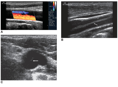
Specialties in Sonography- Vascular Sonography
The sonographer examines the arterial and venous systems of the arms and legs, the intracranial and extracranial blood vessels, and abdominal vasculature.
Many studies are performed with standard equipment and a 5- to 7-MHz linear transducer.
Studies are often performed with a combination of PW spectral and color Doppler.
Angle correction is crucial.
Vascular sonography may be direct or indirect.
Specialties in Sonography- Echocardiography - Hemodynamics
The cardiac sonographer or echocardiography examines and assesses the anatomical structures of the heart as well as its hemodynamics.
The sonographer uses real-time 2D imaging, M-mode, and Doppler echocardiography.
The most common test is a transthoracic echocardiogram.
Typically, low-frequency array transducers are used.
The patient is typically in the left lateral decubitus position. The left arm is raised above the patient’s head.
Several breathing techniques are employed to enhance visualization of the heart and reduce lung movement.
Specialties in Sonography- Echocardiography - Stress Echocardiogram
A stress echocardiogram assesses how the heart functions with exertion.
This may be combined with exercise or may be done pharmacologically.
A pharmacologic stress echocardiogram uses drugs to increase blood flow to the heart, mimicking exercise.
A transesophageal echocardiogram is an invasive procedure.
Sedation must be utilized and the patient may require full anesthesia.
Many TEE transducers utilize 5 MHz of frequency.
Pediatric echocardiography is similar to adult, but patient movement is a special challenge. Sedation may be required.
Additional Technologies and Future Applications- Therapeutic Ultrasound
Therapeutic ultrasound is used to increase blood supply to certain areas by heating the tissue to reduce healing time.
High-intensity focused ultrasound destroys tissues such as fibroids or tumors.
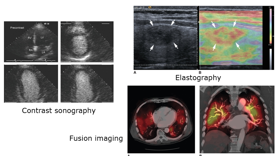
Contrast-enhanced ultrasound enhances the echogenicity of vessels and improves border recognition.
Ultrasound-guided brachytherapy uses ultrasound guidance to treat cancers with radioactive material.
Ultrasound elastography evaluates a mass based on stiffness to predict if the mass is malignant or benign.
Fusion imaging allows the ultrasound machine to communicate with the PACS system to call up previous MRI or CT scans.
Intravascular ultrasound uses a miniature probe to scan the circulatory system.
Additional Technologies and Future Applications- Contrast Sonography, Elastography, Fusion Imaging
…
Additional Technologies and Future Applications- Automated Ultrasound
Automated ultrasound is steered by a computer system.
Focused assessment with sonography for trauma offers the emergency room physician a quick and sensitive method of diagnosing abdominal trauma.
Miniaturization of equipment has lead to higher-definition monitors and smaller computer system housing.
Wireless technology allows the transducer to communicate with the ultrasound machine without a cord getting in the way.
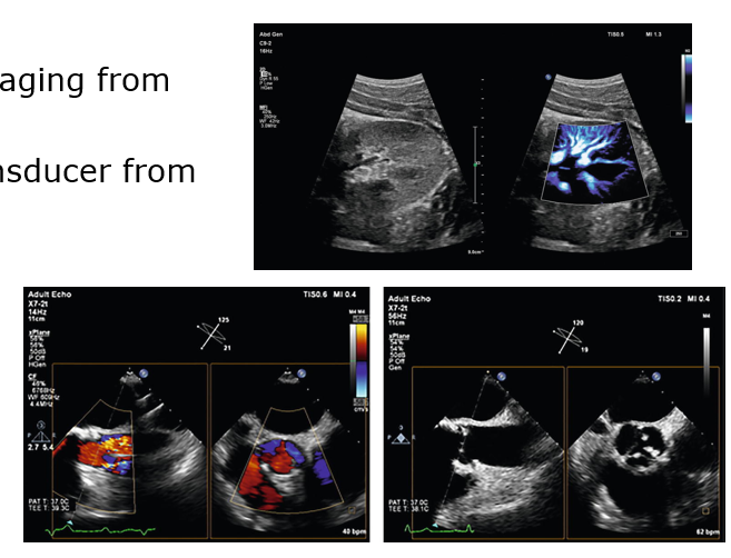
Additional Technologies and Future Applications- MicroFlow Imaging from Phillips
MicroFlow Imaging from Philips
xMATRIX transducer from Philips
Summary for Chapter 3
This chapter is just a starting point for you to begin your studies.
It should be apparent from the reading that our profession is constantly growing.
One vital role of the sonographer is to maintain a thorough knowledge base regarding the sonography profession.
Knowing that the medical uses of ultrasound are constantly evolving should provide you with both enthusiasm about your opportunities going forward and reassurance that you have chosen a wise career path.
What instrument uses ultrasound to detect flaws in metals?
a. Hydrophone
b. Doppler transducer
c. Reflectoscope
d. Continuous-wave transducer
c. Reflectoscope
What instrument was designed to be used underwater for recording or listening to underwater sound?
a. Hydrophone
b. Spectral transducer
c. Reflectoscope
d. Real-time scanner
a. Hydrophone
What mode displays the depth of the returning echo represented on the x-axis and the strength (amplitude) of the reflector represented along the y-axis?
a. B-mode
b. A-mode
c. C-mode
d. M-mode
b. A-mode
What technology is used to analyze the relative stiffness of a mass?
a. Sonohysterography
b. Plethysmography
c. Photoplethysmography
d. Elastography
d. Elastography
Triplex imaging includes which of the following?
a. A-mode, B-mode, and M-mode
b. B-mode, spectral Doppler, and color Doppler
c. M-mode, B-mode, and color Doppler
d. Spectral Doppler, power Doppler, and B-mode
b. B-mode, spectral Doppler, and color Doppler
Which testing method utilizes information gathered from one area of the body to predict disease in another part of the body?
a. Indirect
b. Direct
c. Coherent
d. Incoherent
a. Indirect
A technology that uses a computer system to obtain measurement of images, such as fetal biometry, is said to be
a. Superior
b. Mechanical
c. Automated
d. direct
c. Automated
What is an alteration in the normal structure or number of a chromosome?
a. Congenital defect
b. Fetal aberration
c. Chromosomal anomaly
d. Congenital anomaly
c. Chromosomal anomaly
What mode captures the movement of structures along a single scan line represented over time?
a. B-mode
b. C-mode
c. A-mode
d. M-mode
d. M-mode
Which of the following is not considered a medium through which sound can travel?
a. Vacuum
b. Solid
c. Liquid
d. Gas
a. Vacuum
Who has been referred to as the "father of ultrasound"?
a. Doppler
b. Spallanzani
c. Boethius
d. da Vinci
b. Spallanzani
What is the most common element or crystal used within the ultrasound transducer?
a. Copper titanate
b. Lead iron titanate
c. Iron copper titanate
d. Lead zirconate titanate
d. Lead zirconate titanate
Who established the pebble theory?
a. Doppler
b. Spallanzani
c. Boethius
d. Da Vinci
c. Boethius
What technology does sonography assist in administering radioactive materials close to or within a cancerous tumor?
a. Fusion imagine
b. Hybrid imaging
c. Ultrasound-guided brachytherapy
d. Therapeutic ultrasound
c. Ultrasound-guided brachytherapy
Who first recognized the frequency shift that is created when sound impinges upon a moving object?
a. Christian Doppler
b. Robert Boyle
c. Jacques Currie
d. Charles Langevin
a. Christian Doppler
What is the term for an abnormality with which someone is born?
a. Chromosomal retardation
b. Congenital anomaly
c. Chromosomal fluctuation
d. Fetal karyotyping
b. Congenital anomaly
Which of the following would not be an effective stand-off device?
a. Gel pad
b. Balloon
c. Mound of gel
d. Bag of saline
b. Balloon
What ultrasound mode do we use today to acquire routine grayscale images?
a. Doppler
b. B-mode
c. A-mode
d. M-mode
b. B-mode
What type of scanner allows for constant visual imaging of anatomy as if one were watching a movie?
a. A-mode
b. Continuous wave
c. Real-time
d. M-mode
c. Real-time
Who was the first to use diagnostic ultrasound in the United States?
a. Christian Doppler
b. Joan Baker
c. Jeffrey W. Penny
d. George Ludwig
d. George Ludwig
Introduction
There are many opportunities for you to make your mark on the sonography profession.
Strive for leaving a legacy of excellence behind for future generations.
Sonographers must continually enhance their knowledge.
The healthcare field is always evolving.
Professional Environment
Once you have graduated, a world of opportunity awaits you.
You have the option to join many professional organizations. These have several benefits:
Maintaining competence
Preparing to adjust to the ever-changing healthcare environment and an evolving career
The professional environment includes academic accreditation, national certification, and laboratory accreditation.
Academic Accreditation
This assesses the quality of institutions, programs, and services.
Being accredited demonstrates a program’s desire to maintain high standards and to graduate exceptional entry-level sonographers.
National accreditation is granted through the Commission on Accreditation of Allied Health Education Programs (CAAHEP).
The review process is exceedingly rigorous.
It involves review of materials and personal visits to the institution.
As a student, you may be asked questions during an accreditation site visit.
Accreditation can be obtained for either 5 or 10 years.
Professional Organizations
These organizations provide a means by which you can receive:
The latest information about your occupation
An opportunity to make a personal impact on the profession
There are state-, regional-, and national-level societies.
The Society of Diagnostic Medical Sonography (SDMS) is a nationwide organizations.
Members are at all levels of association with the profession.
Benefits include a national meeting where CME credits can be obtained.
Other national organizations include the Society of Vascular Ultrasound and the American Society of Echocardiography.
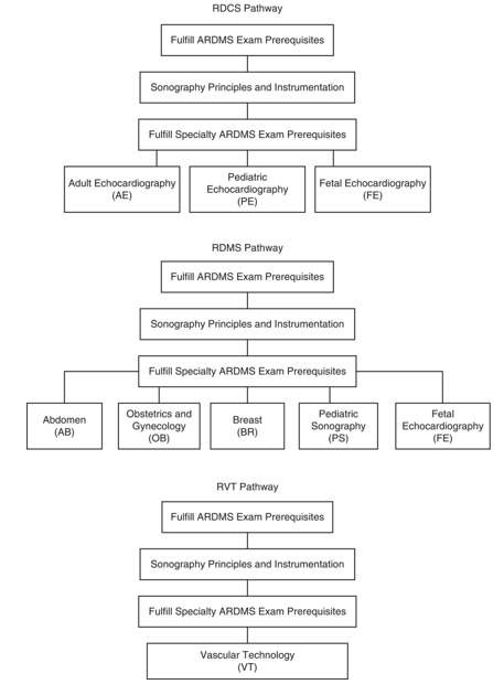
National Certification - American Registry of Diagnostic Medical Sonography Specialties
The American Registry of Diagnostic Medical Sonography offers certification for all sonography specialties:
Registered diagnostic medical sonographer
Registered diagnostic cardiac sonographer
Registered vascular technologist
Certification carries across state borders.
Candidates must pass the Sonography Principles and Instrumentation examination and any specialty examinations.
Testing may be conducted while you are in school, but graduation from your program is required to receive credentials.
National Certification (ARDMS) Pathway Image
…
National Certification- Cardiac and Vascular Specialists
Cardiac and vascular specialists may seek credentials from the Cardiovascular Credentialing International.
The American Registry of Radiologic Technologists also offers credentialing exams for sonographers.
Your state may require you to obtain state licensure in addition to national certification.
Maintaining Certification
This is accomplished through obtaining continuing medical education (CME) credits.
The ARDMS requires sonographers to obtain 30 ARDMS-accepted credits within a 3-year period following initial certification.
CMEs can be obtained through:
Attending meetings
Successfully passing additional certification exams
Publishing original academic papers
Credentials are valid for 10 years.
Laboratory Accreditation
For vascular testing, this is obtained through the Intersocietal Commission for the Accreditation of Vascular Laboratories.
For echocardiography, accreditation is granted through the Intersocietal Accreditation Commission of Echocardiography.
The American College of Radiology is also involved with accreditation of ultrasound practices.
Accreditation processes are rigorous.
Accreditation provides a standard of practice that enhances patient care and ensures uniformity in examinations.
Leadership Roles in Sonography
You are a leader any time you seek to influence the thinking, behavior, or development of another person.
In healthcare, well-informed leaders recognize the patient’s role as a customer requiring satisfaction with your service.
Healthcare delivery and Medicare reimbursement rates are strongly associated with patient satisfaction surveys.
Leadership is the ability to influence others to accomplish a specific goal.
A sonographer demonstrating ethical and appropriate patient care is a leader:
The sonographer influences the patient in a positive manner.
The sonographer is making a personal choice about how and to what extent he or she will use his or her influence.
Leadership Roles in Sonography - Patient Servant
Patient servant is a basic function of all us who work in healthcare.

Essentials of Servantship in Healthcare
Smile
Ensure patient dignity
Respond to patient needs appropriately
Value the uniqueness of the patient (e.g., age, race, gender, emotional state)
Advocate patient rights and safety
Never demonstrate disrespect toward the patient
Thank the patient
Serve the patient with compassion
Help others serve the patient
Identify patient mistreatment and demand improvement
Portray professionalism
Leadership Styles and Followership - Utilize Different Leadership Styles
To be an effective leader, evaluate different leadership styles and learn to use them.
Autocratic leadership style involves giving orders to others.
The work gets done, but this can dampen creativity and motivation.
A transactional leader guides followers to achieve goals through a system of rewards and punishment.
Transformational leaders guide teams and offer inspiration.
They encourage respectful relationships and inspire devotion.
They have a defined vision, take risks, effectively communicate, and have high emotional intelligence.
They provide rewards for good behavior and hard work.
Leadership Styles and Followership - Servant Leadership
Servant leadership involves working collaboratively with employees to make the best decisions possible.
The leader places the needs of others before his or her own.
The leader helps those served to grow as individuals, reach autonomy, and produce to their full potential.
Great leaders tend to be in touch with their own emotions and identify others’ emotional needs.
If a leader is not trusted, the leader will not be effective.
Leaders with high EI cherish 360-degree feedback.
The leader evaluates the employee, and the employee evaluates the leader.
This creates confidence and respect.
Satterlee’s Rules for Followership
Support your immediate leaders in organizational changes as it is most likely not in their control when unpopular decisions or policies are implemented.
Only argue with your leaders when it is necessary and always in private.
Use initiative and make your own decisions, but be sure to explain your reasons to your leaders when clarification is needed.
If you are asked to take a leadership role, take it.
Always tell the truth. A leader cannot lead without honesty from the follower.
Be ready for conflict, and anticipate necessary changes.
When you have a good idea, share it, but you should also be prepared to be the one to take the lead on it.
Openly share the successes of others to leaders, and do not dwell on problems over which you have no control.
If you see a problem, show initiative and fix it yourself.
Always do your best, and put in more than an honest day’s work.
Followership Styles
Resourceful—Will only do enough to keep their job.
Individualistic—Will speak up but is often not taken seriously because they have been identified as a constant complainer.
Implementer—Will do whatever the leader asks of them regardless of consequences and does not challenge the leader.
Partner—Will assume full responsibility for their actions and the leader’s behavior and acts accordingly.
Staff Sonographer
This is the first role for most sonography graduates.
They work on the front lines, serving patients and performing sonographic examinations on a daily basis.
They may be employed in hospital settings, medical offices, and clinics.
Some may be required to take call at night, on the weekends, or both.
They may also be involved in research studies.
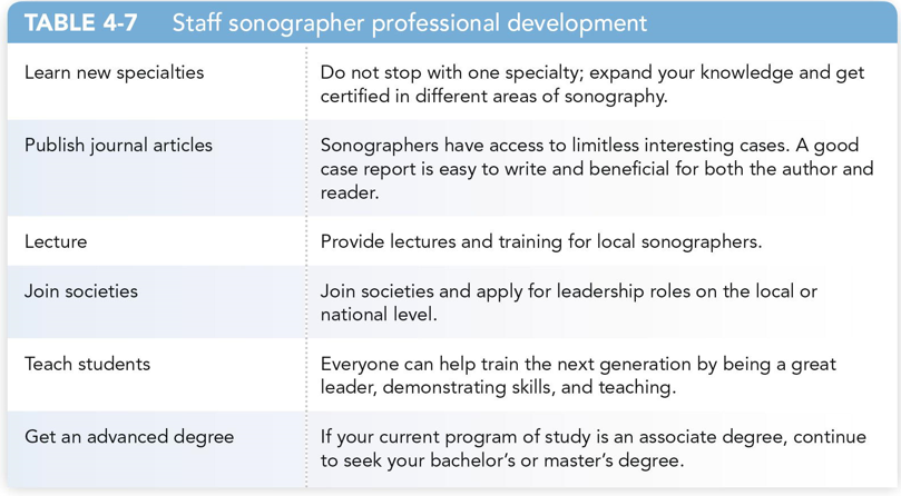
The staff sonographer has essential core responsibilities but still has the capacity to influence the profession in a positive manner through professional development.
Professional Development - Table 4-7 - Staff Sonographer
…
Advanced Practice Sonographer
This is a position the SDMS has been working to create since 1996, a midlevel position also called the ultrasound practitioner.
They would perform and interpret ultrasound procedures in primary or specialty care settings.
A similar position, the advanced cardiovascular sonographer, is a proposal of the ASE task force.
The ultrasound radiologist assistant works between the interpreting physician and the sonographer to refine reports and train residents.
Management in Sonography
A sonography manager or director plans, controls, directs, and organizes the day-to-day functions of the department.
They make rotation schedules, order supplies, work with upper management, attend management meetings, and work with the scheduling department and interpreting physicians to systematize exams and procedures.
Management and leadership are not the same thing, but a manager can be an effective leader.
Such a person would have a better opportunity to progress toward upper management.
Sonography Educators
They can be involved in training generations of sonographers in the classroom and clinical environments.
Prior clinical experience is usually mandatory.
You must be certified in the area in which you teach.
Sonography educators should have a thirst for knowledge, a love of sonography, and passion to make a positive difference in the profession.
Other Opportunities
Travel sonographers are employed by temporary staffing agencies and may be required to travel nationally or internationally.
Ultrasound consultants assist organizations in the process of ultrasound accreditation processes and may help guide changes in practices and trainings.
Sales and application specialists are employed by manufacturers and usually must have a number of years of clinical experience, multiple credentials, and extensive training on machines and equipment.
A sonographer entrepreneur owns part or all of a business related to sales or training.
Career Establishment - Searching and Applying for Jobs
Searching and Applying for Jobs
You may have to travel or take a job to gain experience for your dream job.
Search the Internet for online job search sites and organizations’ human resource pages.
Your educational institution may also have resources.
Word of mouth is one of the best ways to find a job.
Form good relationships with clinical sonographers and managers.
Resume Writing and Social Networking
Adapt your resume to the specific position you are seeking.
Free online assessment tools can grade your resume.
Seek personal and online networking to find people who want to hire you.
Be cautious of what you share on the Internet.
Career Establishment - Interview Tips
Know your resume.
Prepare for common interview questions such as “Tell me about yourself.”
Focus your mind by adding chronology to your answer.
Complete the process of inventorying your personal strengths and weaknesses.
An unwillingness to admit to weaknesses may be interpreted as an unwillingness to learn and improve.
Try identifying a weakness that you can ultimately describe as a strength.
Always seem interested, sit intently, and take your time answering questions.
After your interview, send a thank you note.
Summary of Chapter 4
Though your priority in school is to become clinically competent, you should work toward developing professional relationships with sonographers and managers in the clinical facilities in which you study.
The sonography profession is replete with organizations that allow one to grow as a professional.
Once you have established a career in sonography, you will recognize the need to expand your education, a service that professional organizations readily provide.
Which of the following is not a certification-granting organization?
a. ARDMS
b. CCI
c. ARRT
d. AIUM
d. AIUM
Which of the following is considered the best way to get a job?
a. Face-to-face meeting
b. Online networking
c. Social networking
d. Online searching
a. Face-to-face meeting
Which of the following membership organizations publishes the journal of Diagnostic Medical Sonography?
a. AIUM
b. SDMS
c. SVU
d. JUM
b. SDMS
Which of the following is not a quality of an effective leader?
a. Courageous
b. Oppressive
c. Optimistic
d. Honest
b. Oppressive
To maintain certification through the ARDMS, how many credits must a sonographer obtain in three years?
a. 40
b. 24
c. 16
d. 30
d. 30
The organization that offers the RVS certification is
a. ARDMS
b. ARRT
c. CCI
d. SVU
c. CCI
Which form of leadership places the concerns of others before the self?
a. Transformational leadership
b. Servant leadership
c. Autocratic leadership
d. Transactional leadership
b. Servant leadership
How many days before graduation can a student of an accredited sonography program take the specialty examinations offered by the ARDMS?
a. 30
b. 45
c. 50
d. 60
d. 60
What type of leadership not only guides and motivates employees but exhibits charisma and offers inspiration?
a. Servant leadership
b. Transformational leadership
c. Transactional leadership
d. Autocratic leadership
b. Transformational leadership
Which of the following establishes practice guidelines for each specialty and makes official statements on behalf of the profession on a range of issues, including safe ultrasound practice techniques?
a. SDMS
b. ASE
c. AIUM
d. FFE
c. AIUM
Which national membership organization publishes the Journal of Ultrasound in Medicine?
a. AIUM
b. ARRT
c. SDMS
d. ASE
a. AIUM
What organization is the national organization for echocardiographers?
a. ARRT
b. ASE
c. SDRS
d. SVU
b. ASE
What organization establishes standards by which many different allied health programs are reviewed?
a. AIUM
b. JRC-DMS
c. ACR
d. CAAHEP
d. CAAHEP
Which of the following is the national membership organization for physicians, vascular technologists, students, and other cardiac and vascular professionals?
a. ARRT
b. ASE
c. SDRS
d. SVU
d. SVU
To become a registered sonographer through the ARDMS, one must do which of the following?
a. Pass the SPI examination
b. Pass the SPI examination and a specialty examination
c. Pass a single specialty examination
d. Pass two specialty examinations
b. Pass the SPI examination and a specialty examination