Physiology Exam 1
1/123
Earn XP
Name | Mastery | Learn | Test | Matching | Spaced | Call with Kai |
|---|
No analytics yet
Send a link to your students to track their progress
124 Terms
combinaation biomolecules
lipoprotein: blood transport molecules
glycoprotein: membrane structure
glycolipids: membrane receptors
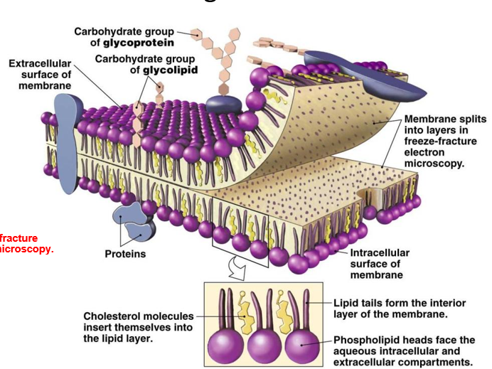
cell membrane
structure: fluid mosaic model, phospholipid bilayer
function
physical barrier
gateway for exchange
communication
cell structure
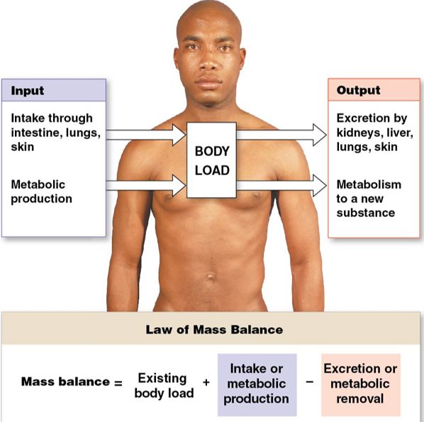
mass balance in the body
(mass balance) = (existing body load)
+ (intake & metabolic production)
- (excretion & metabolic removal)
\
**homeostasis**: the ability of the body to maintain a relatively constant internal environment
\
equilibrium in cells:
* __osmotic equilibrium__: water goes in and out
* __chemical disequilibrium__: differential solute concentration
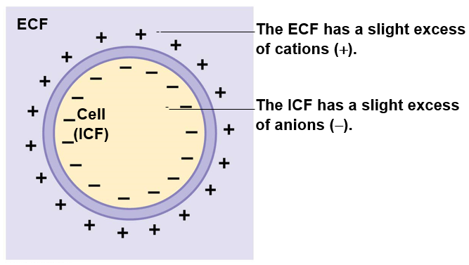
* __electrical disequilibrium__: inside of cells is negative to extracellular fluid
\
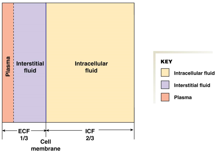
**Extracellular Fluid (ECF)** = 1/3 of fluid volume
* **Plasma** = blood, 75% of ECF
* **Interstitial fluid** = between cells, 25% of ECF
\
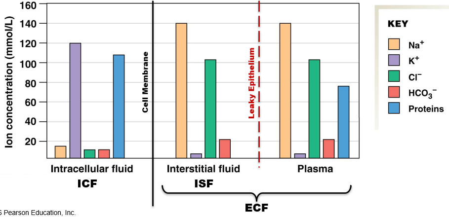
barrier between ICF and ECF: cell membrane
barrier between plasma and interstitial: “leaky” epithelium
chemical disequilibrium in ICF vs ECF
ECF
high Na+ (~145 mM)
low K+ (~5 mM)
high Cl- (~108 mM)
proteins only in plasma, not interstitial fluid
ICF
low Na+ (~ 15 mM)
high K+ (~150 mM)
high Cl- (~10 mM)
high proteins
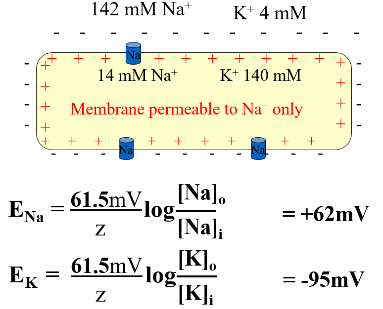
E for each ion at resting:
Na+: +60 mV
K+: -90 mV
Cl-: -63 mV
* aided by aquaporins
\
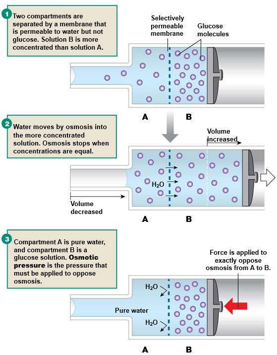
**osmotic pressure**: pressure required to exactly oppose a given concentration gradient
\
**osmolarity**: concentration of solute in particles per liter of solution
tonicity
tonicity: volume change of a cell placed in solution
based on concentration of non-penetrating solutes
hypotonic solution = cell swells
isotonic solution = cell remains the same
hypertonic solution = cell shrinks
\
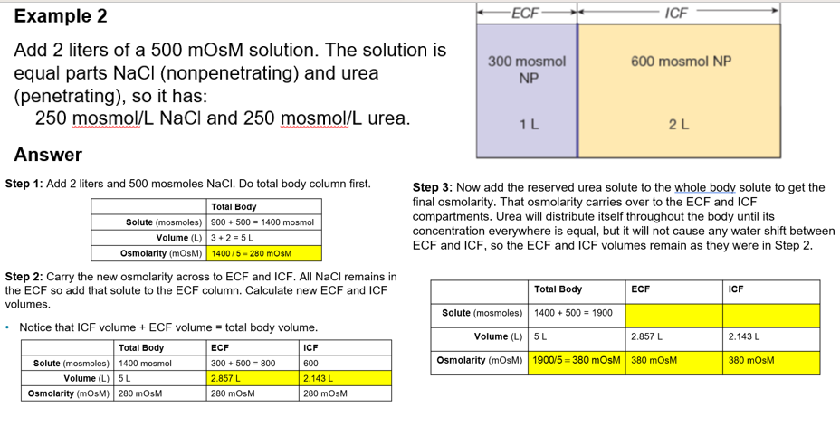
Work out total body first. Remember that osmolarity of ECF and ICF will be equal to total body.
\
If working with both non-penetrating and penetrating solutes, start with non-penetrating solutes to figure out water movement (volume of ICF and ECF).
\
Then add penetrating solutes back in to figure out solute (keep volumes the same).
common body fluid scenarios
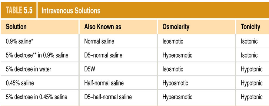
IV
IV fluid enters plasma (ECF)
0.9% NaCl is isosmotic to cells
NaCl is non-penetrating, glucose (dextrose) is penetrating
sweating
lose water by evaporation, leaves salt behind
sweat comes from plasma (ECF)
increases osmolarity of the ECF so that water leaves the cells
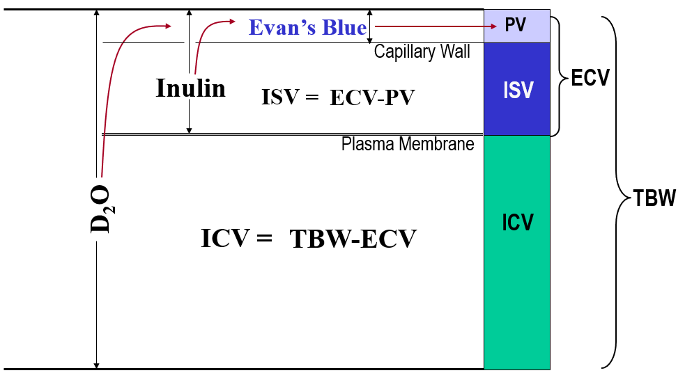
measurement of body fluid
dilution method: add a known amount of dye/molecule and then measure concentration when diluted in whole body
use c1v1 = c2v2
measurements:
plasma volume: Evan’s blue
ECF volume: inulin
total body volume: D20 (heavy water)
interstitial volume: ECF - plasma
ICF volume: total body - ECF
\
**dehydration**: loss of fluids in excess of 1% or more of body weight
* __caused by__: excess sweating, evaporation from burn surface, chronic hyperventilation, vomiting/diarrhea, etc.
* __leads to__: ECF hypovolemia, **hypotension**, inability to sweat (hyperthermia)
* high to low concentration
* net movement until concentration is equal
\
__faster when__:
* short distance
* higher temperature
* smaller molecular size
* more permeable membrane
* larger surface area of membrane
* thinner membrane
* larger concentration gradient
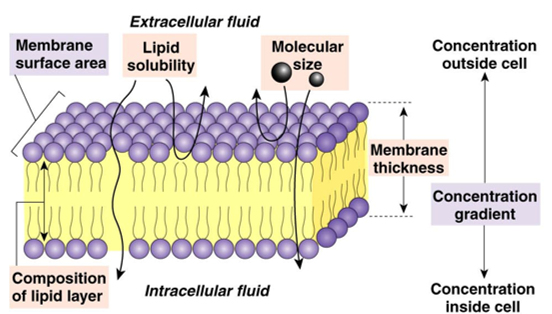
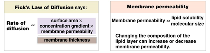
Fick’s law of diffusion
rate of diffusion ∝ (membrane surface area)(membrane permeability)(concentration gradient)(membrane thickness)⁻¹
membrane permeability ∝ (lipid solubility)/(molecular size)
1. structural proteins
2. enzymes
3. membrane receptor proteins
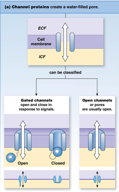
4. transporters (channel or carrier)
* open channel
* gated channel
* **mechanical**: responds to pressure changes (blood pressure)
* **voltage**: responds to electrical state (nerve/muscle contraction)
* **chemical**: responds to molecules that bind to channel protein
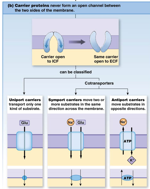
carrier membrane proteins
carrier proteins have specificity to their substrates
uniport: 1 substrate, 1 direction
symport: 2 substrates, same direction
antiport: 2 substrates, opposite directions (ATP)
* glucose goes into cell through GLUT transporter (down concentration gradient)
* glucose is phosphorylated to glucose-6-phosphate
* keeps concentration of glucose inside cell low so that glucose continues to flow in
\
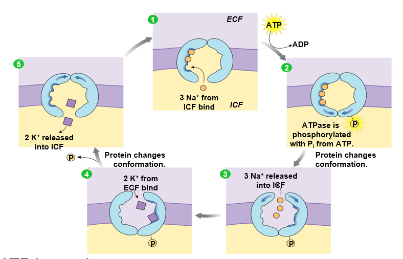
__ex__: Na+ K+ ATPase antiporter
* maintains high Na+ outside cell and high K+ inside
* 3 Na+ go out, 2 K+ go in
* inside of cell is slightly more negative than outside
\
__mechanism__:
1. 3 Na+ from inside cell bind
2. ATPase is phosphorylated
3. Protein changes conformation
4. 3 Na+ released into ECF
5. 2 K+ from ECF bind
6. Protein changes conformation (dephosphorylated)
7. 2 K+ released inside cell
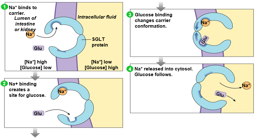
secondary active transport
uses potential energy of Na+ gradient to transport a different molecule against its concentration gradient
ex: sodium glucose transporter (SGLT)
Na+ in ECF binds to carrier, creating a binding spot for glucose
Glucose from ECF binds to carrier, changing carrier conformation
Na+ and glucose are released inside cell
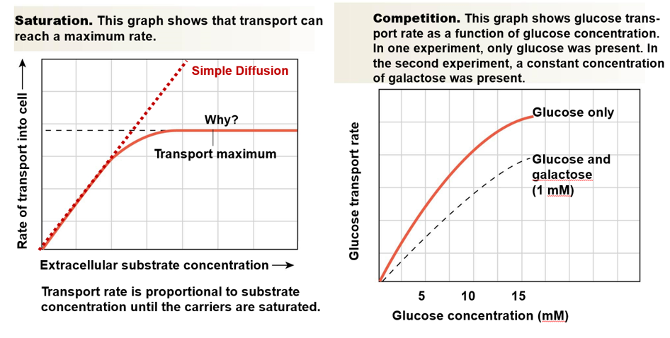
* transport reaches a maximum rate
* once all carriers are full, a higher concentration of substrate doesn’t increase rate
\
**competition**:
* ex: maltose or galactose can compete with glucose
* inhibitor can get to active site first
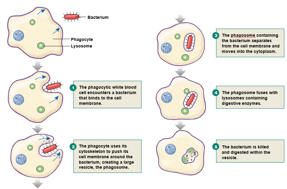
1. bacteria binds to cell membrane
2. cell uses **cytoskeleton** to extend membrane around bacteria
3. bacteria is engulfed into **phagosome**
4. phagosome fuses with lysosomes
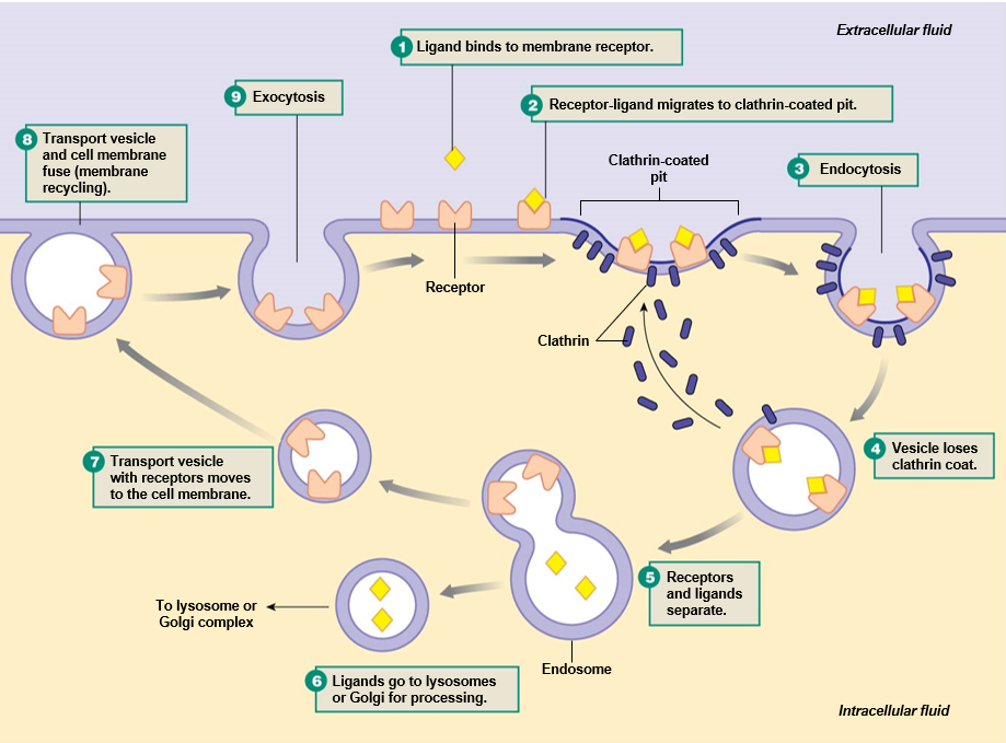
* **receptor-mediated** uses **clathrin**-coated pits
* **caveolae** use **lipid rafts** for endocytosis without clathrin
\
__receptor-mediated process__:
1. ligand binds to receptors on cell membrane and migrate to clathrin-coated pit
2. vesicle forms and is pinched off
3. clathrin coat is lost
4. receptors are recycled to cell surface
5. ligands go to lysosome
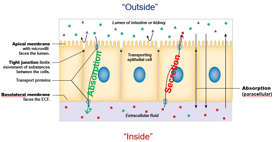
* **apical membrane**: faces the lumen of intestine/kidney (“outside”)
* **basolateral membrane**: faces the ECF (“inside”)
* **tight junctions**: form connections between cells and limits movement of materials across cell layer
\
__processes__:
* **absorption**: movement from lumen to ECF
* **secretion**: movement from ECF to lumen
\
__types of transport__:
* **paracellular**: through tight junctions
* **transcellular**: through epithelial cells themselves
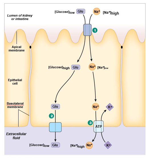
1. **Na+ glucose symporter**: apical, secondary active transport
2. **GLUT transporter**: basal, facilitated diffusion
3. **Na+ K+ ATPase**: basal, primary active transport
\
Na+ is high outside cell, low inside cell
glucose is low outside cell, high inside cell
* the body is overall neutral
* the ICF is slightly negative and the ECF is slightly positive
\
The resting membrane potential of a cell is relative to the outside of the cell
* outside of cell is 0mV
* inside is usually around **-70 mV**
* opposite charges attract
* **conductor**: material that allows electrons to travel through
* **insulator**: matierial that does not allow free movement of electrons
\
**Ohm’s law**: I = Vg
* I is current
* V is voltage
* g is conductance
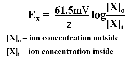
* specific to a particular ion
* at what charge inside cell will the ion stop diffusing across membrane
\
z is the charge on the ion
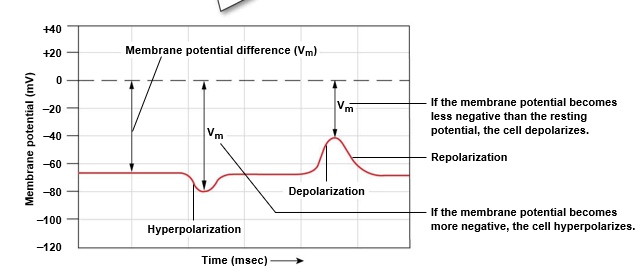
changes in membrane potential
*outside of cell is always reference point (0 mV)
hyperpolarization: membrane potential becomes more negative than baseline (resting)
depolarization: membrane potential becomes more positive than resting
* E(Na) will approach **+62 mV** inside cell
\
**K+**: assume only K+ can cross membrane
* E(K) will approach **-95 mV** inside cell
\
**overall membrane potential**: both K+ and Na+ cross
* membrane potential is about **-70 mV** inside cell
* mostly due to K+ (cell more permeable to K+)
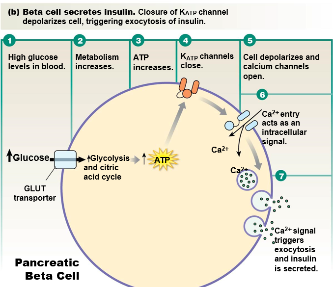
* **ATP-gated K+ channel**: closes with ATP
* **voltage-gated Ca2+ channel**: opens with more positive membrane potential
\
__with high glucose levels__:
* glucose flows into cell and metabolism increases
* increase in ATP causes K+ channel to close
* K+ no longer leaves cell
* membrane potential **depolarizes** (more positive)
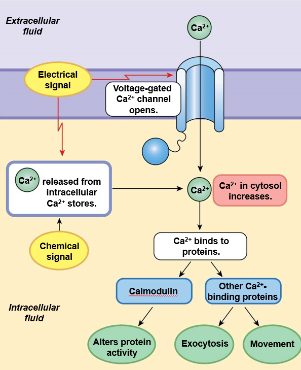
* voltage-gated Ca2+ channel opens
* Ca2+ enters cell and triggers exocytosis of insulin
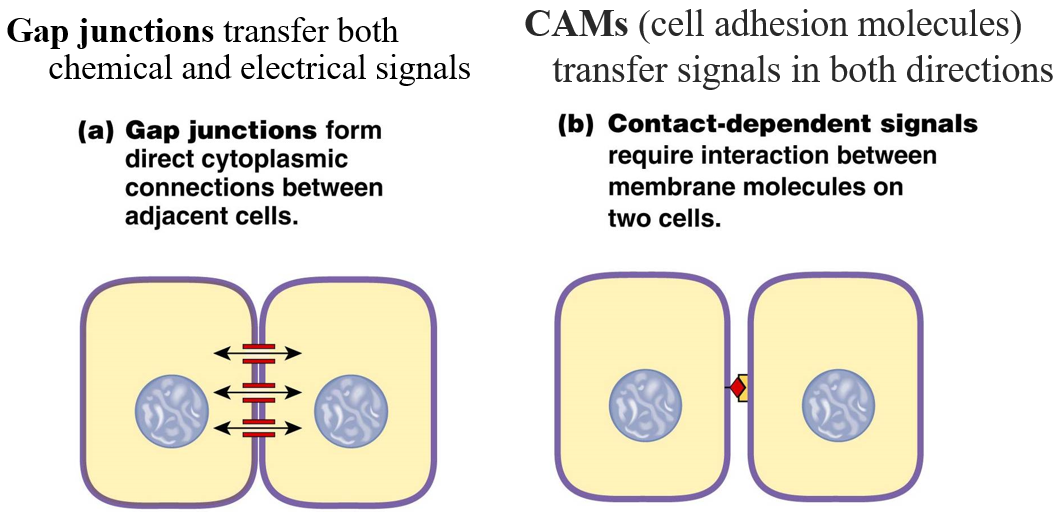
1. **Gap junctions**: connect cytoplasm of neighboring cells
2. **CAMs (cell adhesion molecules)**: interaction between membrane molecules on two cells
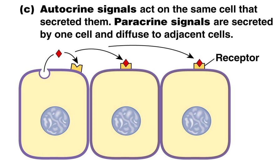
1. **paracrine signals**: secreted by one cell and diffuse to adjacent cells
2. **autocrine signals**: act on the same cell that secreted them
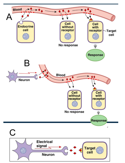
1. **Hormones**: released into bloodstream by endocrine cells
2. **Neurohormones**: released into bloodstream by neurons
3. **Neurotransmitters**: sent from neuron directly to target cell
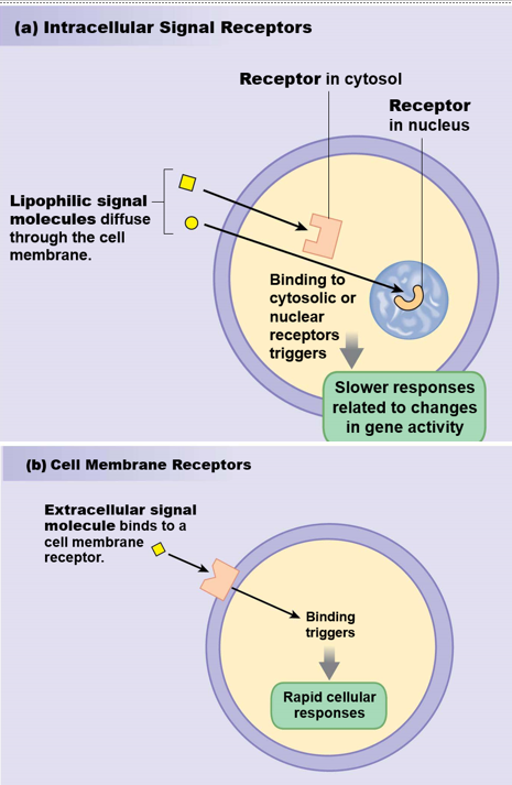
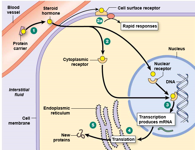
intracellular and extracellular signal receptors
lipophilic molecules can cross the cell membrane
bind to cytosolic or nuclear receptors
extracellular signal molecules bind to the cell membrane receptor and trigger a response
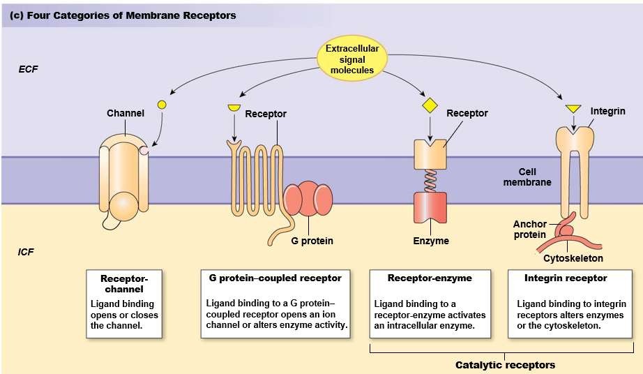
1. receptor-channel (fast-acting)
2. G-protein coupled receptor
3. receptor-enzyme
4. integrin receptor (cytoskeletal response)
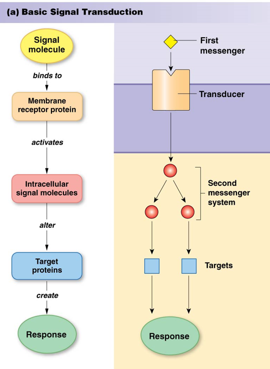
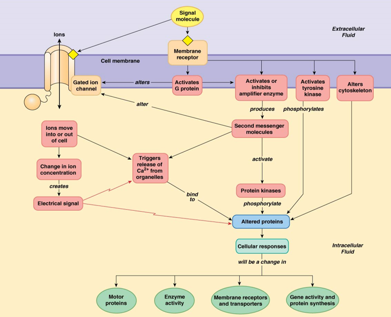
signal transduction
basic pattern:
Signal molecule (first messenger) binds to…
Membrane receptor protein (transducer) activate…
Intracellular signal molecules (second messenger) alter…
Target proteins create…
Response
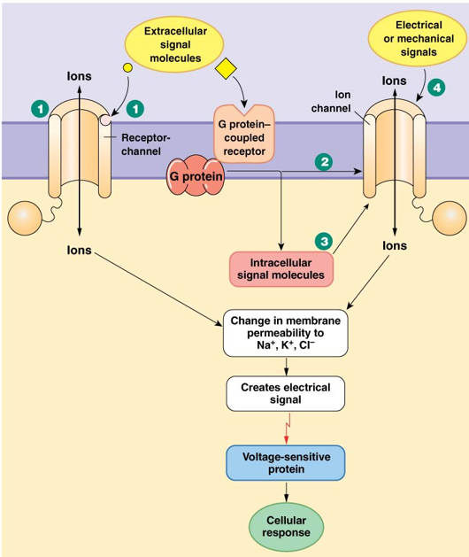
ion receptor channels
Four ways they work:
Receptor channels open or close in response to signal molecule binding
Some channels are directly linked to G proteins (when ligand binds G protein couple receptor, ion channel opens/closes)
Some channels respond to intracellular second messengers
Electrical or mechanical signals open or close ion channels
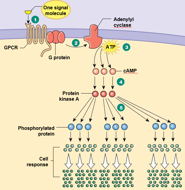
G protein coupled receptor example: adenylyl cyclase
Signal molecule binds to GPCR
G protein is activated, activates adenylyl cyclase
Adenylyl cyclase converts ATP to cyclic AMP (cAMP)
cAMP activates protein kinase A
Protein kinase A phosphorylates other proteins
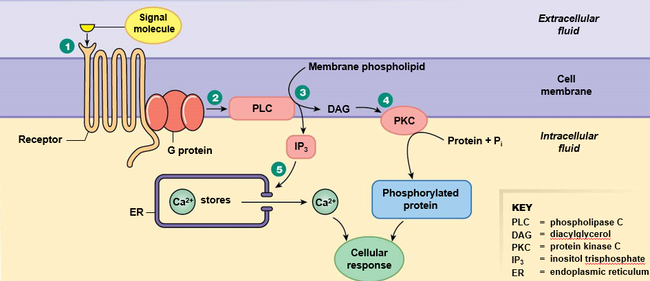
G protein couple receptor example: phospholipase C
Signal molecule binds to GPCR
G protein is activated, activates phospholipase C (PLC)
PLC cuts membrane phospholiplids into a head group (IP3) and diacyl glycerol (DAG).
IP3 opens a channel in the ER, releasing Ca2+
DAG activates protein kinase C
Protein kinase C phosphorylates other proteins
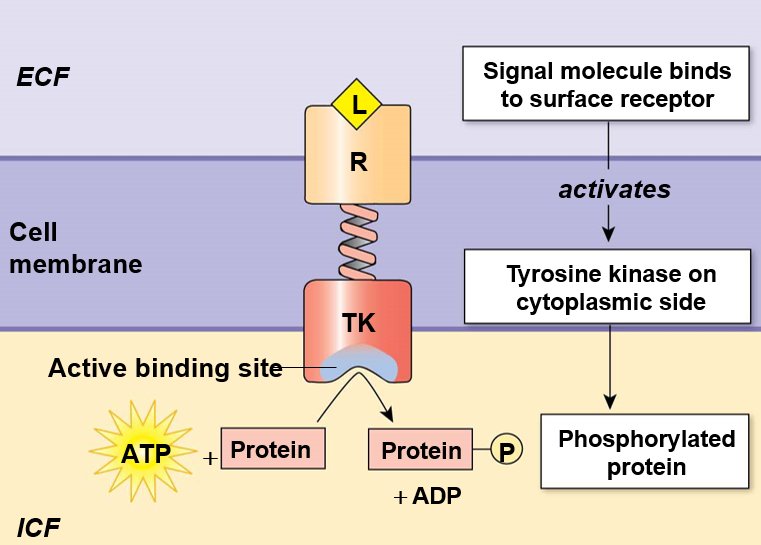
receptor-enzymes
A ligand binds the receptor portion
The attached enzyme (cytoplasmic side) changes conformation to carry out its enzymatic function
usually kinase or guanylyl cyclase
example: tyrosine kinase
integrin receptor
outside cell: bind to ligands or extracellular matrix proteins
inside cell: integrins attach to cytoskeleton through anchor proteins

signal transuction summary
intracellular signals: Ca2+
inside the cell - stored in endoplasmic reticulum
released from intracellular compartments through second messengers
enters the cell through gated channels
binds to other proteins - calmodulin, etc.
intracellular signals: gases
three main ones: nitric oxide (NO), carbon monoxide (CO), and hydrogen sulfide (H2S)
Nitric Oxide
produced by endothelial cells, diffuses into smooth muscle cells
causes dilation of blood vessels
reactive with H20 and O2, short half-life = local control
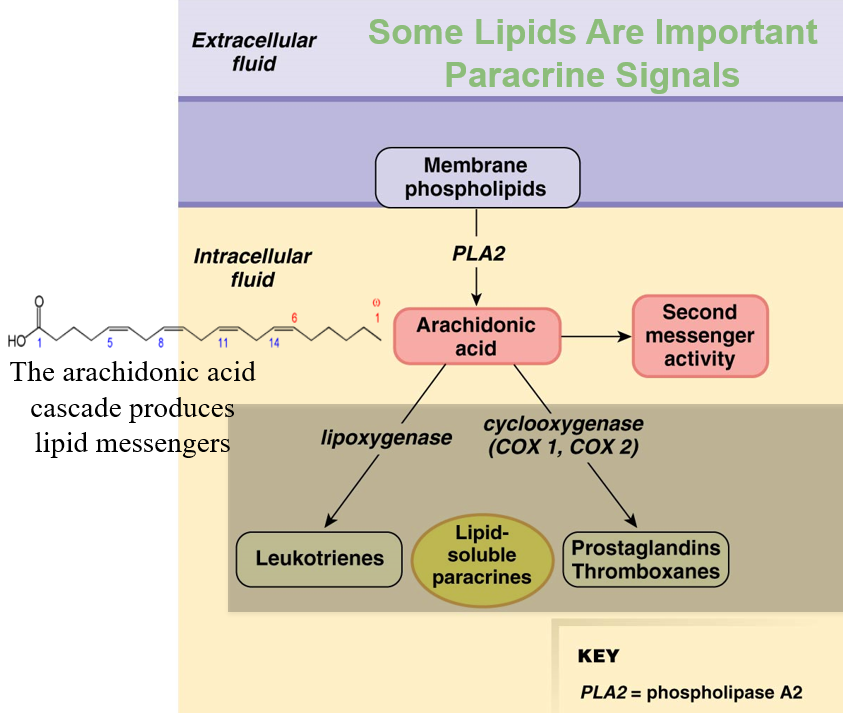
intracellular signals: lipids
phospholipase 2: cuts off 2nd fatty acid to make arachidonic acid (2nd messenger)
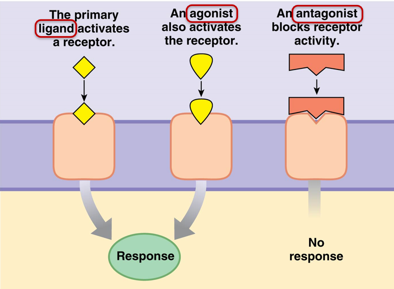
signal pathway modulation: receptor ligands
ligand: activates receptor
agonist: activates receptor, competes with ligand
antagonist: blocks receptor activity
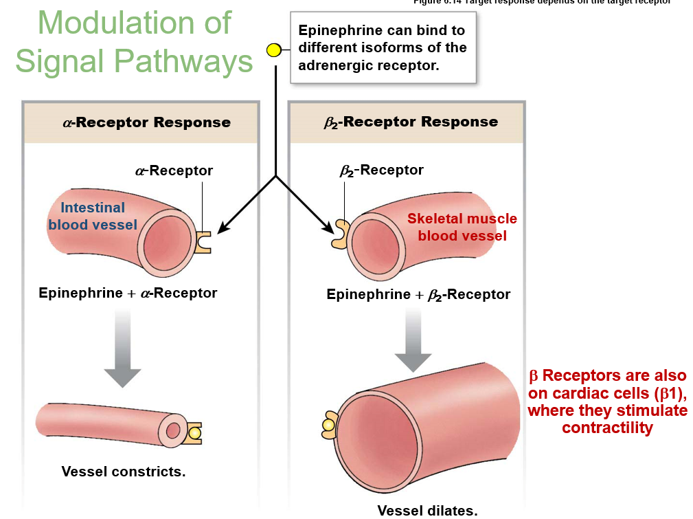
signal pathway modulation: receptors
one ligand can bind different receptors with different responses
epinephrine binds alpha receptor in intestine → blood vessel constricts
epinephrine binds beta2 receptor in skeletal → blood vessel dilates
epinephrine binds beta1 receptor in cardiac → stimulate contractility
downregulation: decrease number of receptors
desensitization: binding to chemical modifier
upregulation: increase number of receptors
ways to terminate signal: transport away, breakdown signal, endocytosis
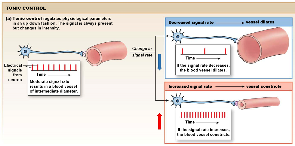
tonic control
signal is always present, but changes in intensity
intermediate signal, can go up or down
ex in neurons: increase signal rate means constriction of blood vessel, decrease signal rate means dilation
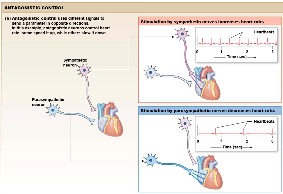
antagonist control
different signals for two different directions
like gas pedal and brake pedal
faster change in response
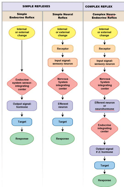
simple and complex reflex pathways
simple endocrine
simple neural
complex neuro-endocrine
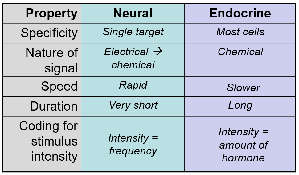
nueral vs endocrine control
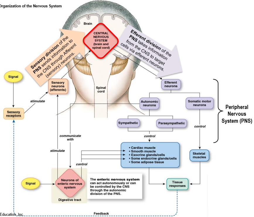
nervous system overview
Central Nervous System (CNS): brain and spinal cord
Peripheral Nervous System (PNS):
sensory (afferent) neurons: send signals to brain
efferent neurons: receives signals from brain
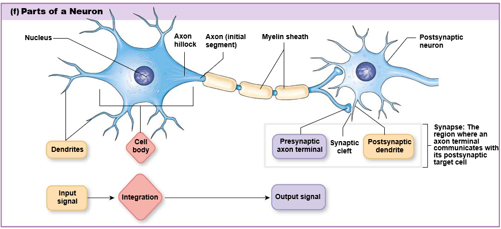
neuron cell structure
dendrites: receive input signals
cell body: integration center
axon: carry outgoing information
synapse: output signal
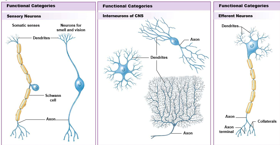
types of neurons
sensory: both dendrites and axon separated from cell body
interneurons: CNS neurons, highly branched
efferent: single long axon, dendrites as branches from cell body (classic neuron shape)
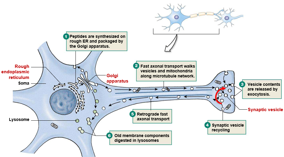
fast axonal transport
Protein synthesis in ER and packaging in Golgi
Motor proteins walk vesicles and mitochondria down axon along microtubule network
Exocytosis of vesicles
Synaptic vesicle recycling
Retrograde fast axonal transport
Old membrane components digested in lysosome
Glial cells
astrocytes: help create healthy environment for neurons in CNS
take up ions, water, neurotransmitters
help form blood brain barrier
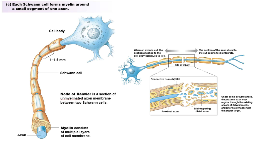
myelin sheath: oligodendrocytes in CNS, Schwann cells in PNS
multiple layers of phospholipid membrane surrounding axon
acts as insulation, speeds up signal transmission
node of Ranvier: section of unmyelinated axon between two Schwann cells
sometimes after an axon injury axon can reform under myelin sheath
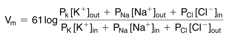
Goldman-Hodgkin-Katz (GHK) equation
membrane potential that results from the contribution of all ions that can cross the membrane
Na+, K+, and Cl- have the most influence
P = relative permeability of ion
denominator and numerator of Cl- is reversed due to negative charge on ion
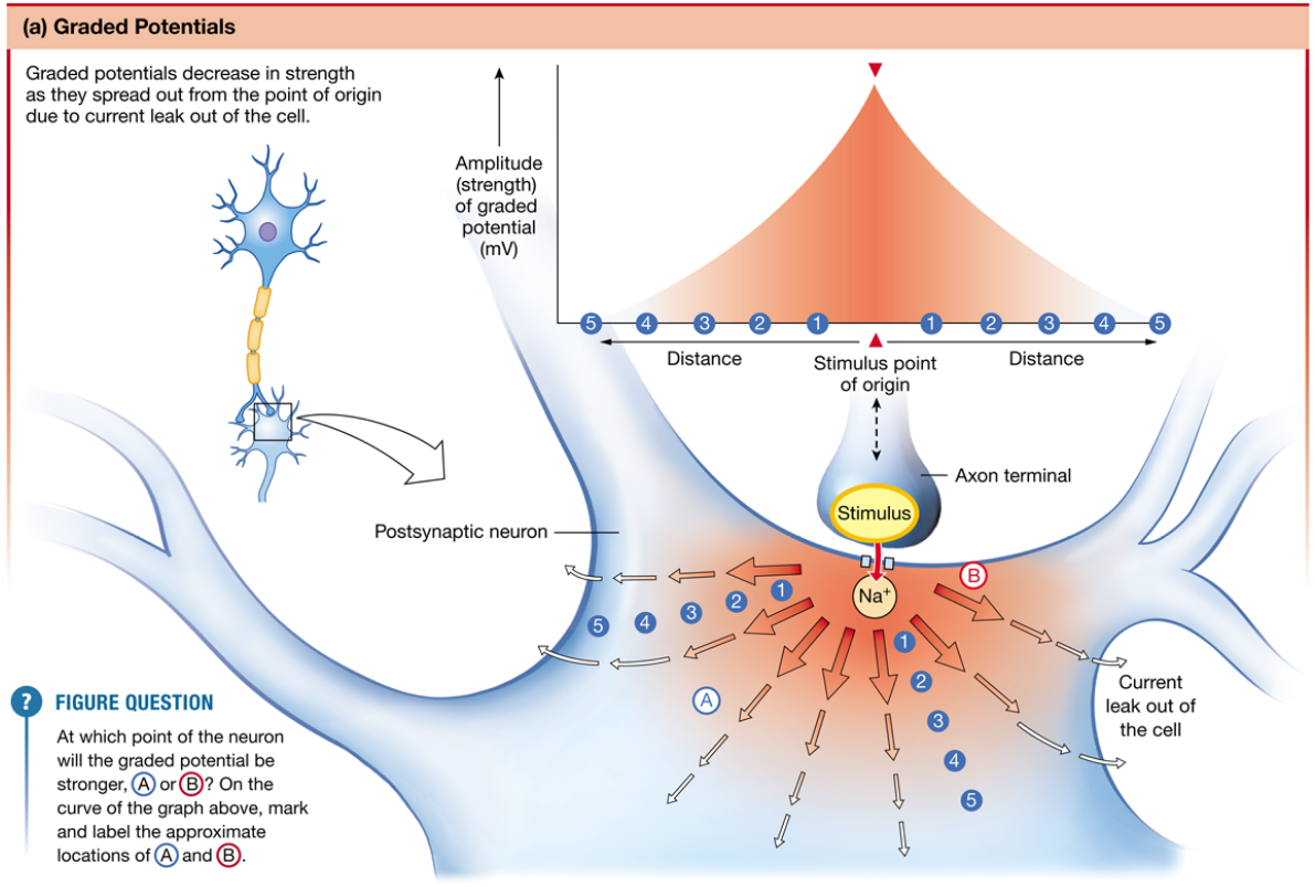
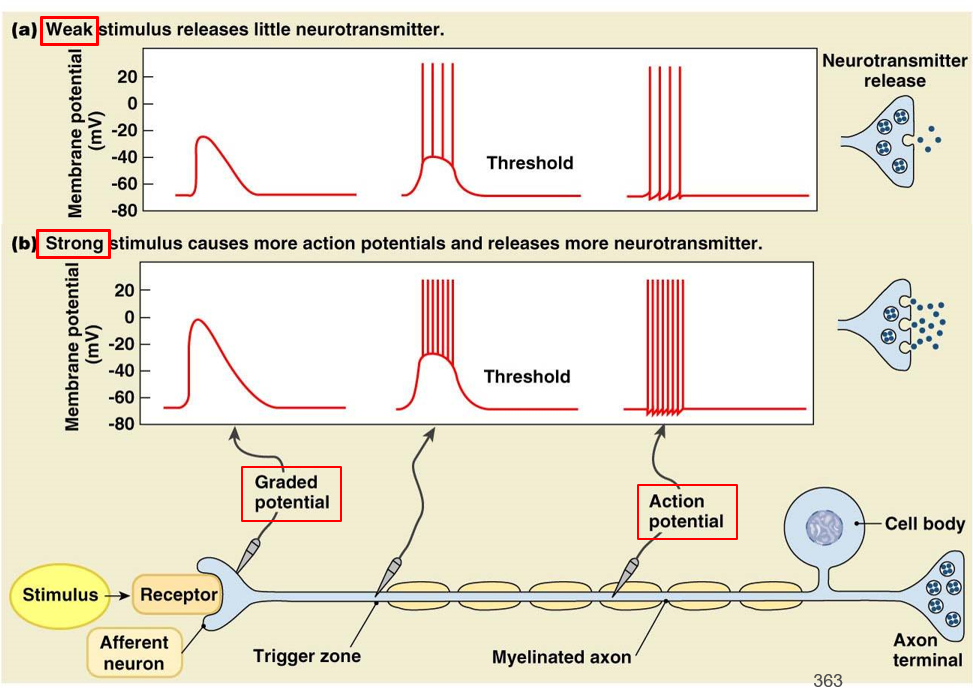
graded potential
decreases in strength as it spreads out from a point of origin
variable, can be different levels (intensity)
occurs in dendrites, cell body
usually receives a chemical signal
if signal is above a threshold when it reaches the ‘trigger zone’, it causes an action potential
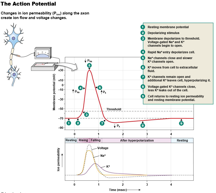
action potential
depolarizations that travel long distances down axon
all-or-nothing (frequency)
occurs in trigger zone through axon
uses voltage-gated channels (electrical signal)
fires over and over until graded potential fades
conduction: high speed movement of action potential along axon
action potential (steps)
Depolarization:
An Na+ gated channel is opened. Na+ enters the cell.
Voltage-gated Na+ and K+ channels begin to open.
Even more Na+ enters the cell (positive feedback loop).
Repolarization:
Delayed opening of K+ voltage-gated channels. K+ exits the cell.
Inactivation gates close and stop Na+ from entering cell.
Hyperpolarization:
K+ leaving the cell.
Na+ channels reset (voltage gate closes, inactivation gate opens)
Resting:
both K+ and Na+ channels closed.
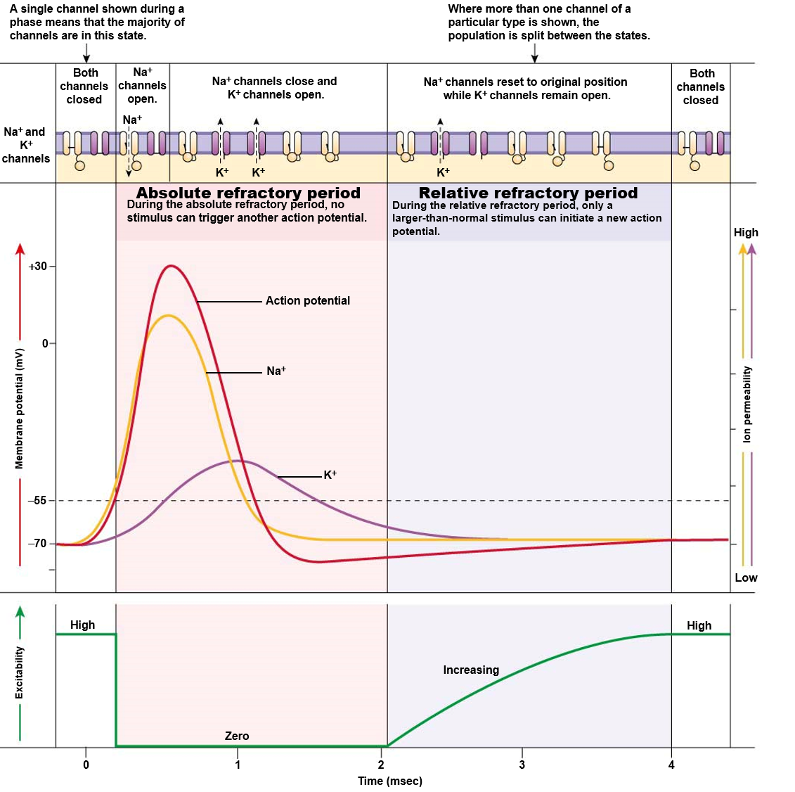
refractory period
absolute refractory period
can’t fire another action potential during this time
reset of Na+ gates (inactivation gate opens, voltage gate closes)
prevents action potential from going backward or overlapping
relative refractory period
action potential can fire but requires larger stimulus
some but not all Na+ gates are reset
K+ gates are still open
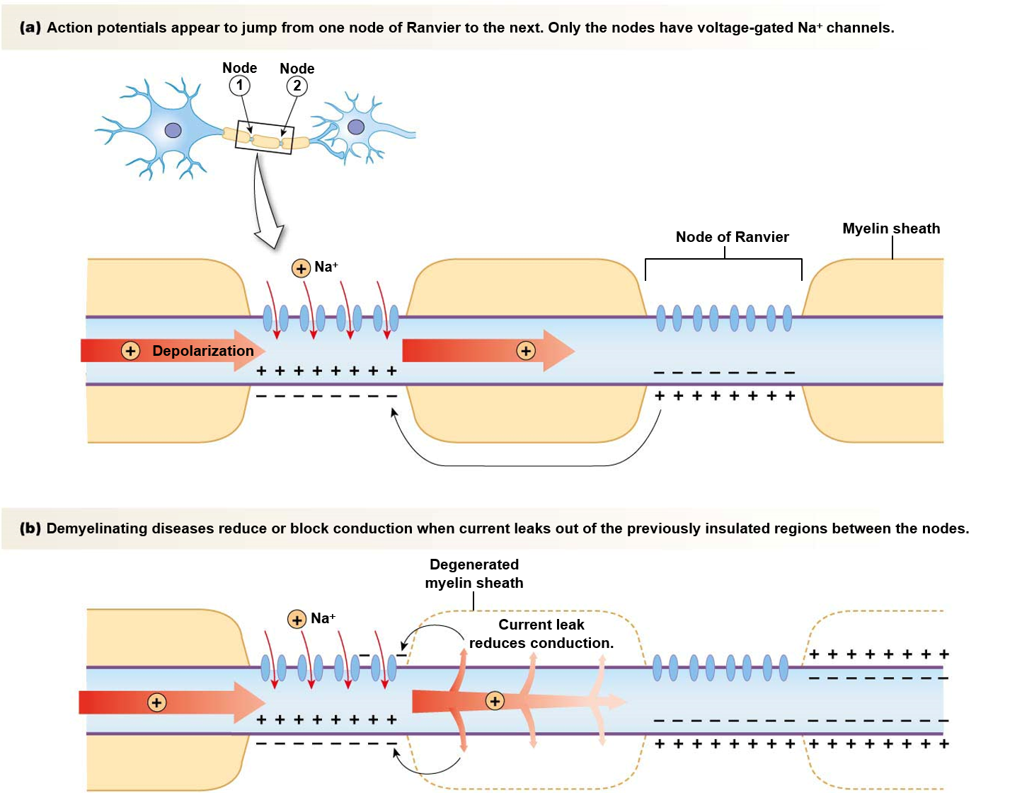
action potential speed
speed of action potential influenced by:
diameter of axon (larger = faster)
resistance of axon membrane to ion leakage (myelinated = faster)
only Nodes of Ranvier have channels - action potentials “jump” between nodes.
saltatory conduction
electrical vs chemical synapses
electrical synapse
pass electrical signals through gap junctions
can be bidirectional
synchronizes activity of a network of cells
chemical synapse
neurotransmitters cross synaptic cleft
unidirectional
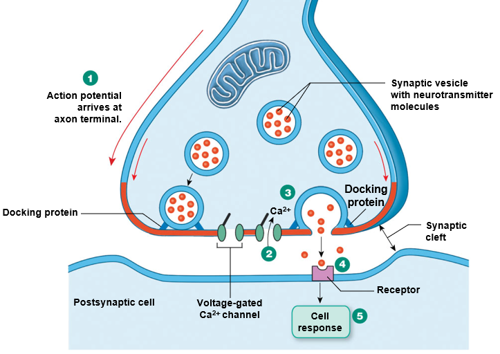
neurotransmitter release
action potential reaches axon terminal
depolarization of axon terminal opens voltage-gated Ca2+ channels
Ca2+ enters cell, interacts with docked synpatic vesicles to trigger exocytosis
short diffusion of neurotransmitters across synpatic cleft
neurotransmitters bind receptors (ligand-gated) in postsynaptic cell
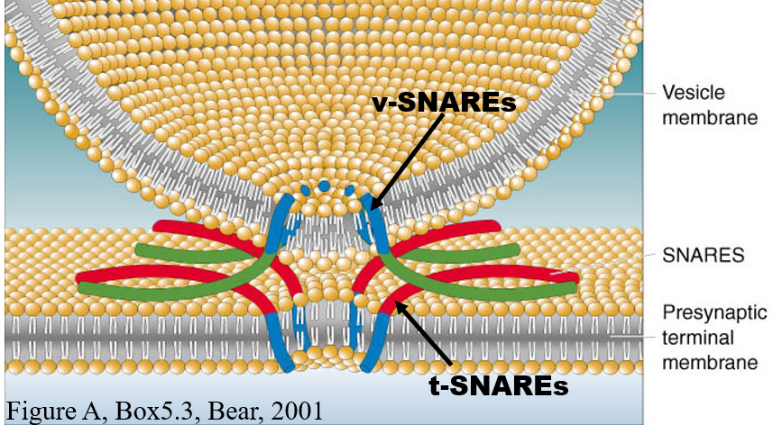
synaptic vesicles and docking proteins
SNARE proteins drive fusion
tSNARE and vSNARE wind together
SNAP-25 and other proteins make 4 helices
bacterial toxins can target these proteins (botulism, botox, tetanus)
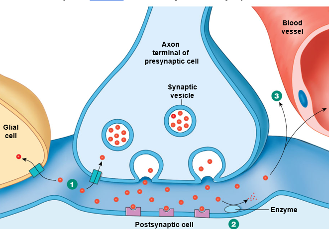
termination of neurotransmitter activity
three ways:
Take neurotransmitter back up into axon terminal
Break neurotransmitter down with an enzyme
Neurotransmitters diffuse out of synaptic cleft
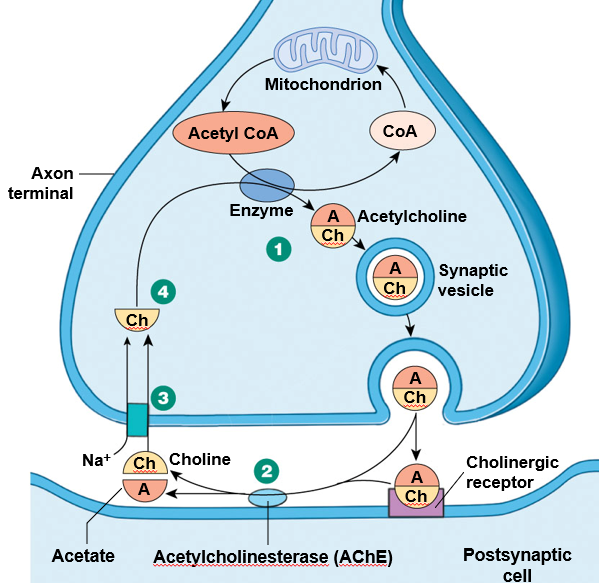
synthesis and recycling of acetylcholine
mechanism:
acetylcholine is synthesized in pre-synpatic cell
in the synaptic cleft, acetylcholinesterase breaks down acetylcholine
choline taken back up into pre-synaptic cell by cotransport with Na+
choline is recycled to make more acetylcholine
affected by:
nerve gas: inactivates acetylcholinesterase, muscles constantly activated (spasms)
curare: antagonist to receptors, die of paralysis
convergent and divergent neuron pathways
divergent: one neuron sends signal to many other neurons
convergent: many neurons sends signals to one neuron

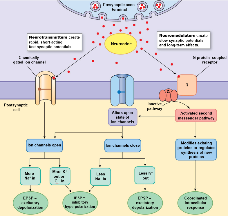
postsynaptic responses
excitatory depolarization (EPSP): makes an action potential more likely
open Na+ channels (more Na+ in)
close K+ channels (less K+ out)
inhibitory hyperpolarization (IPSP): makes an action potential less likely
close Na+ channels (less Na+ in)
open K+ channels (more K+ out)
open Cl- channels (more Cl- in)
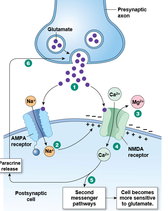
long-term potentiation and depression
long-term potentiation (LTP): permanent increase in connection
long-term depression (LTD): permanent decrease in connection
glutamate is key:
has receptors AMPA and NMDA
NMDA is blocked by a gate and Mg2+ ion
Mg2+ ion is released when depolarization occurs through AMPA channel opening
when MG2+ ion os released, Ca2+ flows in and activates second messenger pathway (LTP)
sensitivity to glutamate is increased
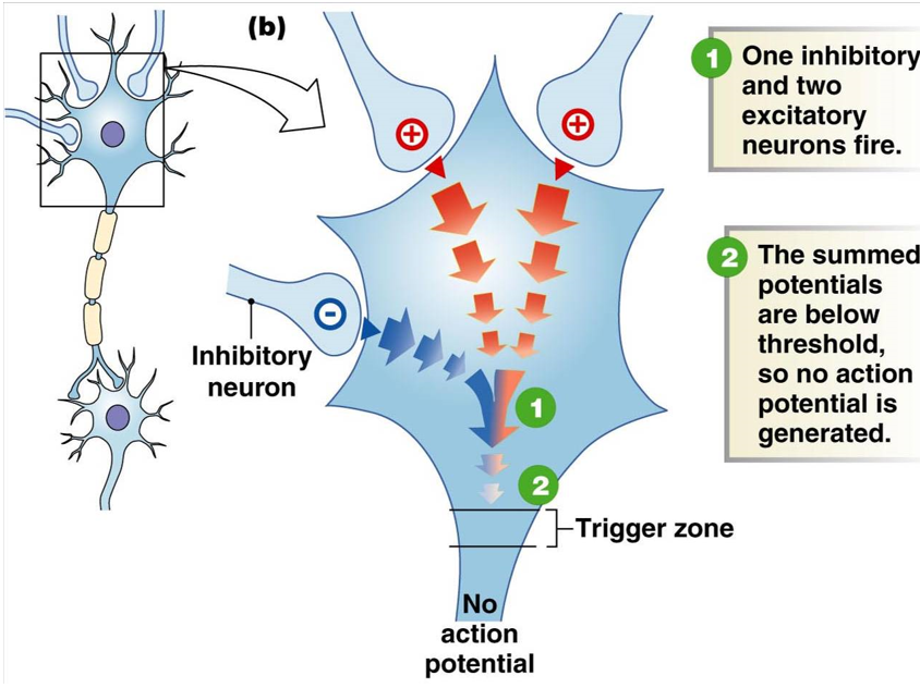
spatial summation
additive effect of multiple neurons sending a signal to another neuron at the same time (more than one graded potential)
if several excitatory neurons fire at the same time, they might not individually reach the threshold for an action potential, but together they do
if excitatory and inhibitory neurons fire at the same time, their effects can ‘cancel out’
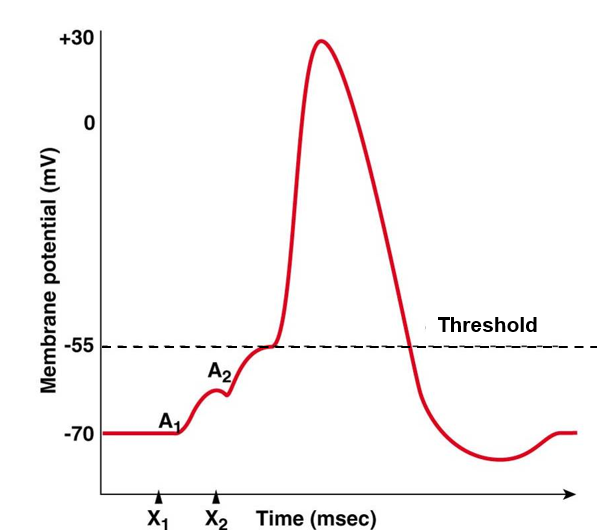
temporal summation
additive effect of multiple neurons sending a signal to another neuron close together in time (more than one graded potential)
if graded potentials are too far apart in time, then no summation occurs
if graded potentials are close together in time & arrive at trigger zone in short period of time, they may sum and create an action potential
hormones
hormone: chemical signal that goes throughout the body
half-life = length of activity
classification:
Peptide hormones
Steroid hormones
Amino-acid derived hormones
peptide hormones
storage: made in advance, store in secretory vesicles
release from parent cell: exocytosis
transport in blood: dissolved in plasma
half-life: short
receptor location: cell membrane
response: seccond messenger system, fast
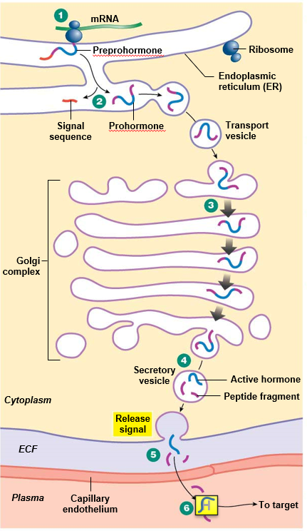
peptide hormone synthesis
proteolytic, post-translational modification
preprohormone: large, inactive precursor
cleaved in ER to become prohormone
prohormone: smaller, inactive precursor
cleaved in secretory vesicle to become active hormone
steroid hormones
cholesterol-derived, lipophilic
storage: synthesized on demand, made in adrenal glands and gonads
release from parent cell: simple diffusionbinds
transport in blood: carrier proteins
half-life: long
receptor location: usually cytoplasm & nucleus, sometimes cell membrane
response: usually gene activation (slow), sometimes nongenomic (fast)
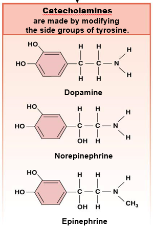
amino-acid derived hormones: catecholamines
modify tyrosine R group, behave like peptides
storage: made in advance, store in secretory vesicles
release from parent cell: exocytosis
transport in blood: dissolve in plasma
half-life: short
receptor location: cell membrane
response: seccond messenger system, fast
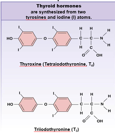
amino-acid derived hormones: thyroid
made from 2 Tyr and iodine, behave like steroid
storage: made in advance, store in secretory vesicles
release from parent cell: transport protein
transport in blood: carrier proteins
half-life: long
receptor location: nucleus
response: gene activation, slow
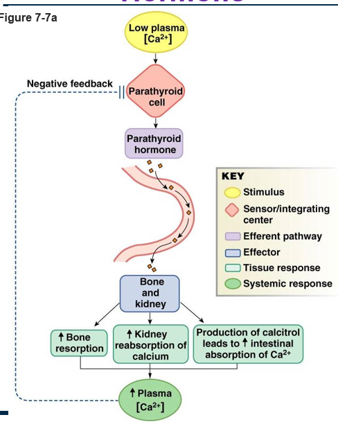
example: parathyroid simple endocrine reflex
input: low concentration of blood Ca2+
integrating center: parathyroid cell
efferent pathway: parathyroid hormone released into bloodstream
effector: bone and kidney
response:
increase bone resporption (release Ca2+)
increase kidney reabsorption of Ca2+
produce calcitrol → increase intestinal absorption of Ca2+
increase concentration of blood Ca2+ (negative feedback)
neurohormones
Catecholamines (from adrenal medulla)
Hypothalamus
Pituitary gland (anterior and posterior)
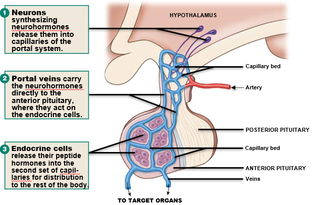
anterior pituitary
endocrine tissue - secretes 6 hormones
release is controlled by neurohormones from hypothalamus
process:
hypothalamus neurons realease neurohormones into capillaries of portal system
portal veins carry neurohormones directly to anterior pituitary
endocrine cells release their peptide hormones into capillaries for distribution to rest of body
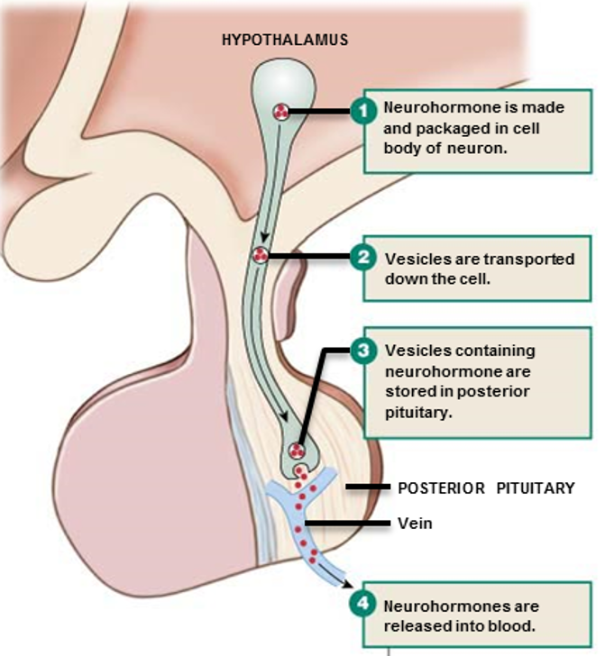
posterior pituitary
neural tissue -secretes 2 neurohormones
process:
neurohormone made and packaged in cell body of neuron (hypothalamus region)
vesicles transported down the cell
vesicles containing neurohormone are stored in posterior pituitary
neurohormones released directly into the blood
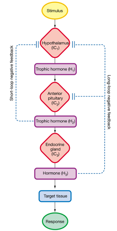
endocrine control
Three integrating centers of hypothalamus-pituitary
Hypothalamus: stimulated by CNS
Anterior pituitary: stimulated by hypothalamic hormones that travel thru portal system
Endocrine gland: stimulated by anterior pituitary hormones
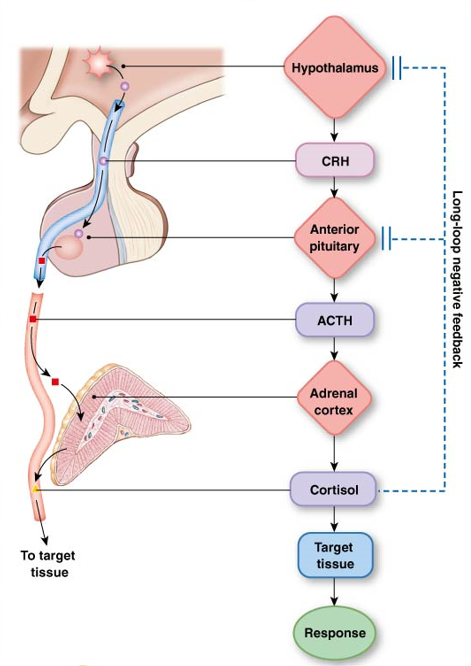
short-loop and long-loop negative feedback
short-loop: a pituitary hormone feeds back to decrease hormone secretion by the hypothalamus
long-loop: the hormone secreted by the peripheral endocrine gland feeds back to suppress secretion of its anterior pituitary and hypothalamic hormones
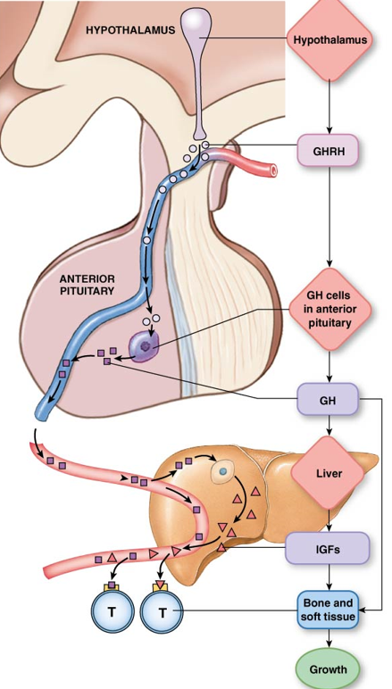
example: growth hormone
Hypothalamus releases growth hormone-releasing hormone (GHRH) into portal system.
GHRH acts on anterior pituitary growth hormone cells to release growth hormone (GH) into bloodstream.
GH acts on liver cells to release insulin-like growth factors (IGFs) into bloodstream.
IGFs act on bone and soft tissue to stimulate growth.
example: cortisol
Hypothalamus releases corticotropin-releasing hormone (CRH) into portal system.
CRH acts on anterior pituitary to release adrenocorticotropin (ACTH) into bloodstream.
ACTH acts on the adrenal cortex to release cortisol into bloodstream.
Cortisol feeds back to suppress secretion off CRH and ACTH. (long-loop negative feedback)
exogenous cortisol: will supress secretion in hypothalamus and anterior pituitary, but stimulate target tissue
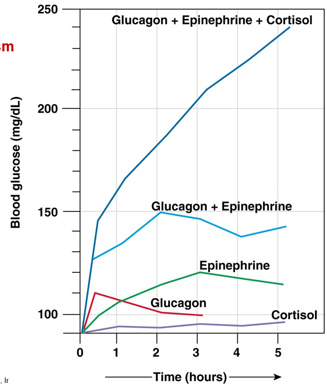
hormone interactions
synergism: combined hormone effect is greater than sum of the individual effects
permissiveness: need a second hormone for full effect
antagonism: one hormone opposes the action of the other
hormone pathologies terms
hypersecretion: excess hormone production
hyposecretion: insufficient hormone production
hypertrophy: enlargement of organ/gland
atrophy: decrease in size of organ/gland
down-regulation: decreased number of receptors
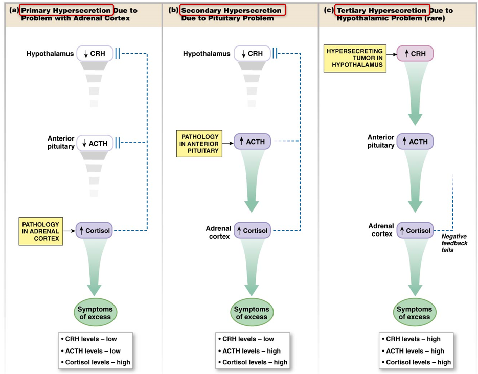
hormone pathologies diagnosis
diagnose based on relative hormone levels
primary: issue is with last endocrine gland in pathway
secondary: issue with pituitary gland
tertiary: issue with hypothalamus
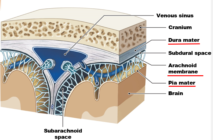
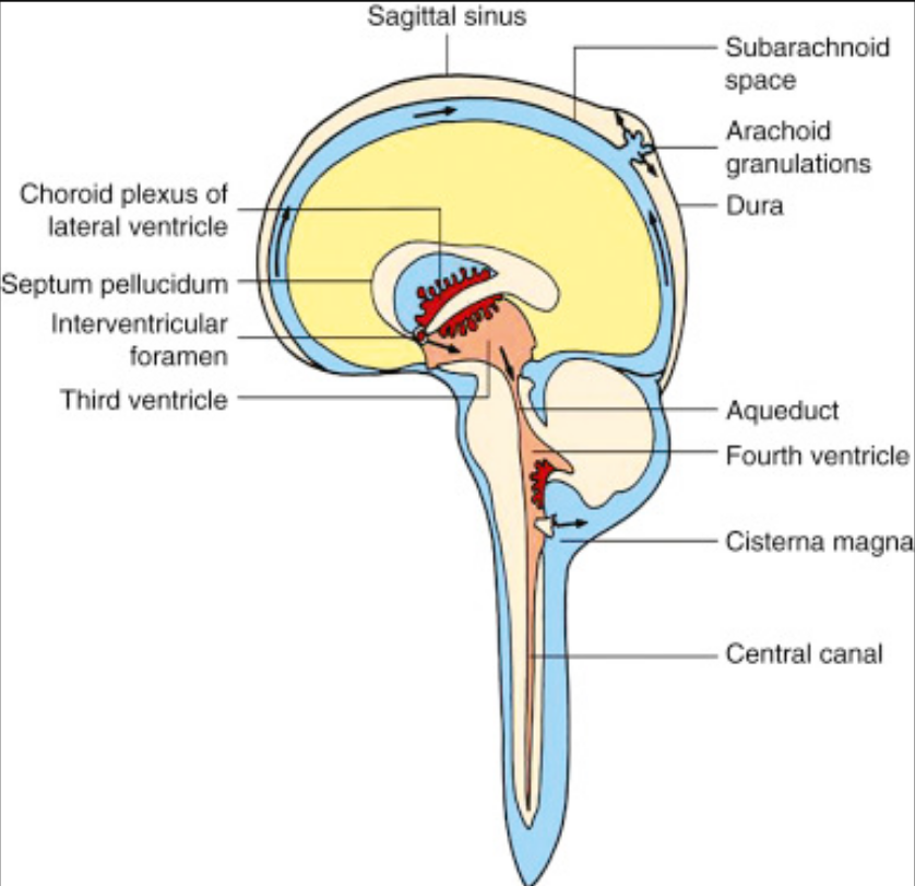
CNS: meninges
membranes that provide protection and cushioning for brain
dura mater (outer)
arachnoid membrane
pia mater (inner)
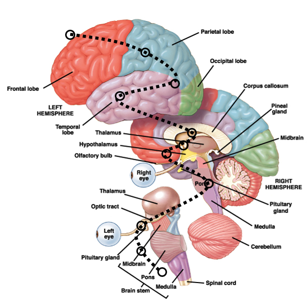
CNS: anatomy
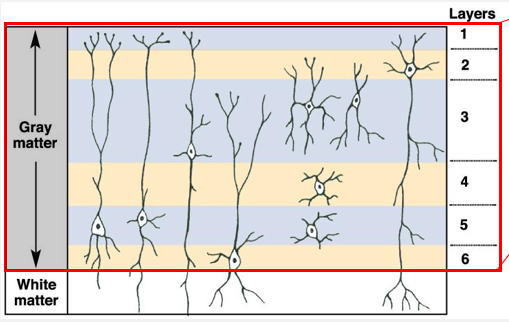
CNS: white and gray matter
gray matter: cell bodies
white matter: myelinated axons
ex: cerebral cortex cell layers
CNS: cerebrospinal fluid
water & materials from blood transported through layer of endothelial cells to cerebrospinal fluid (CSF)
CSF circulates through CNS
fluid returns to veins through arachnoid villi
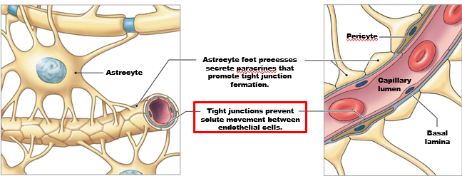
blood-brain barrier
usually endothelial cells are ‘leaky’, but in the brain tight junctions prevent solute movement between endothelial cells
astrocytes surrounding blood vessel promote tight junction formation
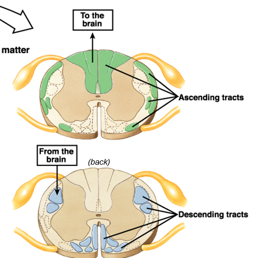
spinal cord
afferent tracks go up toward brain: dorsal/back
efferent tracks go down away from brain: ventral/front
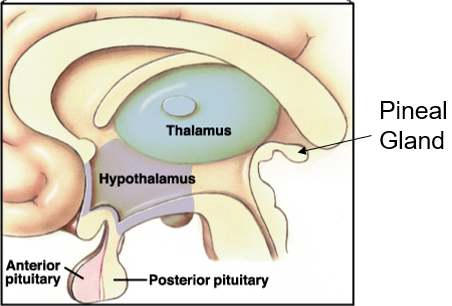
diencephalon
diencephalon = hypothalamus + thalamus
thalamus: relay station, senses that are coming in
hypothalamus: homeostasis control center, subconscious
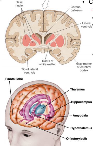
basal nuclei, corpus callosum, & limbic system
basal nuclei (ganglia): control of movement
corpus callosum: communication between hemispheres
limbic system:
cingulate gyrus: emotion
hippocampus: learning and memory
amygdala: emotion, reflexive memory, fear
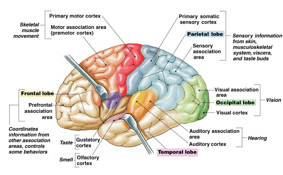
cerebral cortex
frontal lobe:
gustatory cortex
behavior, coordinate info from other association areas
parietal lobe: sensory info from skin, muscluloskeletal system, viscera, taste buds
occipital lobe: vision
temporal lobe: auditory cortex, olfactory cortex
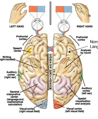
lateralization and crossover
visual field crossover: optic chiasmus
sensory & motor crossover: spinal cord
lateralization: certain functions on one side of brain
geometry and music on right
language and symbols on left