Microlab Lab Review:
1/56
There's no tags or description
Looks like no tags are added yet.
Name | Mastery | Learn | Test | Matching | Spaced | Call with Kai |
|---|
No analytics yet
Send a link to your students to track their progress
57 Terms
Plate Streak Types
Used to isolate individual bacterial colonies from a mixed culture. Isolation is necessary so that a pure culture can be obtained for identification or further experiments.
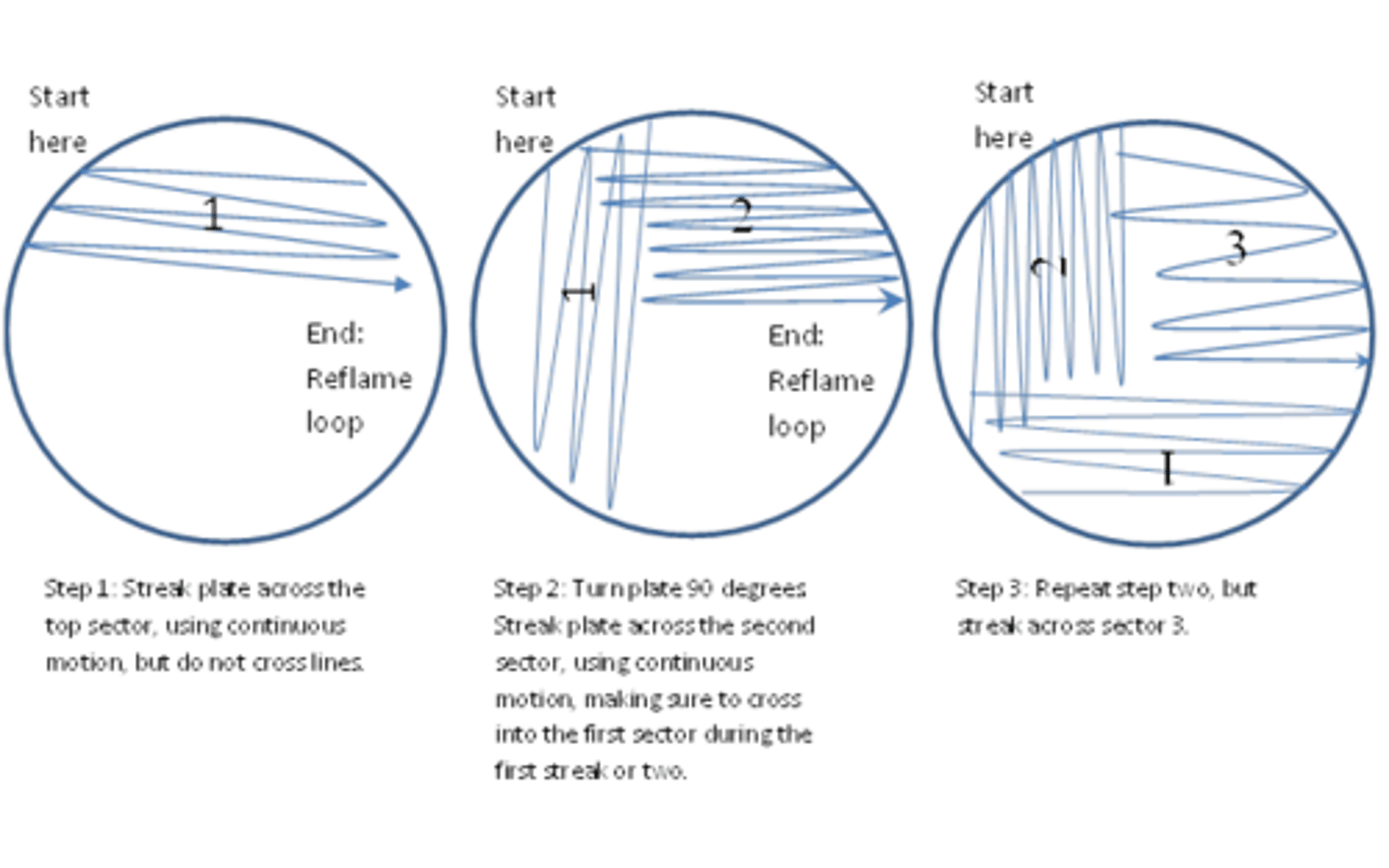
Quadrant Streak
Plate divided into 4 sectors
Loop sterilized between streaks
Each streak spreads fewer cells
Final quadrant produces isolated colonies
T-Streak
Plate divided into 3 sections
Streaked in a T pattern
Used when moderate dilution is needed
Zig-Zag Streak
Continuous streak across plate
Not ideal for isolation
Used to simply distribute bacteria
Why streaking works
Each new streak dilutes the bacterial population, allowing single cells to grow into individual colonies.
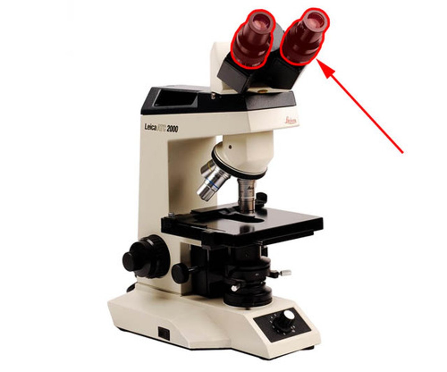
Parts of the Microscope: Ocular lens
Eyepiece used to observe specimen.
Magnification = 10x
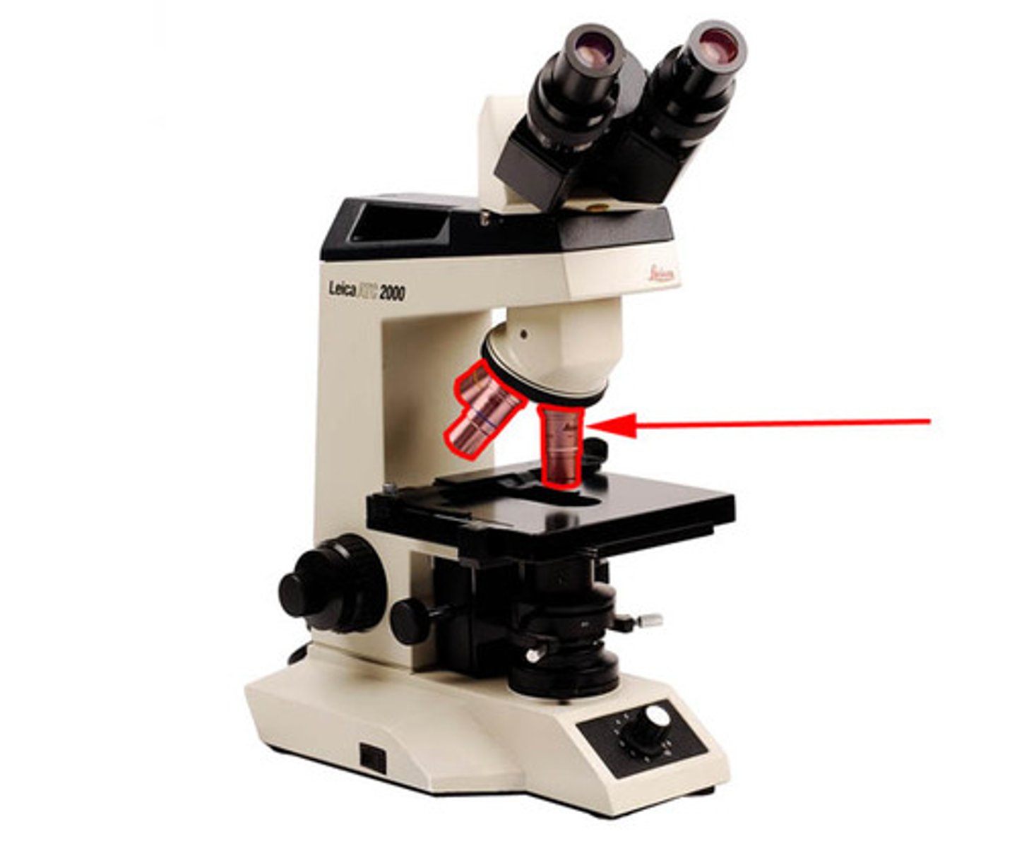
Objective lenses
Objective Magnification
Scanning 4x
Low power 10x
High power 40x
Oil immersion 100x
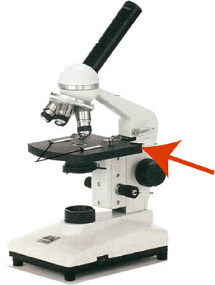
Stage
Platform where slide sits.
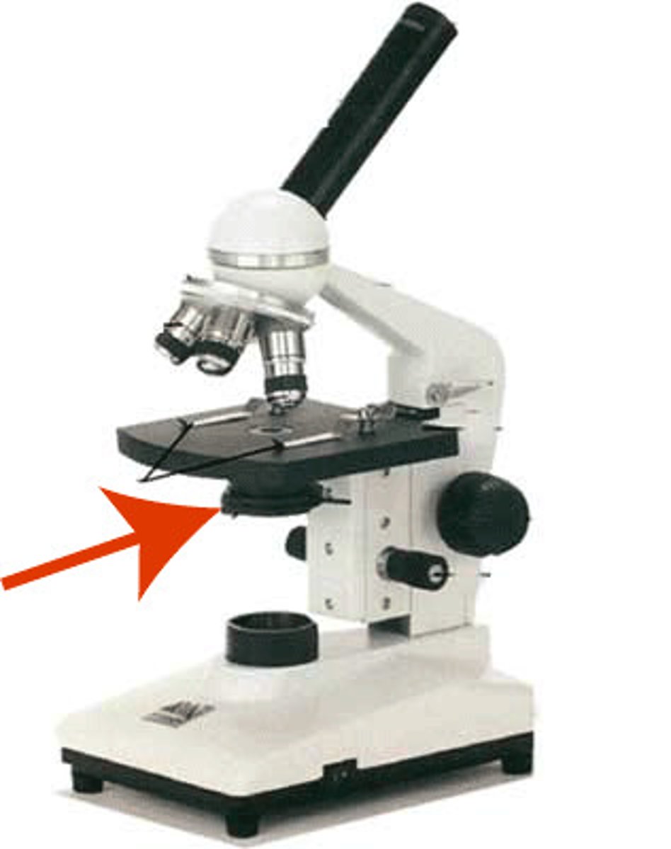
Condenser
Focuses light onto specimen.
Controls brightness and resolution.
Coarse focus knob
Moves stage quickly for initial focus.
Used only with low magnification.
Fine focus knob
Used for precise focusing, especially at high magnification.
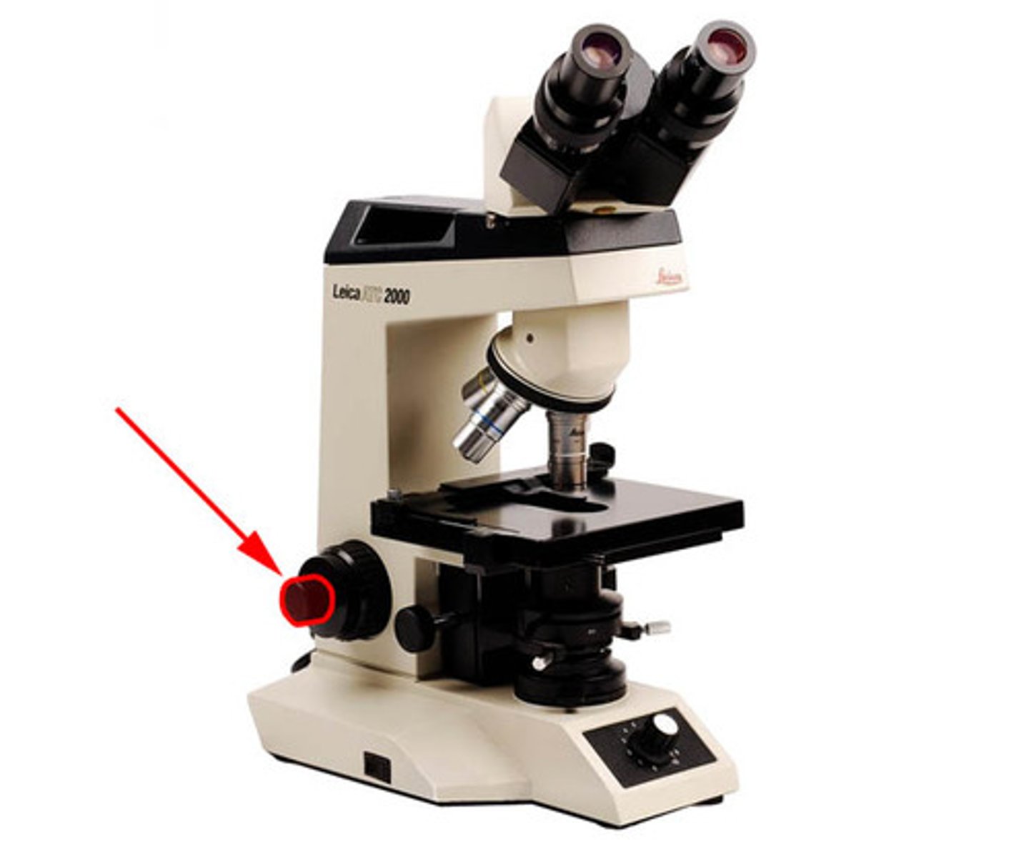
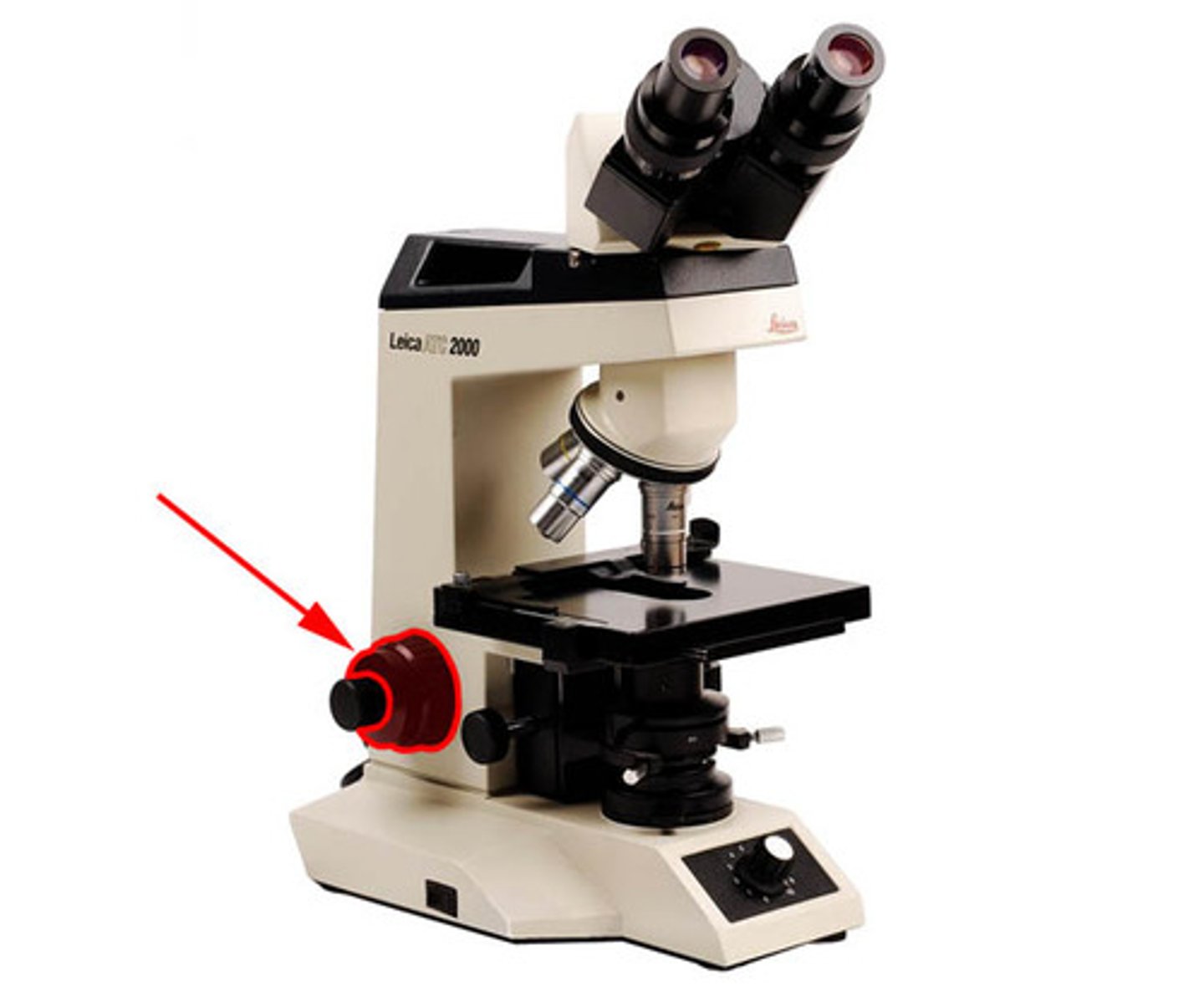
Light source
Illuminates specimen.
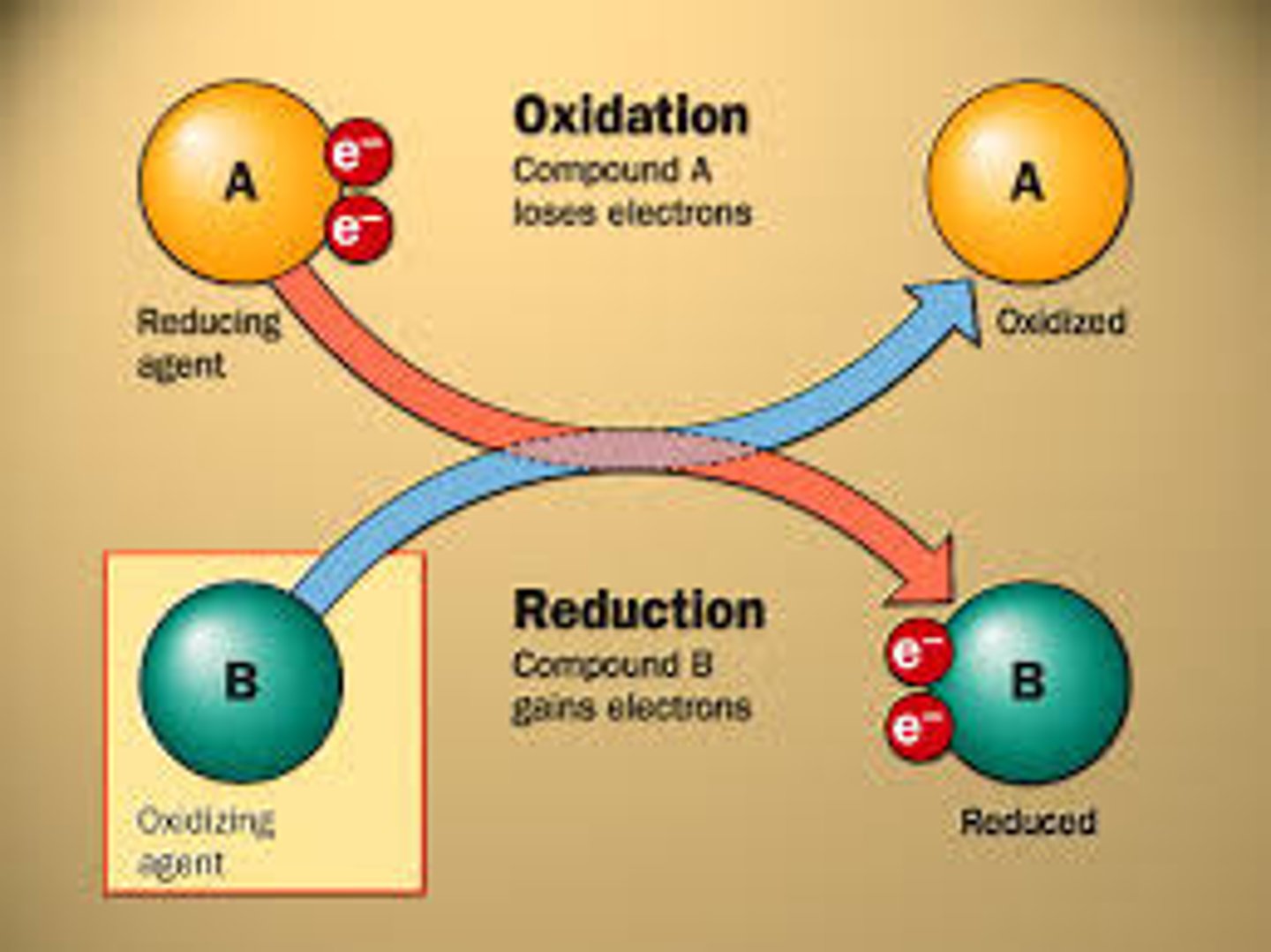
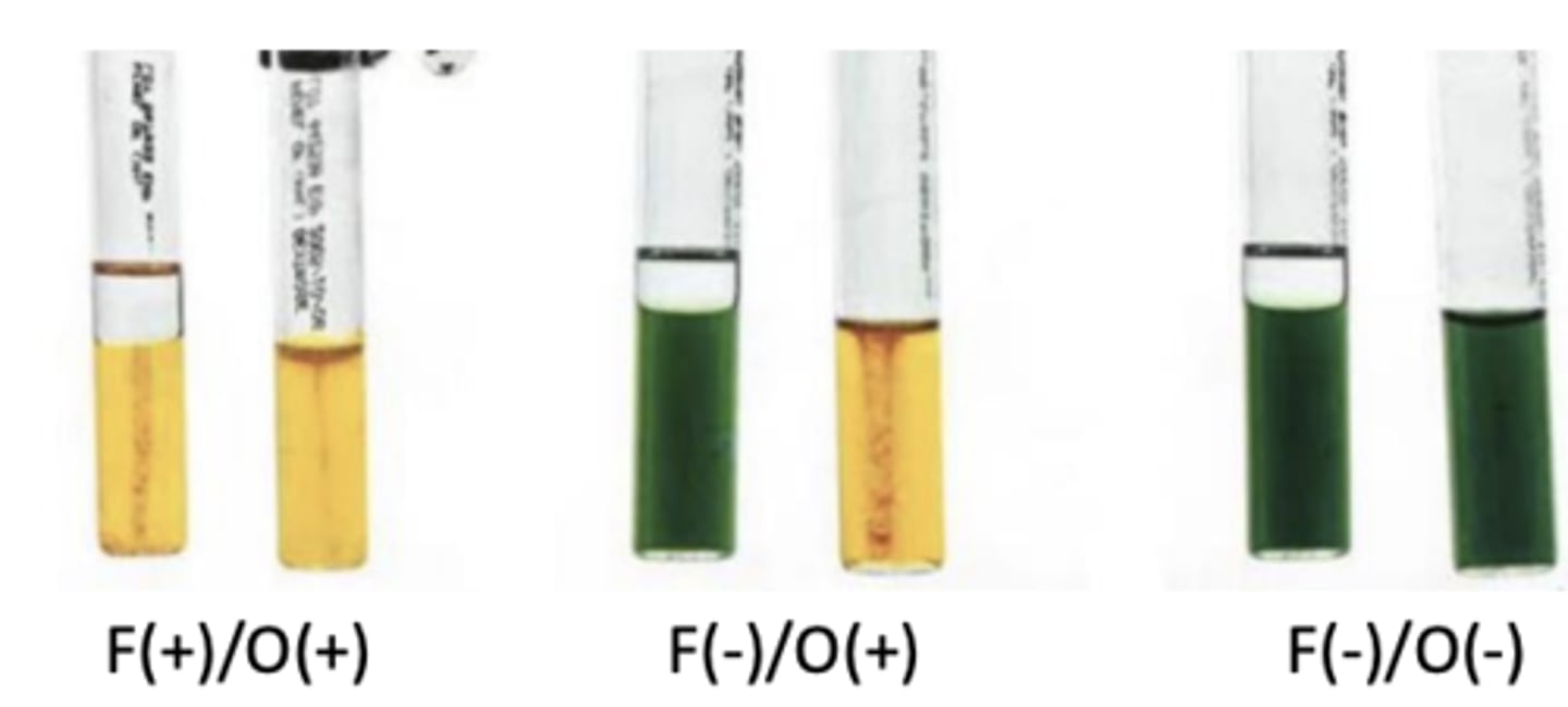
Oxidation vs Fermentation (O‑F test)
This test determines how bacteria metabolize glucose.
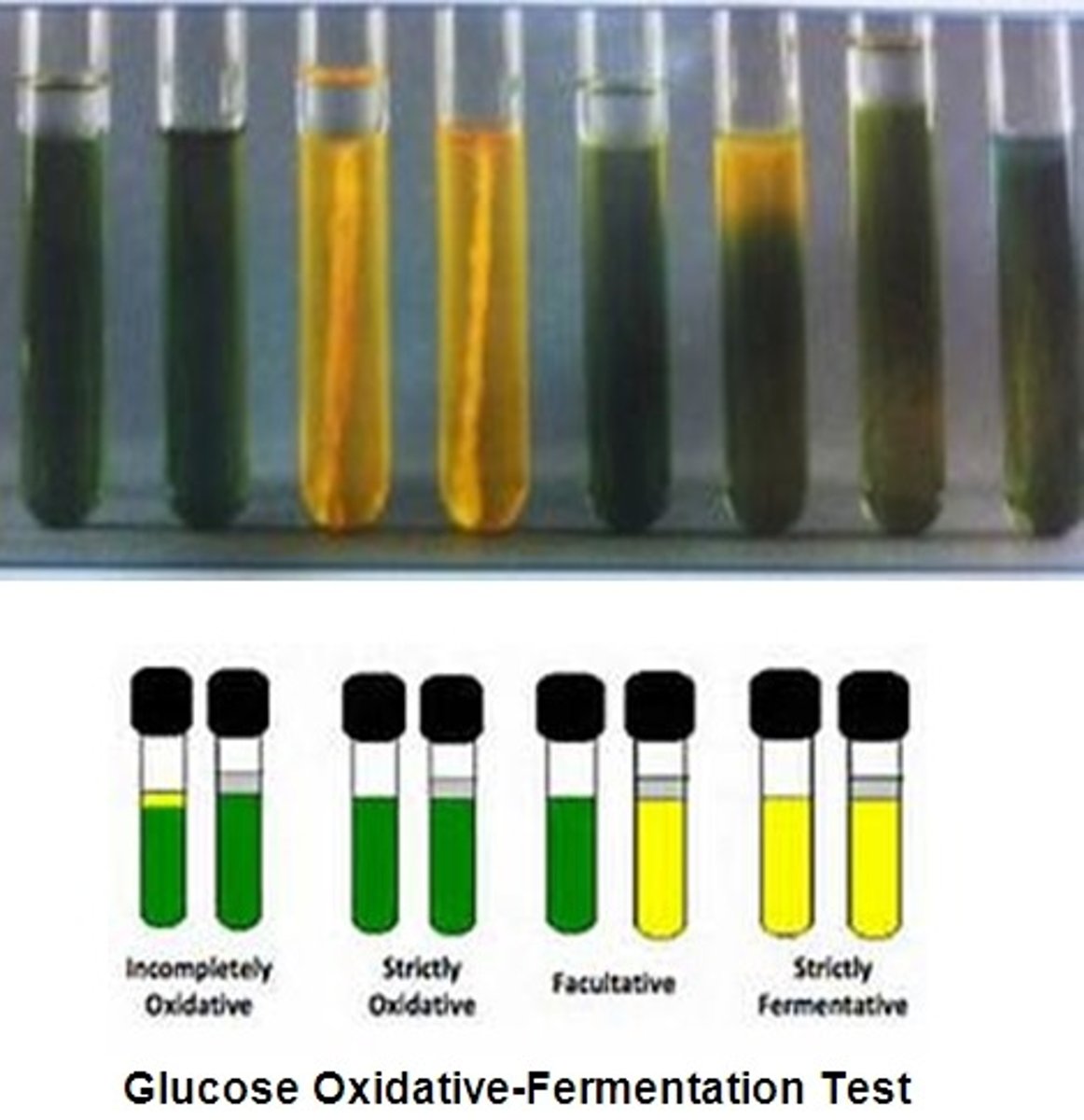
Oxidation
Requires oxygen
Glucose broken down using aerobic respiration
Oxidative Positive
Only open tube turns yellow
Fermentation
Does not require oxygen
Energy produced anaerobically
Fermentation Positive
Both tubes turn yellow
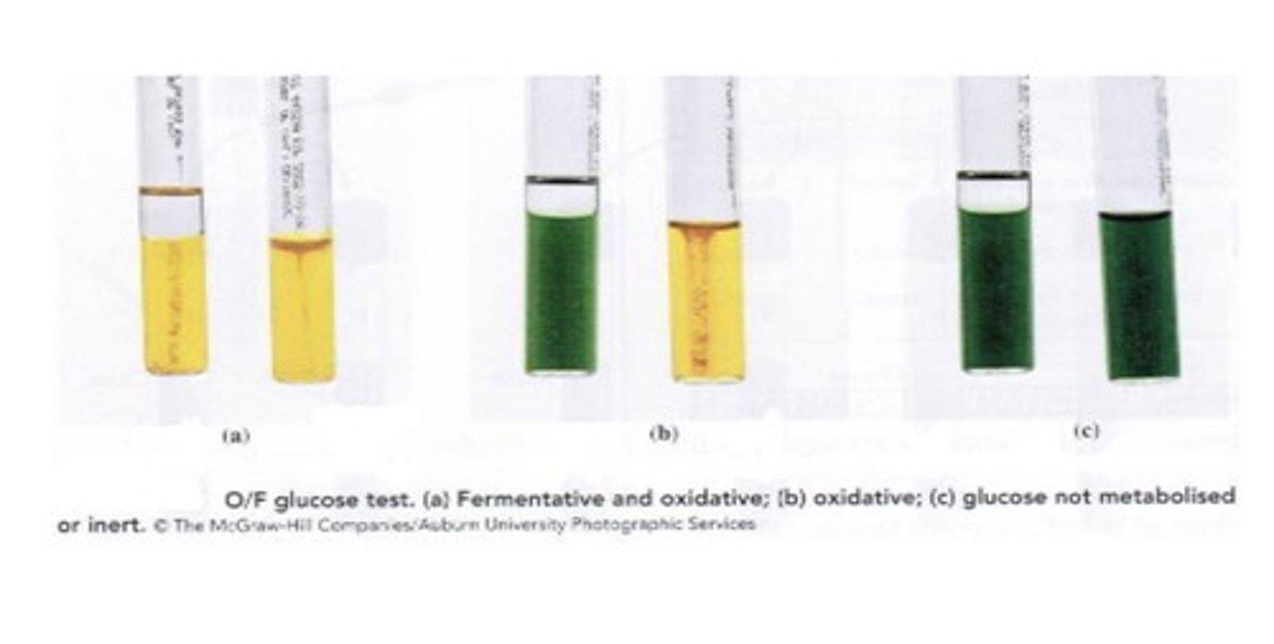
No metabolism
Medium remains green
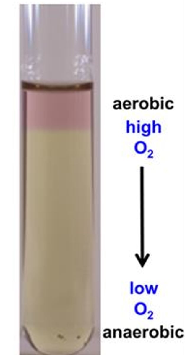
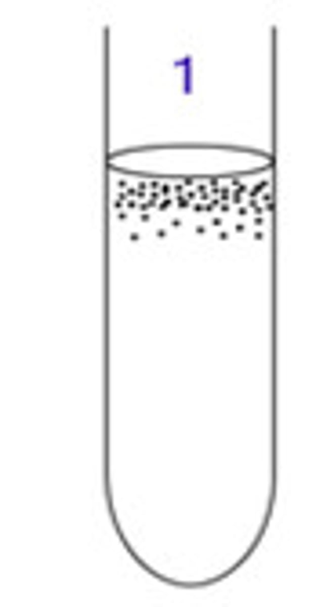
Obligate Aerobe
Requires oxygen.
Growth pattern: Top of tube only
Example: Pseudomonas
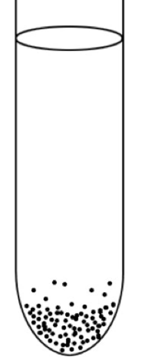
Obligate Anaerobe
Oxygen is toxic.
Growth pattern: Bottom of tube
Example: Clostridium
Facultative Anaerobe
Can grow with or without oxygen.
Growth pattern: Throughout tube but heavier at top
Example: E. coli
Aerotolerant Anaerobe
Does not use oxygen but tolerates it.
Growth pattern: Even growth throughout

Broth Growth Types: Uniform turbidity
Even cloudy growth.
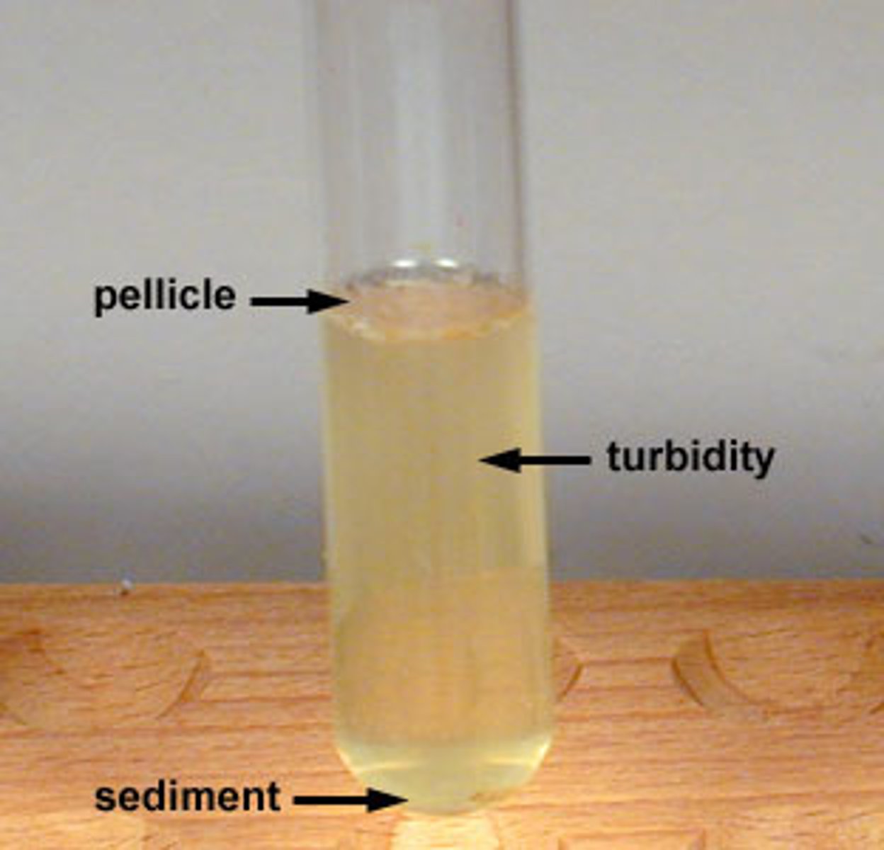
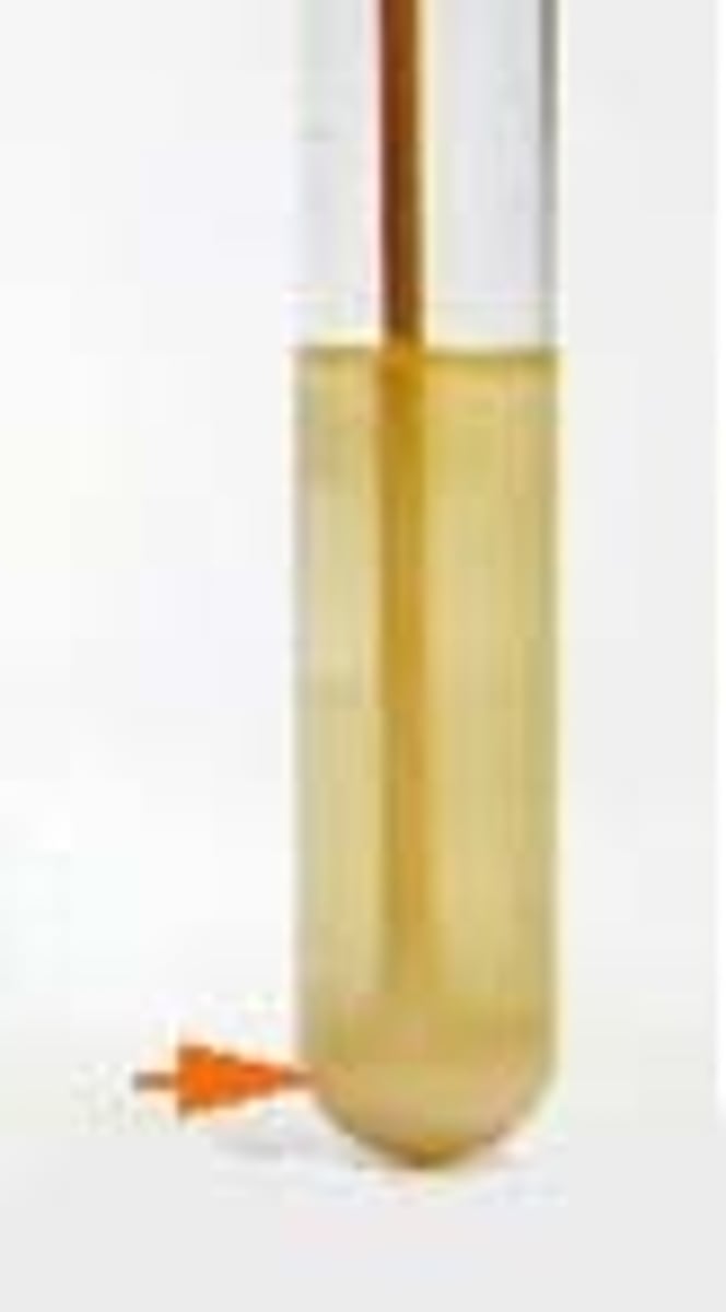
Broth Growth Types: Sediment
Cells settle at bottom.
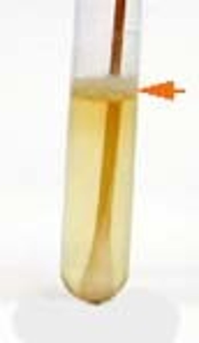
Broth Growth Types: Pellicle
Growth forms film on surface.
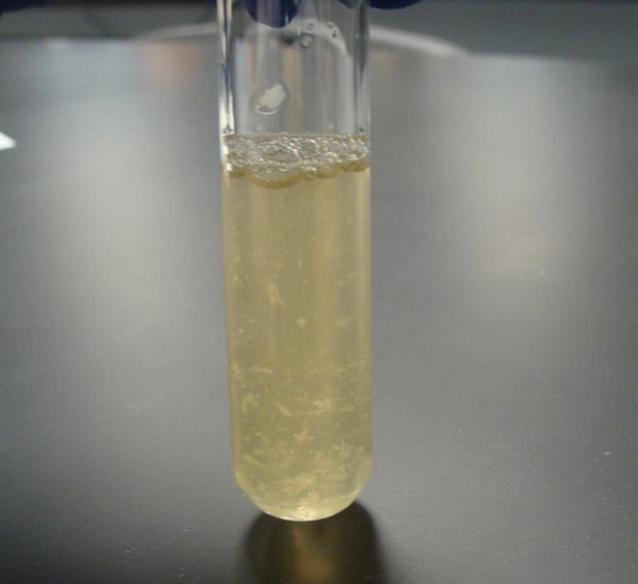
Broth Growth Types: Flocculent
Clumps floating in broth.
Slant Growth
Slants are used to maintain bacterial cultures.
Characteristics to Describe Bacteral Morphology
Shape
Margin
Texture
Pigment
Elevation
Example description:
"Raised, smooth margin, creamy texture, opaque white colonies."
Aperture Number (Numerical Aperture)
Numerical aperture measures the ability of the objective lens to collect light.
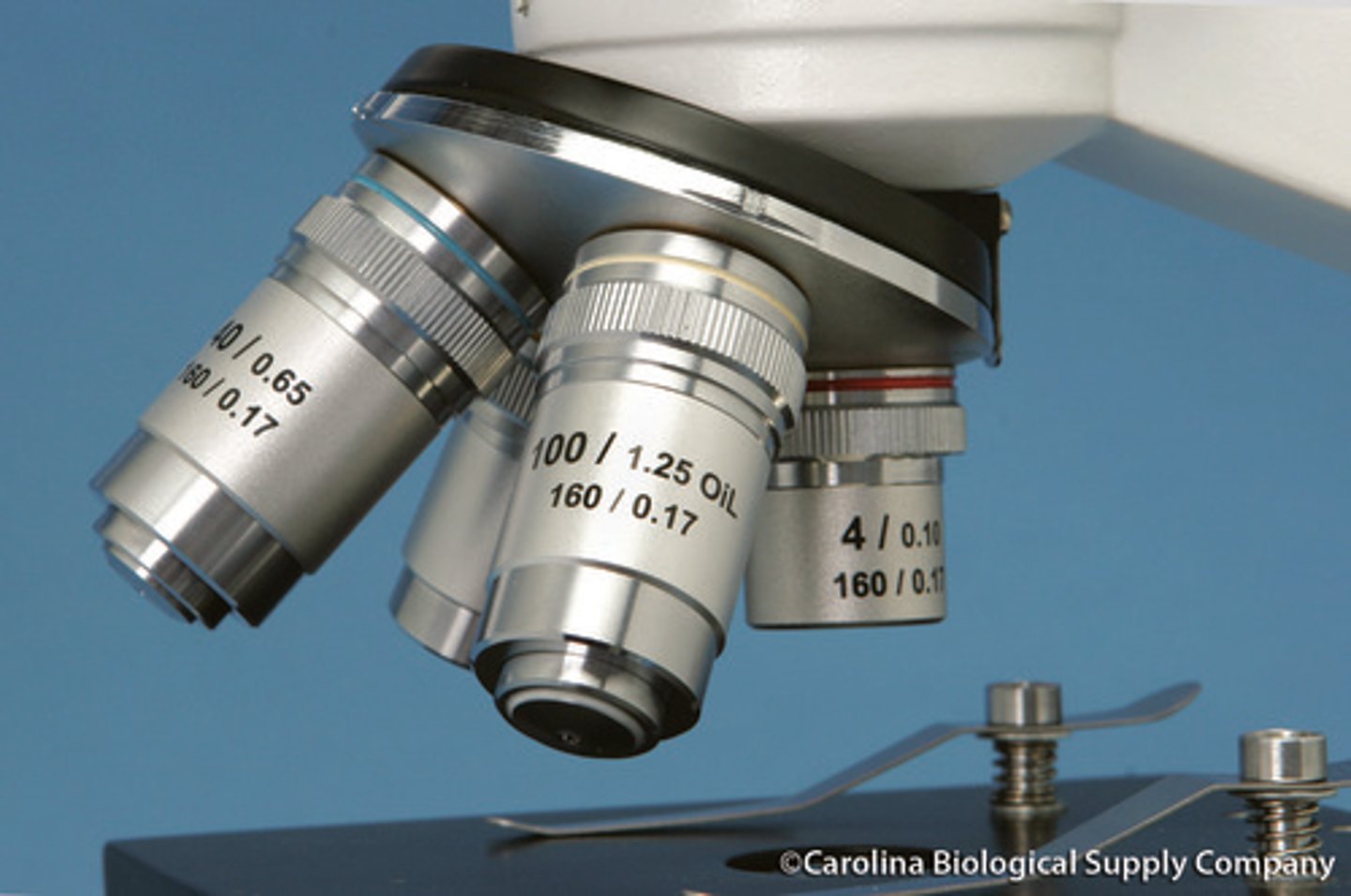
Higher NA =
Better Resolution
Where NA is found?
Printed on the objective lens.
Example of NA
40x / 0.65
0.65 = numerical aperture.
No growth in lab experiments
culture dead
inoculation failed

Contamination in experiment
Poor aseptic technic
Incorrect color change in lab
Incorrect incubation
Wrong reagent order
Simple stain
Uses basic dye, positive charge, hence why it stains negatively charged cells.
Simple stain examples
crystal violet
methylene blue
safranin
Purpose of Simple Stain
Observe cell shape, size, arrangement
Heat-fixing may distort cells
Negative stain
uses acidic dye (negatively charged), which is repelled by (negatively charged0 cell walls, leaving clear cells on a dark background
Negative stain example
nigrosin
Negative stain Purpose
Stains background instead of cells
Cells appear clear
no heat-fixing so cells stay in their original state, to see bacteria in their natural size, shape and arrangement
Differential stains
Used to distinguish bacteria. (multiple stains involved)
Differential stains examples
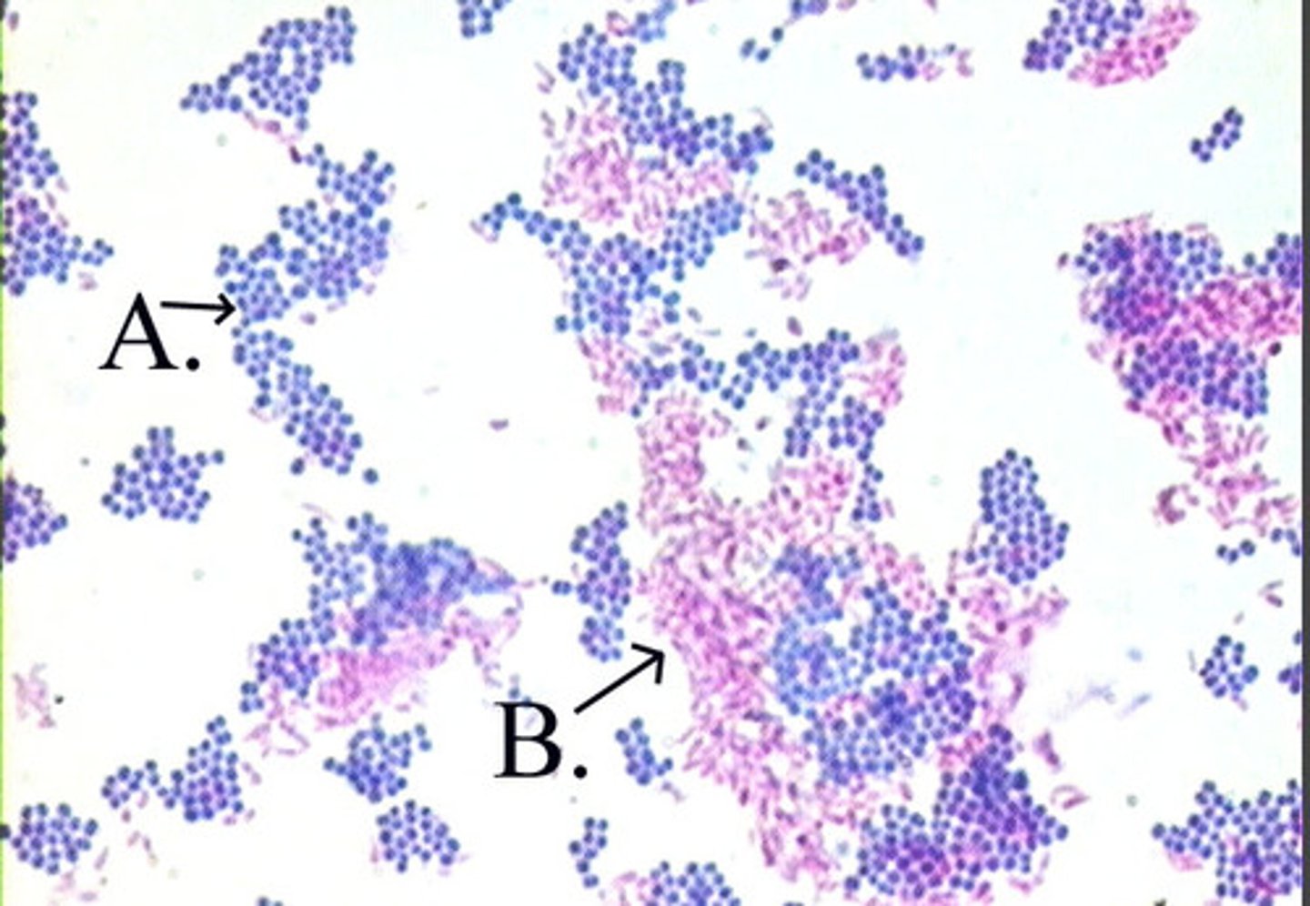
Gram stain
Acid fast stain
Endospore stain
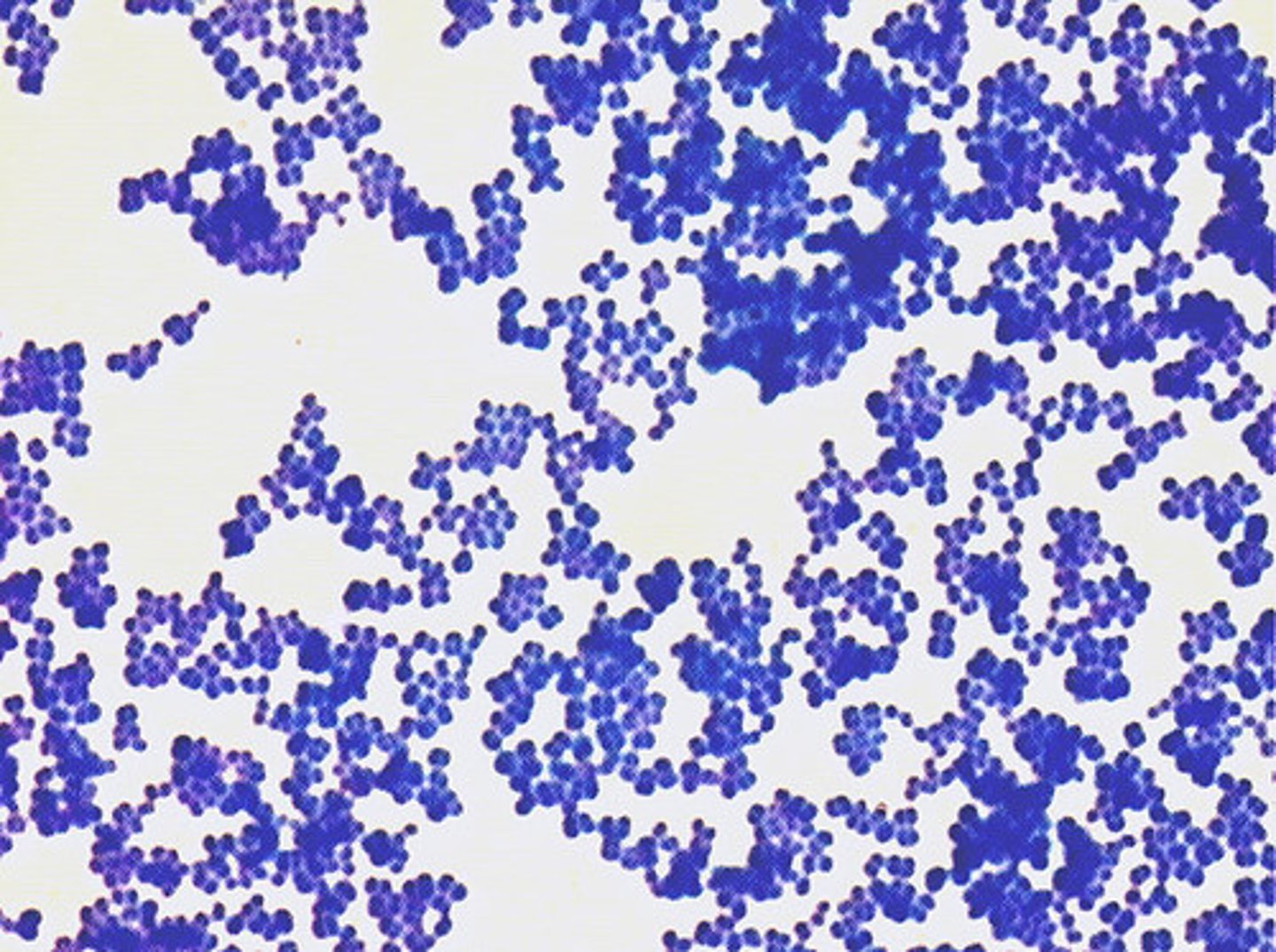
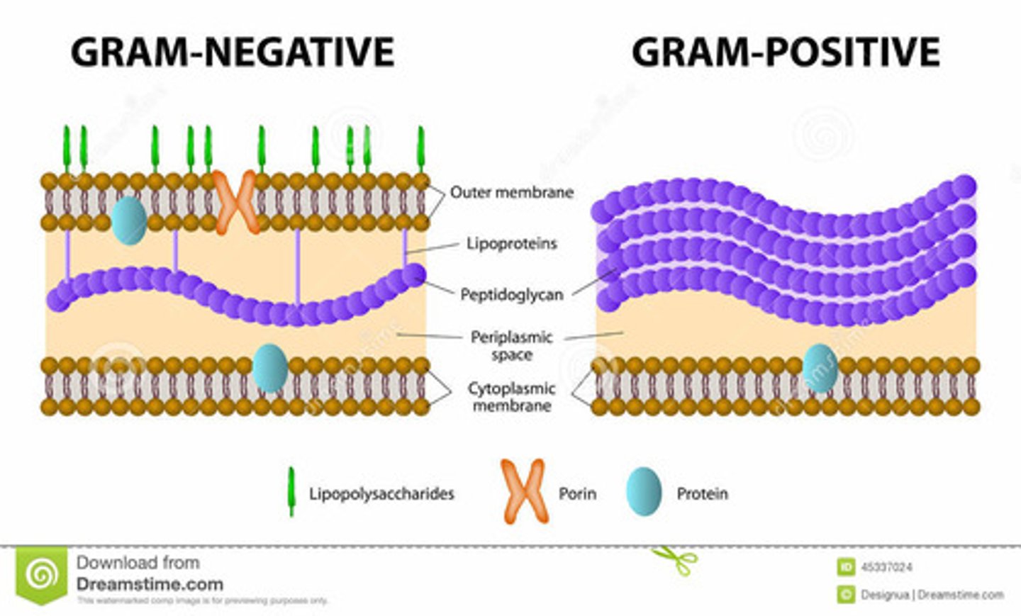
Gram‑positive bacteria
Have 1 lipid layer, thick peptidoglycan, and teichoic acids
Result of Gram + Stain
Retain crystal violet.
Color: Purple

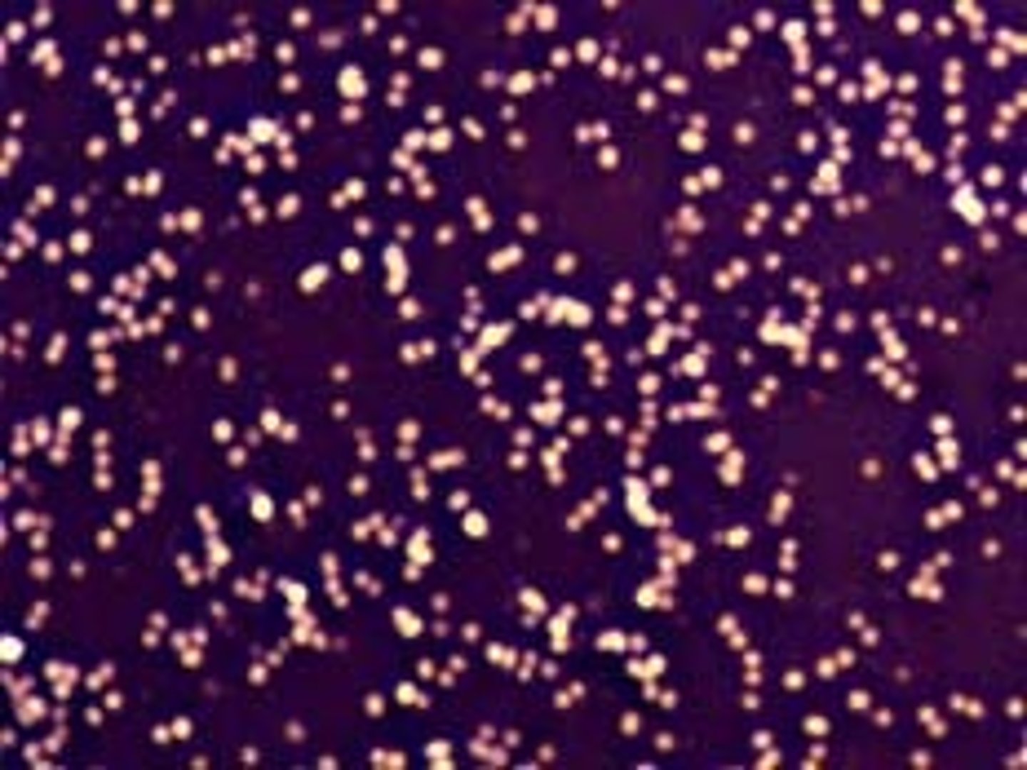
Gram‑negative bacteria
Have 2 lipid layers and thin peptidoglycan and LPS
Gram - Stain
Alcohol removes stain.
Counterstain (safranin) colors them:
Pink
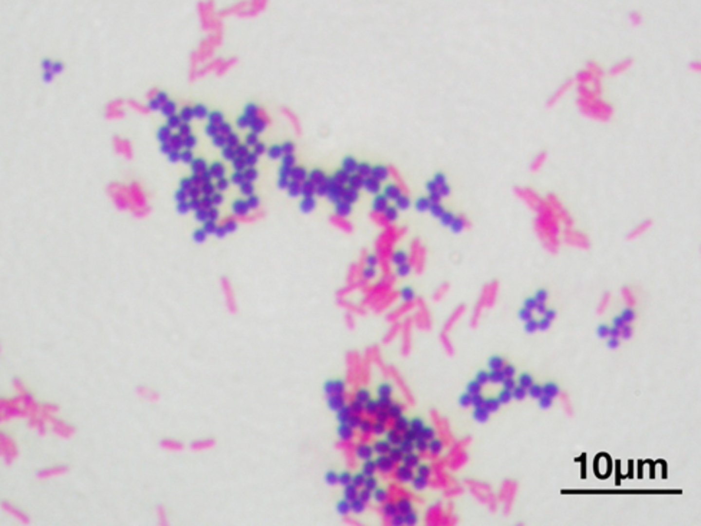
Gram Stain
Crystal violet
Iodine (mordant)
Alcohol (decolorizer)
Safranin (counterstain)
Simple stain Procedure
basic dye
stains bacteria
heat fixed
Result: cells colored
Negative stain Procedure
acidic dye
stains background
no heat fixation
Result: cells appear clear
Advantage: Cells remain true size and shape.
Principle of Acid‑Fast Stain
Used to detect bacteria containing mycolic acid.
Acid Fast Stain
Contain mycolic acid, have a waxy, hydrophobic wall, and that the test is meant for the Mycobacterium genus, including organisms like M. leprae and M. tuberculosis.
What is the principle of Acid-Fast Stain?
Normal water-soluble stains do not penetrate the waxy mycolic acid-rich wall well.
What type of stain is used in Acid-Fast Staining?
Carbolfuchsin, a phenolic stain.
How does steam heat affect the mycolic acid in Acid-Fast Staining?
Steam heat melts the mycolic acid and creates pores that let the stain enter.
Acid fast bacteria: Red
Non acid fast bacteria: Blue
What is in the Cell Membrane
Cell membrane contains:
phospholipids
proteins
enzymes
transport proteins
Function: Controls movement of substances in and out of cell.
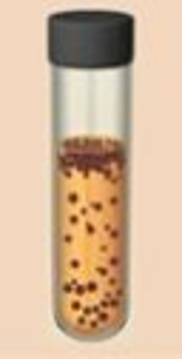
Fluid Thioglycollate Medium (FTM)
FTM is used to determine oxygen requirements of bacteria.
The medium contains chemicals that remove oxygen, creating an oxygen gradient.