Looks like no one added any tags here yet for you.
What is blood?
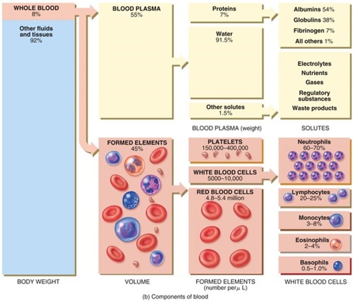
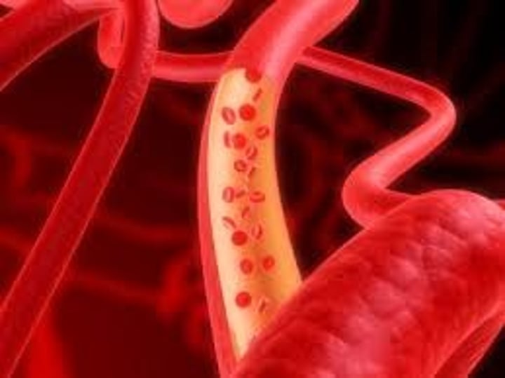
Blood is a fluid connective tissue; contains cells surrounded by a liquid extracellular matrix (plasma)
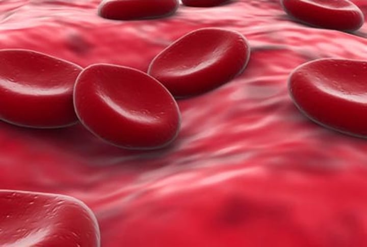
Erythrocytes
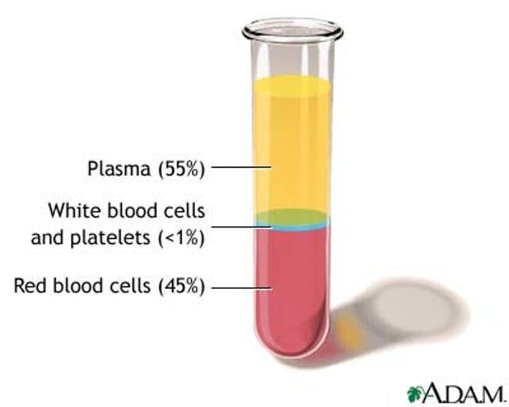
Red blood cells; they make up ~45% of blood
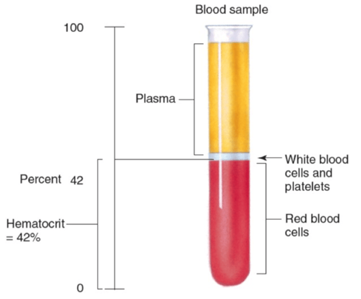
Hematocrit
The percent of blood volume that is composed of red blood cells (normal is around 45%-50%)
How is blood composition measured?
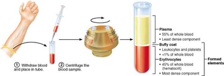
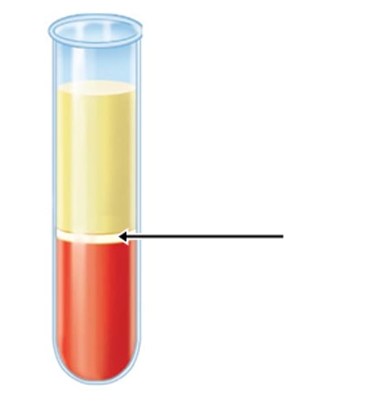
After blood is drawn, a centrifuge separates the blood into three components based on density: RBCs, WBCs, and plasma
Buffy coat
White blood cells (leukocytes) and platelets; this makes up 1% of blood
Blood plasma
The pale yellow fluid portion of blood, contains water/proteins/solutes; this makes up 55% of blood
Proteins in blood plasma (3)
Albumin, globulins, fibrinogen
Albumins (protein)
Most abundant plasma protein, transports lipids/hormones and helps regulate osmotic pressure of the blood
Globulins (protein)
Plasma protein that form antibodies
Fibrinogen (protein)
Plasma protein that is converted to fibrin in the clotting process
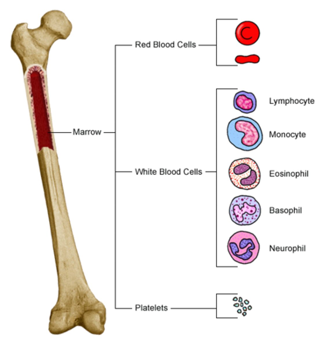
What are the cellular components of blood?
Erythrocytes (Red Blood Cells), Leukocytes (White Blood Cells), and Thrombocytes (Platelets)
What are the three major functions of blood?
Transportation, regulation, protection
What does blood transport?
Blood transports oxygen, carbon dioxide, nutrients, hormones, heat, and waste products
How does blood regulate homeostasis?
1. pH regulation via buffers
2. Temperature regulation by absorbing/releasing heat
3. Maintains fluid balance via platelets
How does blood protect the body?
Blood protects against excessive blood loss by clotting, and uses white blood cells to protect against infections
How long do blood cells live?
Lymphocytes (T and B cells) are able to live for years; most other blood cells live for hours/days/weeks
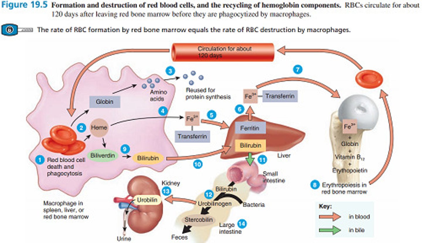
How long do RBCs live?
120 days (4 months)
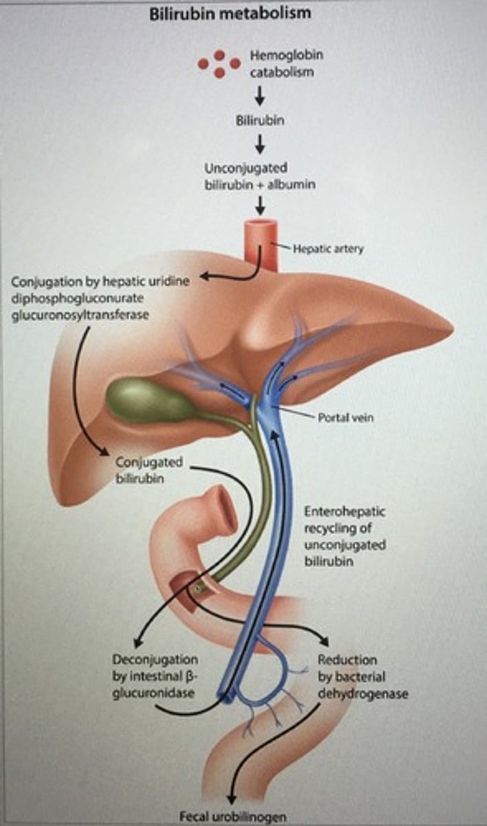
Where does the recycling of hemoglobin take place?
In the liver and spleen
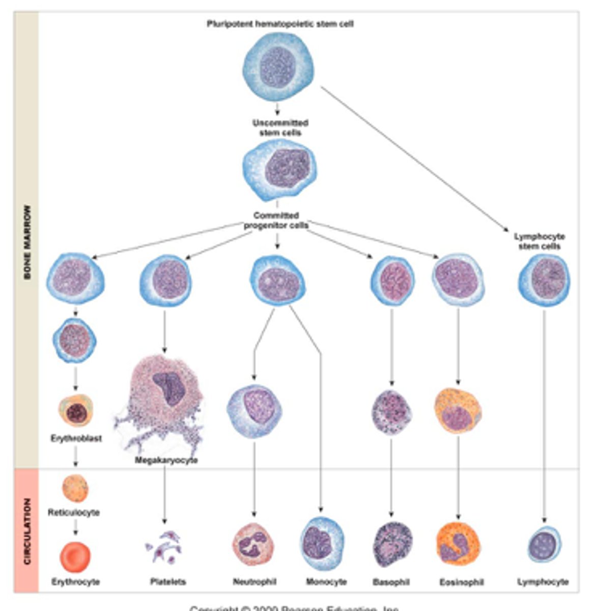
Pluripotent stem cells
Stem cells located in red bone marrow; they give rise to all the different types of blood cells
Hematopoiesis
The process of blood cell formation; occurs in the red bone marrow
Hematopoietic cells
Also called hemocytoblasts (meaning "blood cell bud"); these stem cells differentiate into the 3 types of blood cells
What are colony-stimulating factors?
A type of glycoprotein that stimulates the differentiation of stem cells into specific white blood cells
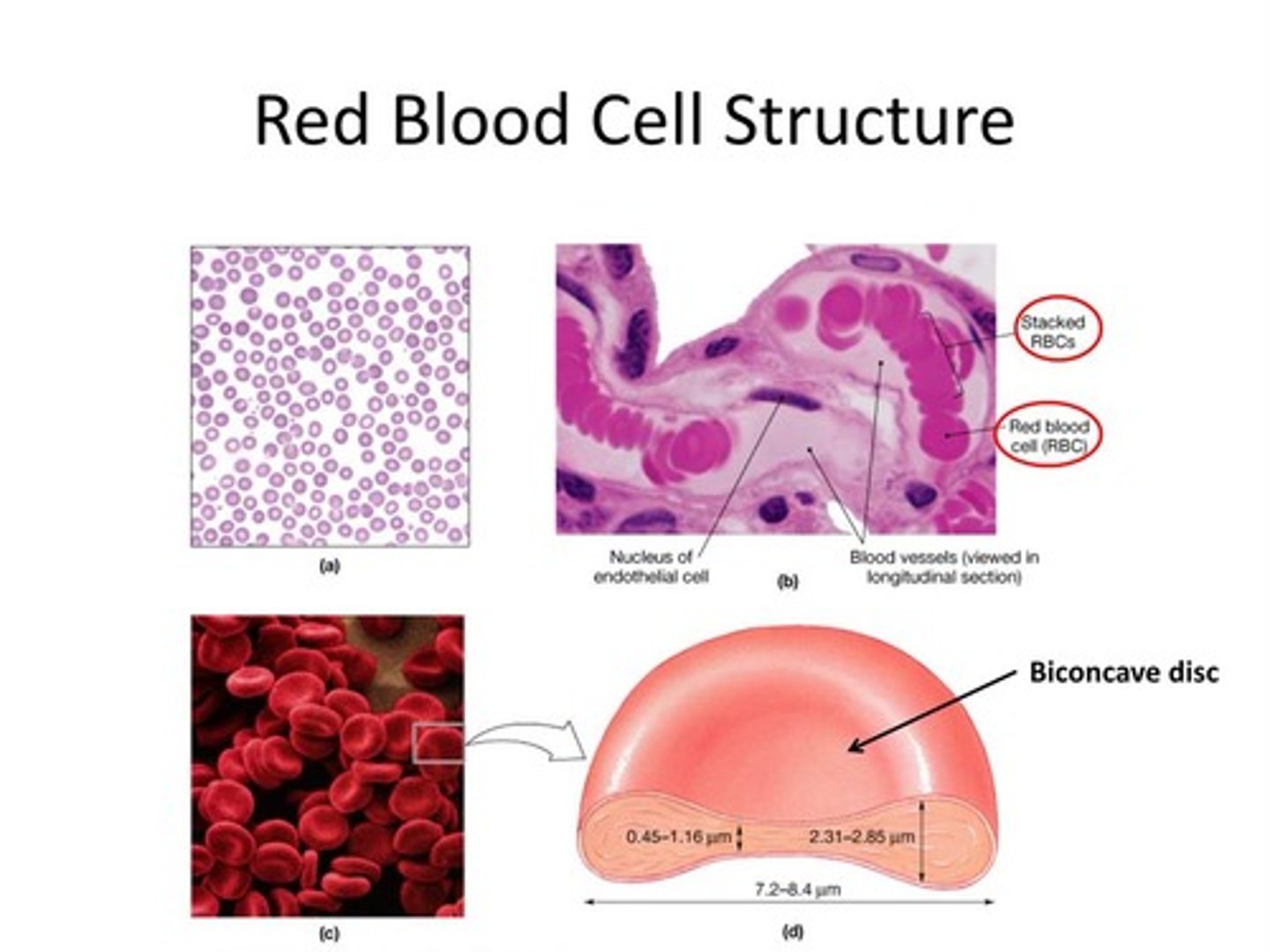
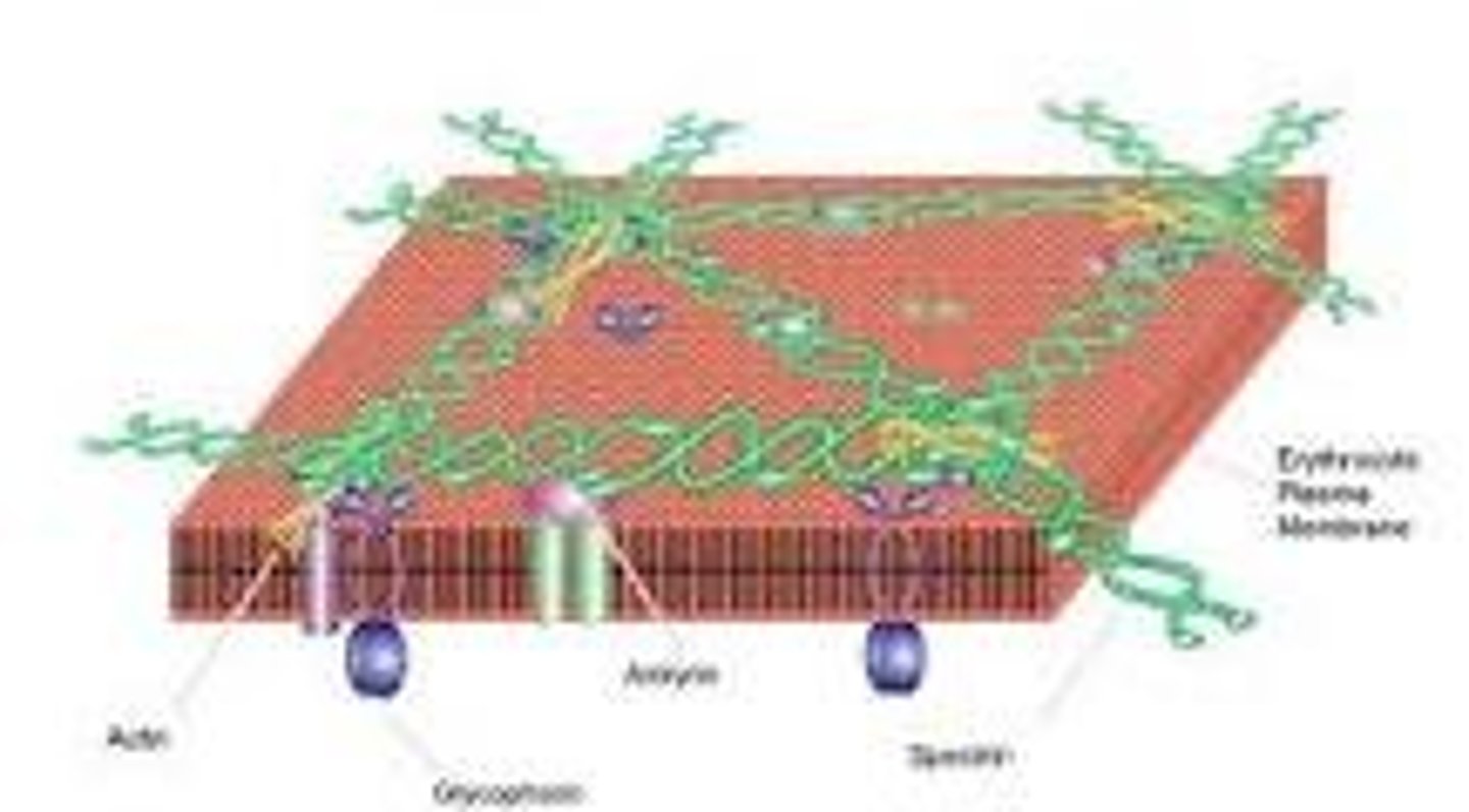
What is unique about the structure of red blood cells?
RBCs are biconcave discs (allows increased surface area), and they do NOT have nuclei or mitochondria (allows for more space for hemoglobin)
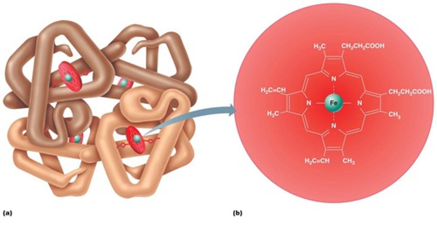
Hemoglobin
A protein in red blood cells that contains 4 iron molecules (Fe) and carries oxygen
Heme group
Disks that contain an iron molecule in the middle; the disks are the sites of oxygen binding
How many hemoglobin molecules are in each RBC?
A single RBC contains 250 million Hb molecules
Spectrin
A protein in the plasma membrane of RBCs which allows it to change shape
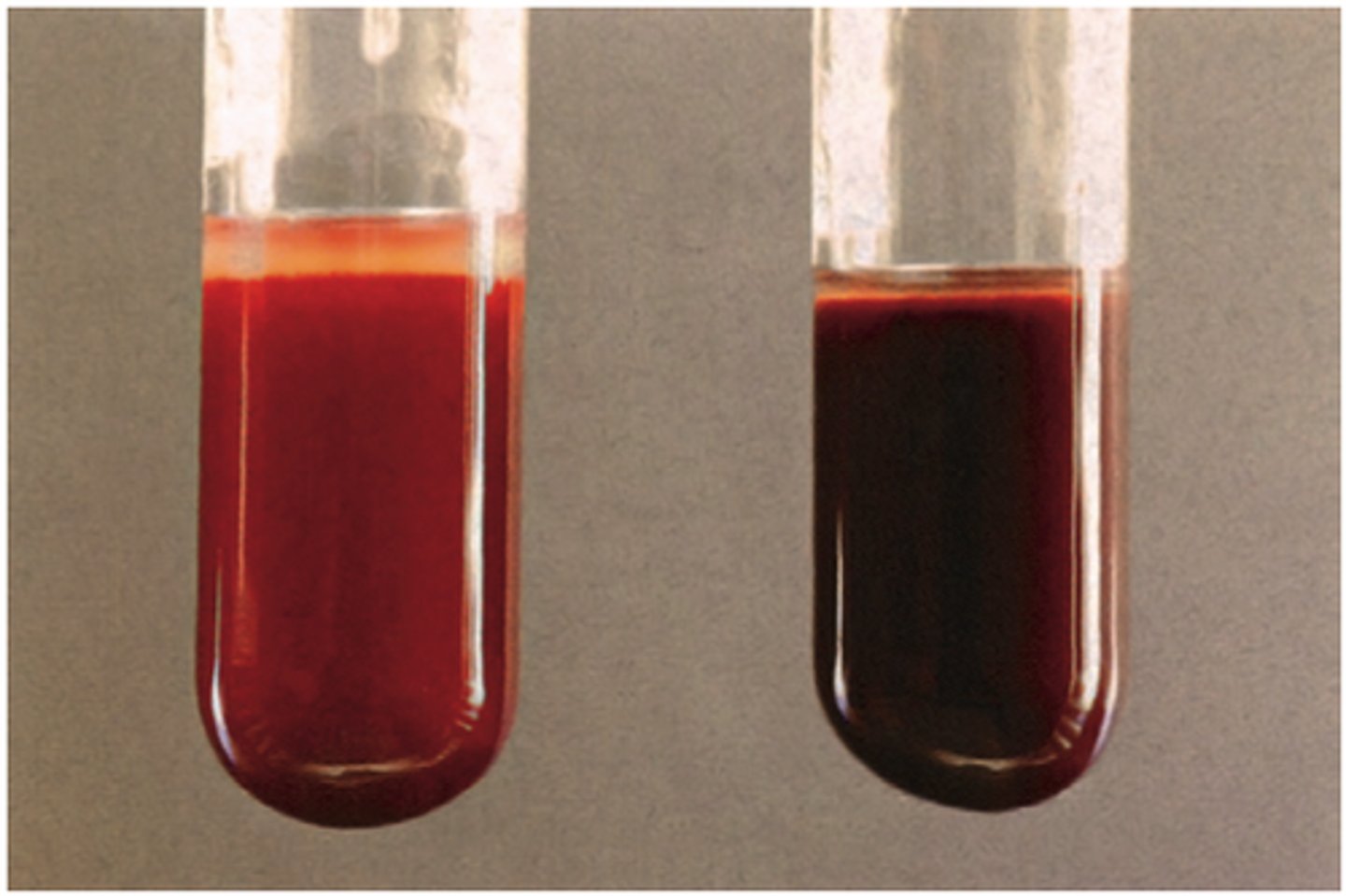
Oxyhemoglobin
Hemoglobin loaded with oxygen, produces a bright red color
Deoxyhemoglobin
Hemoglobin without oxygen, produces a dark blue-ish/red color
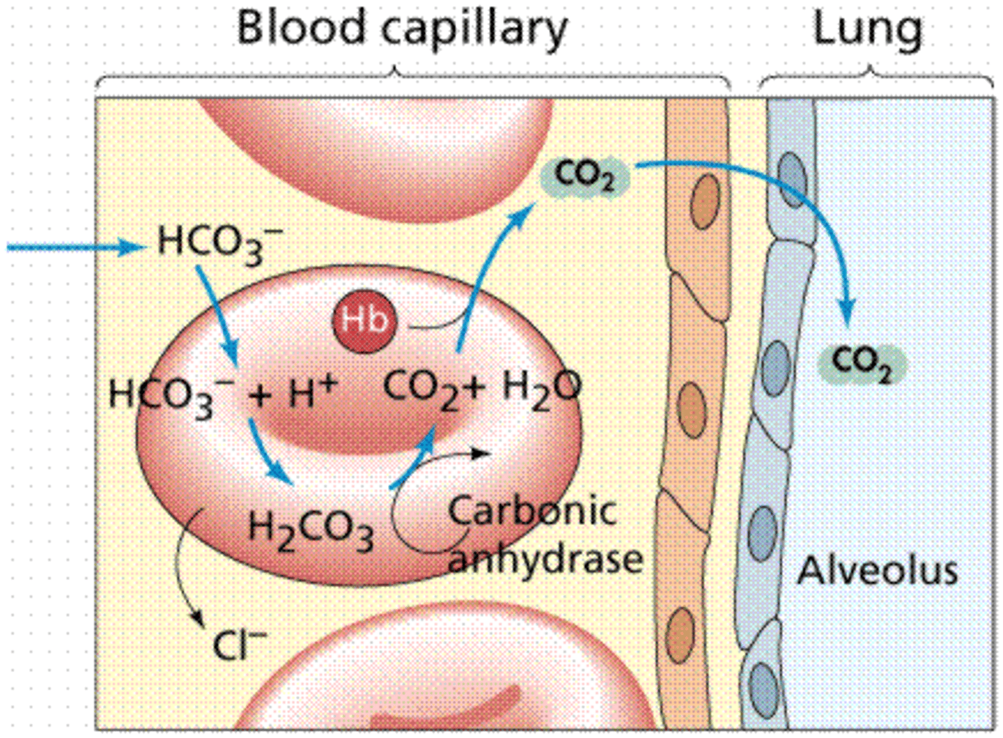
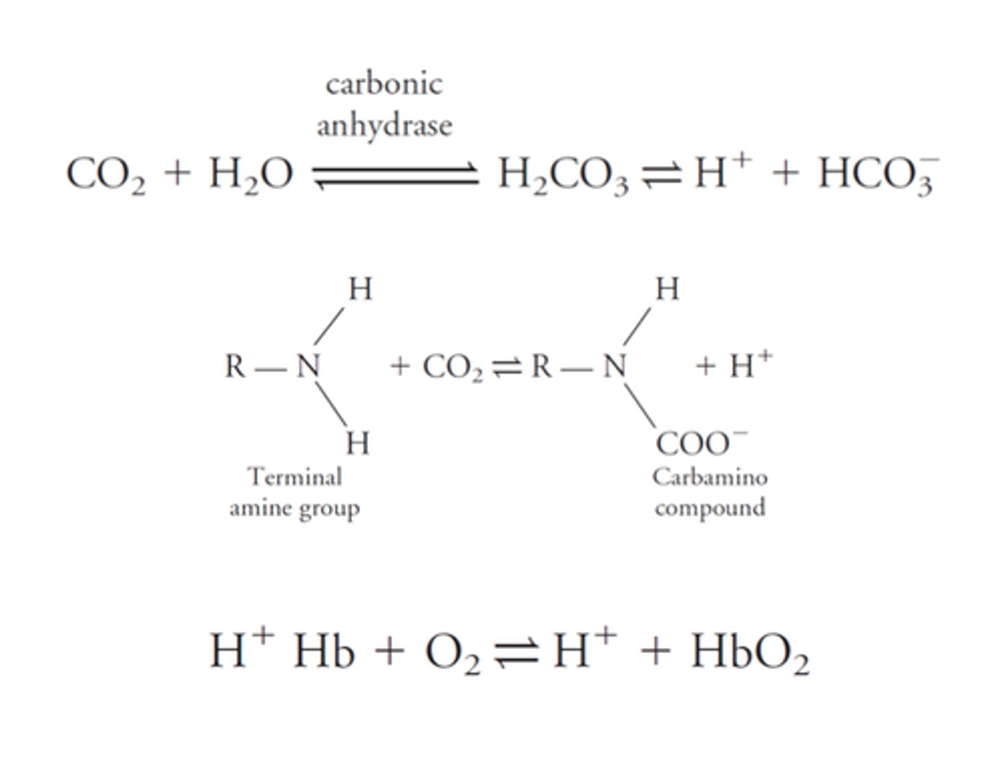
Carbaminohemoglobin
Hemoglobin bound to carbon dioxide
How does hemoglobin regulate blood pressure?
Hemoglobin releases nitrous oxide (NO), which stimulates vasodilation --> improves blood flow, enhances oxygen delivery
Carbonic anhydrase
An enzyme present in erythrocytes that catalyzes the conversion of CO2 and H2O into carbonic acid (H2CO3)
Bilirubin
An orange/yellow pigment in bile that formed by the breakdown of hemoglobin during the recycling of RBCs
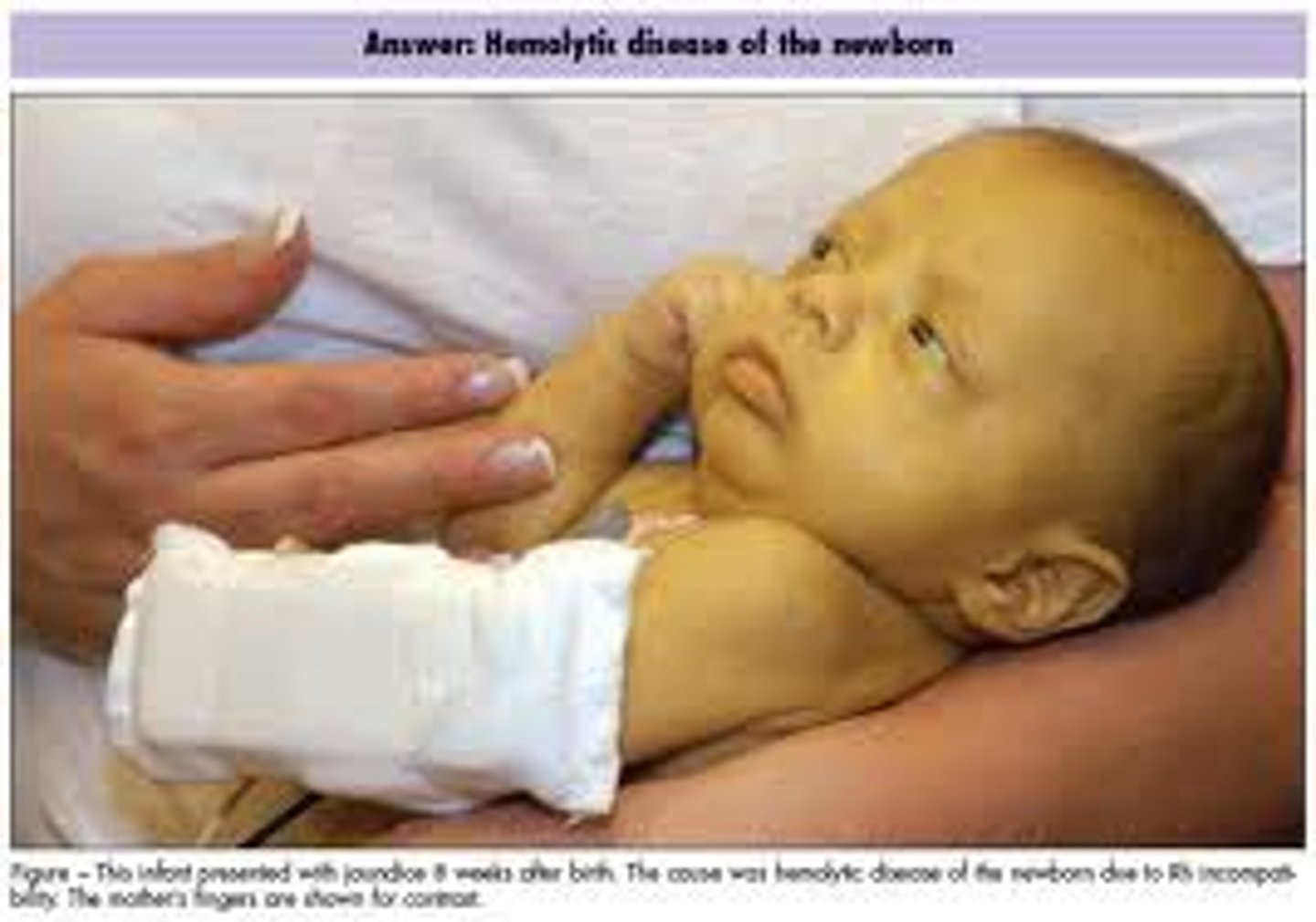
Jaundice
Symptoms: Yellowing of the skin and the whites of the eyes
Cause: Caused by an accumulation of bile pigment (bilirubin) in the blood
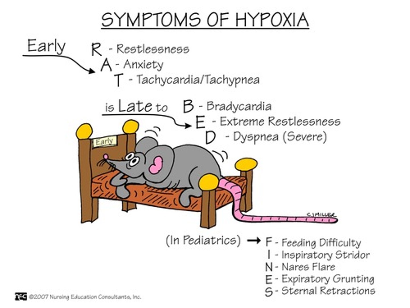
Hypoxia
Low oxygen in the blood
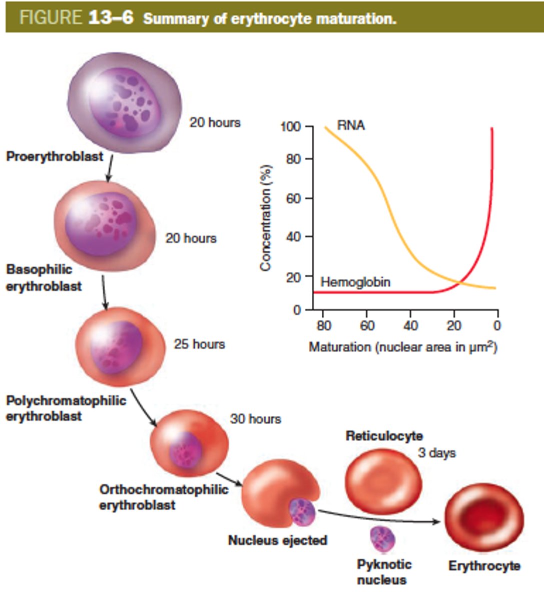
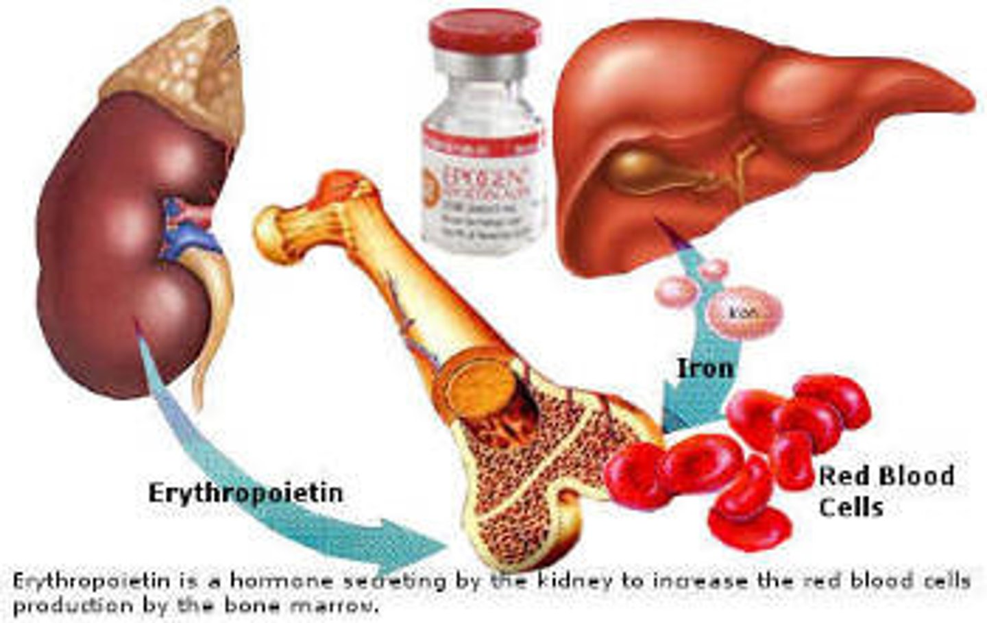
Erythropoiesis
The formation of red blood cells; occurs when a hormone (erythropoietin) is released in response hypoxia (low O2 in the tissues)
Reticulocytes
Immature red blood cells in bone marrow; they enter the circulation and mature in 1 to 2 days
What are possible consequences of increased hematocrit?
Increased RBCs in the blood causes it to become dehydrated and increases the risk clotting/stroke/heart failure
Anemia
Definition: A condition in which the blood does not contain enough oxygen to support all of the body's tissues
Symptoms: Fatigue, weakness, shortness of breath
Causes: Blood loss, not enough RBCs being produced, too many RBCs being destroyed
Sickle cell anemia
A type of genetic anemia caused by a mutation to the hemoglobin protein --> RBCs are misshapen --> they are unable to carry O2 properly
Iron-deficiency anemia
Inadequate iron supply --> hemoglobin cannot be created --> RBCs are small and pale without hemoglobin --> anemia
What is the main function of leukocytes?
Fighting infections and diseases
What is unique about the structure of leukocytes?
Leukocytes contain a nucleus and organelles, but they do not have hemoglobin
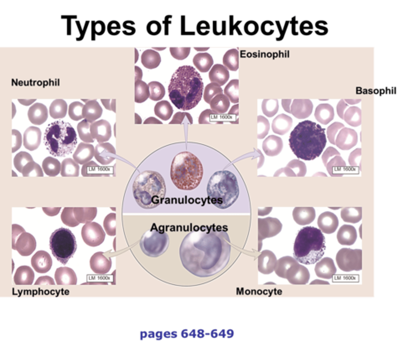
Granular leukocytes (contains granules)
Neutrophils, eosinophils, basophils
What are the "granules" in leukocytes?
Tiny sacs; they contain enzymes/compounds that are used to fight pathogens and reduce inflammation
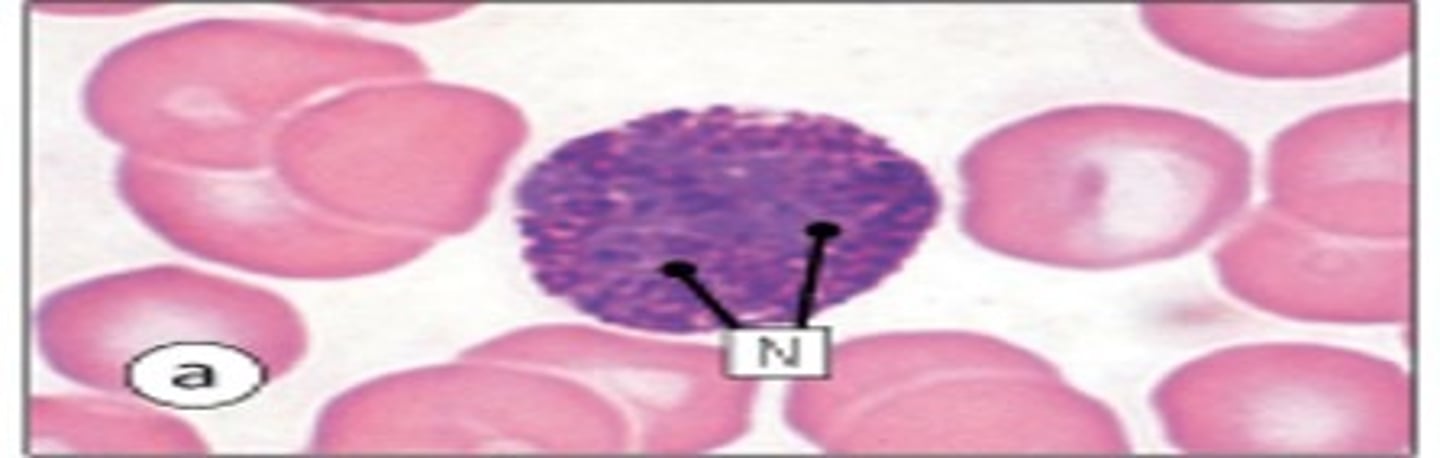
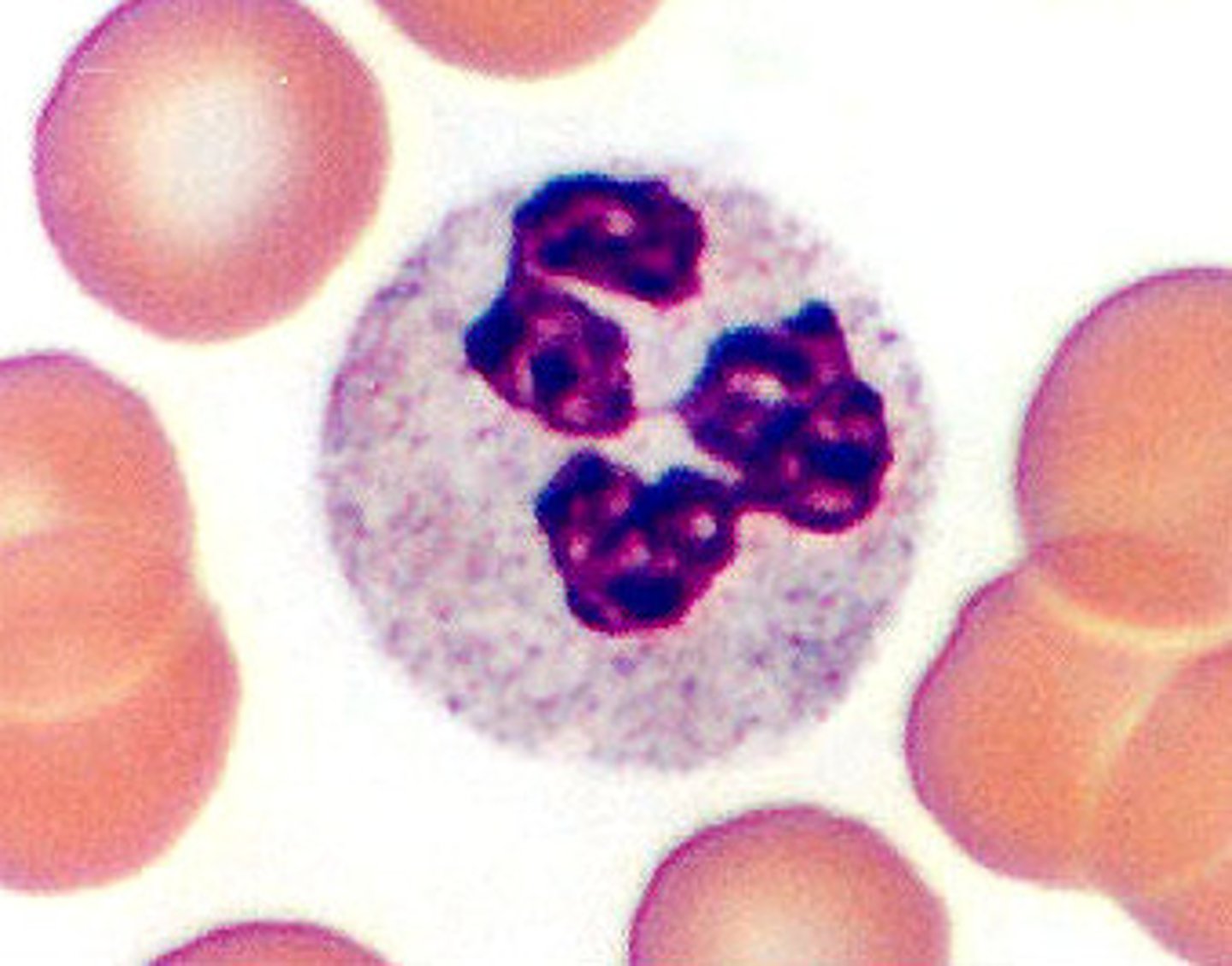
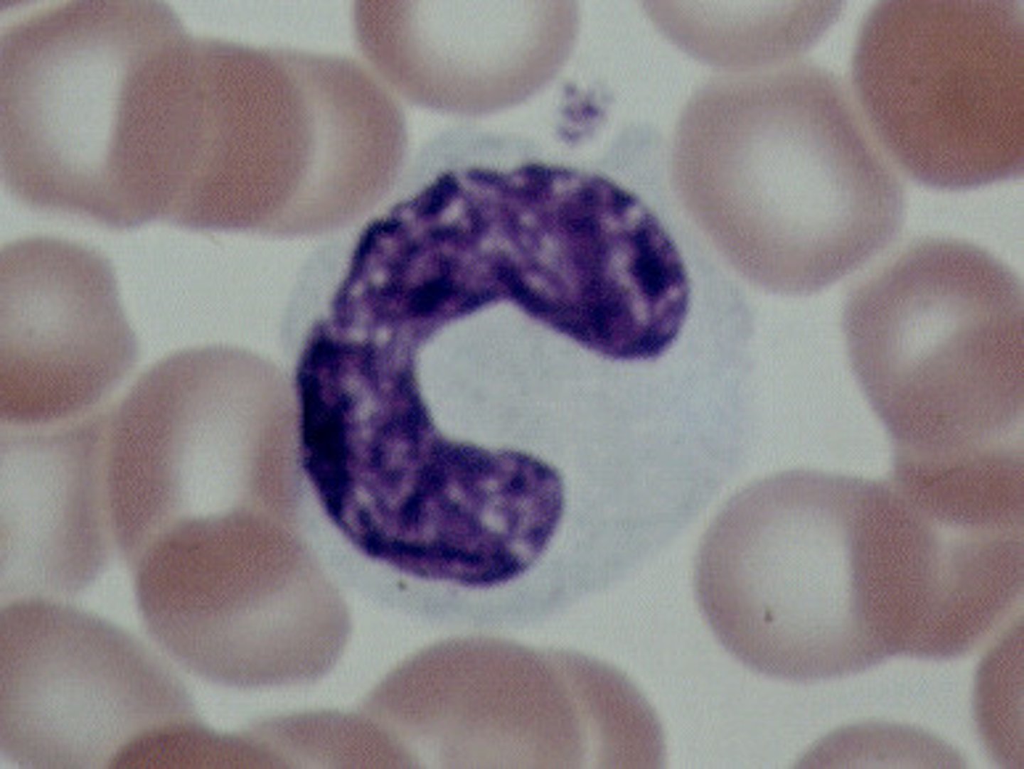
Neutrophils
Structure: Nuclei has three or more lobes
Function: The most common WBC; fights bacterial infections by engulfing bacteria by phagocytosis
Eosinophils
Structure: Nuclei has two lobes, they stain orange/red
Function: A WBC that digests and destroys parasitic worms, they also play a role in allergy and immune response
Basophils
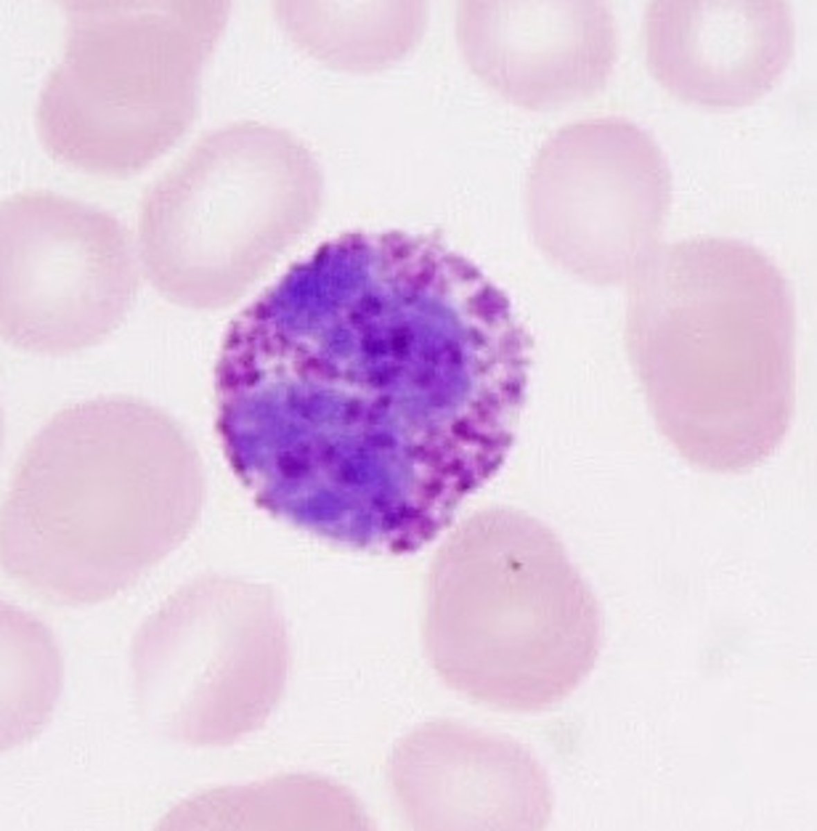
Structure: Nuclei is U or S shaped but is obstructed by HUGE purple/black granules
Function: A WBC that produces histamine --> causing inflammation during an immune response
Agranular leukocytes (contains NO granules)
Lymphocytes, monocytes
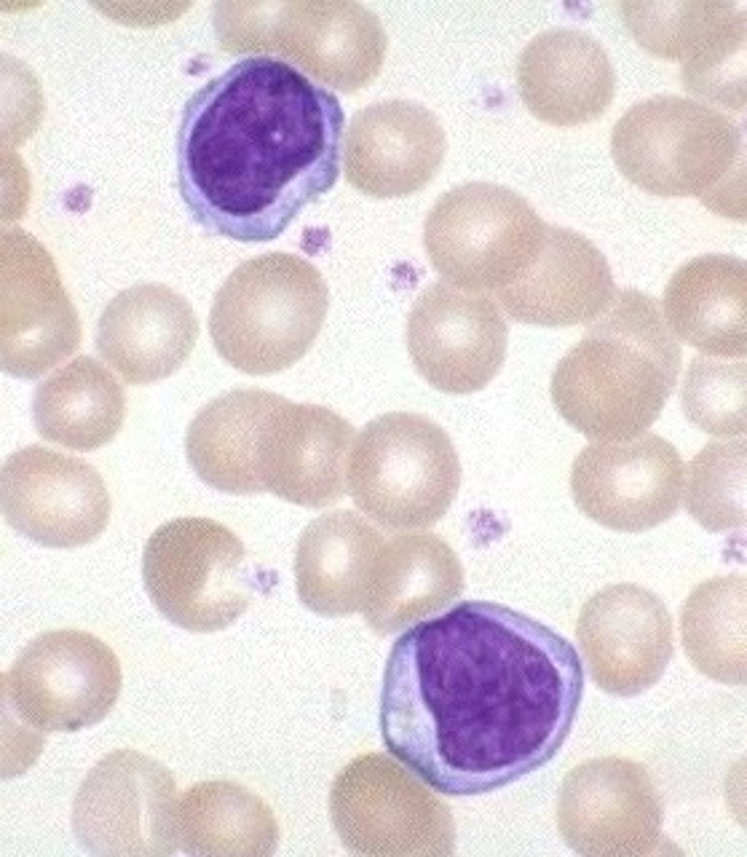
Lymphocytes
Structure: Has a large, dark purple nucleus that takes up most of the cell volume
Function: Two WBCs (B and T cells) make antibodies and fight off viral infections
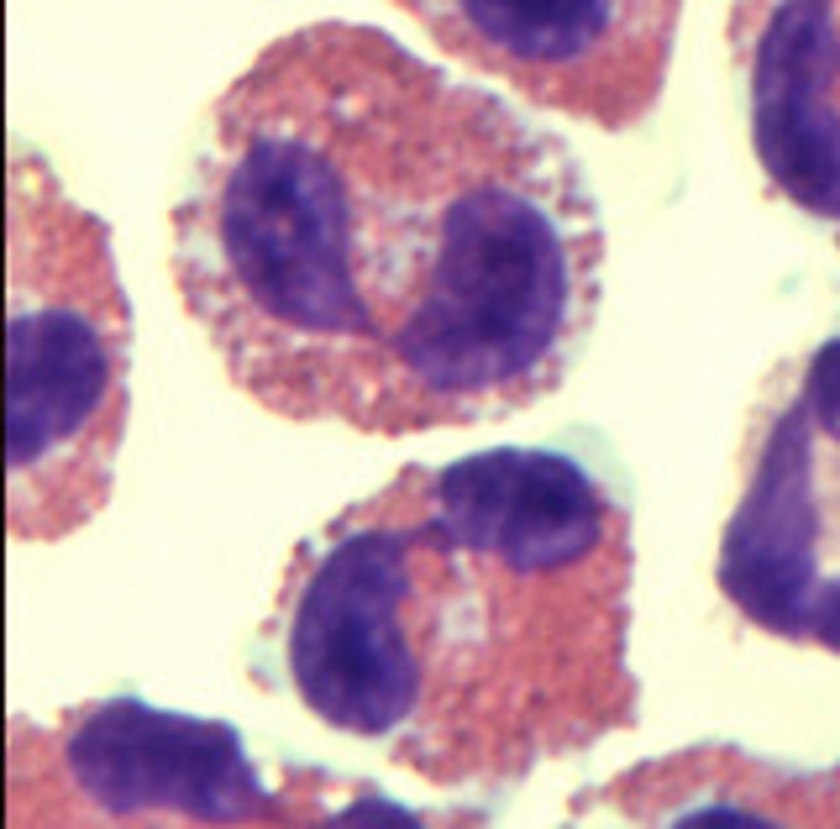
Monocytes
Structure: U shaped nucleus, pale blue cytoplasm
Function: A large WBC that transform into macrophages to fight off viruses and chronic infections
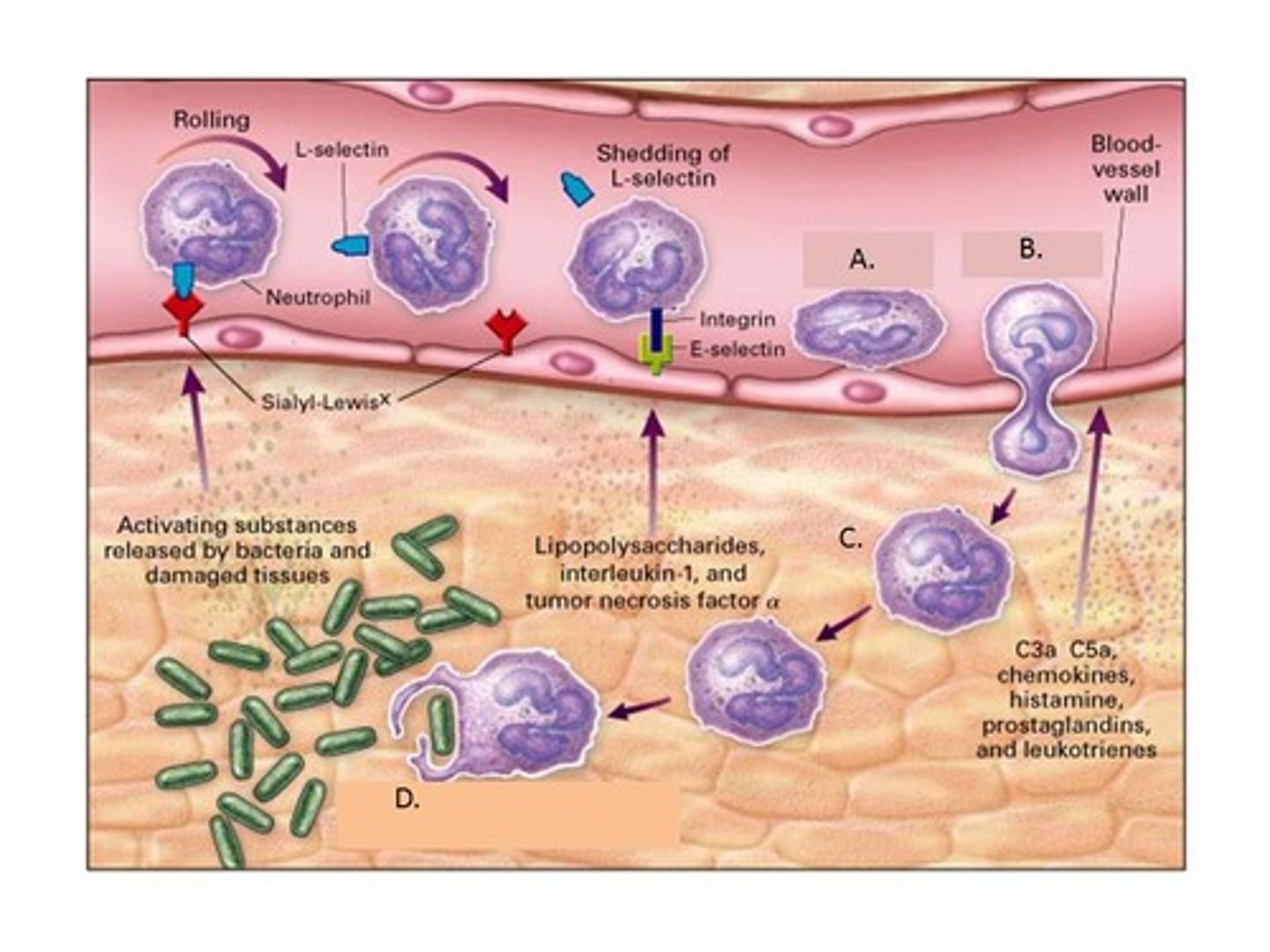
Emigration (diapedesis)
During an invasion, white blood cells leave the bloodstream by squeezing through the arterial walls and collect at sites of invasion
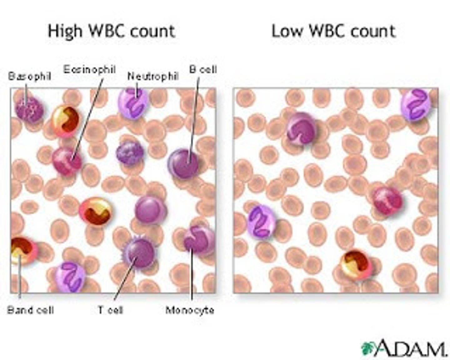
What does white blood cell concentration tell us?
High WBC count indicates infection/inflammation, low WBC count can be caused by drugs (glucocorticoids and anticancer drugs)
Leukopenia
Abnormally low white blood cell count
Leukemias
Cancers of the blood, caused by the overproduction of abnormal WBCs
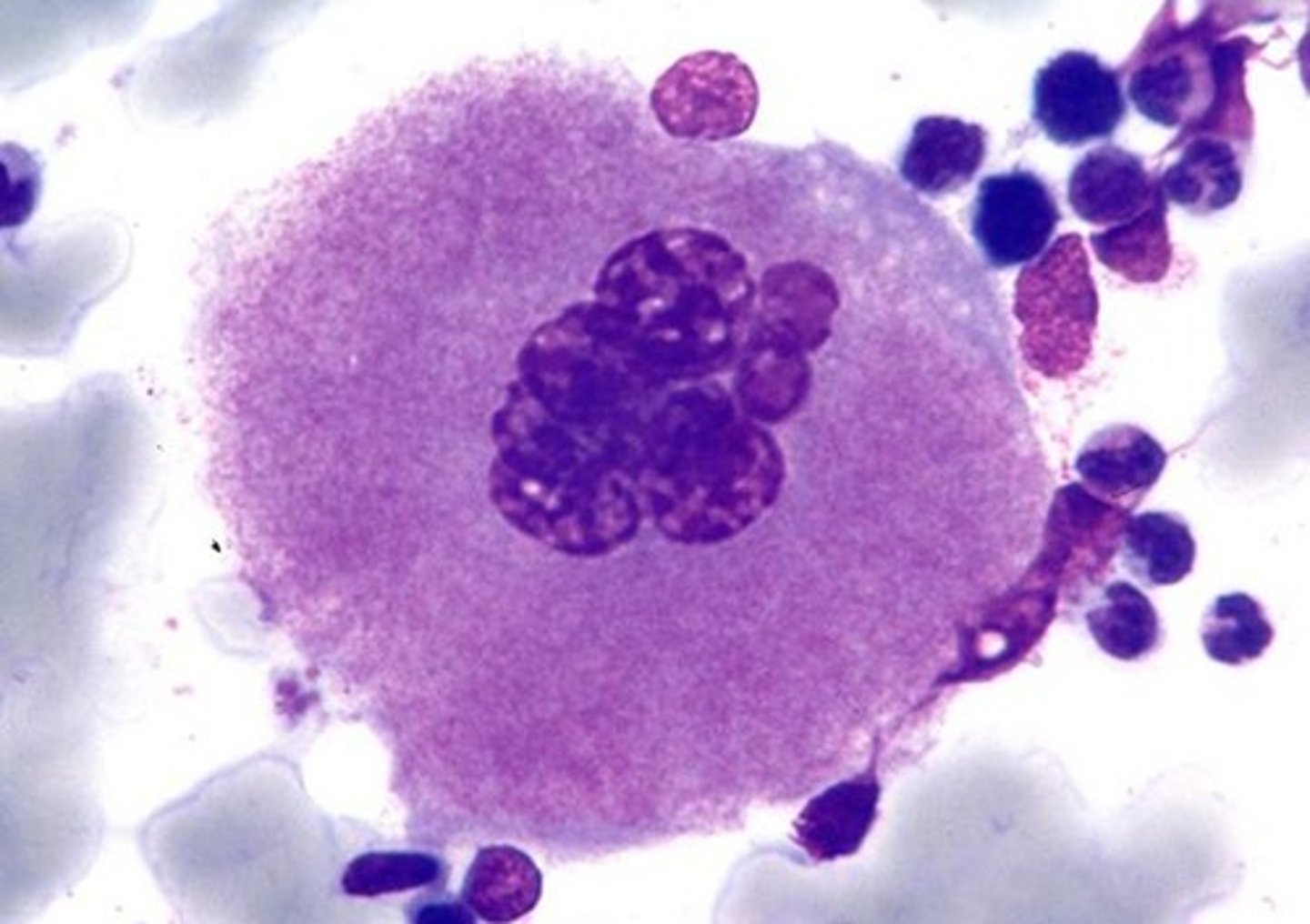
Infectious mononucleosis
Epstein-Barr virus aka mono aka the kissing disease --> causes an LARGE amount of BIG lymphocytes --> the disease was named mono bc they originally thought the giant lymphocytes were monocytes
(common in young adults bc we're all hoes)
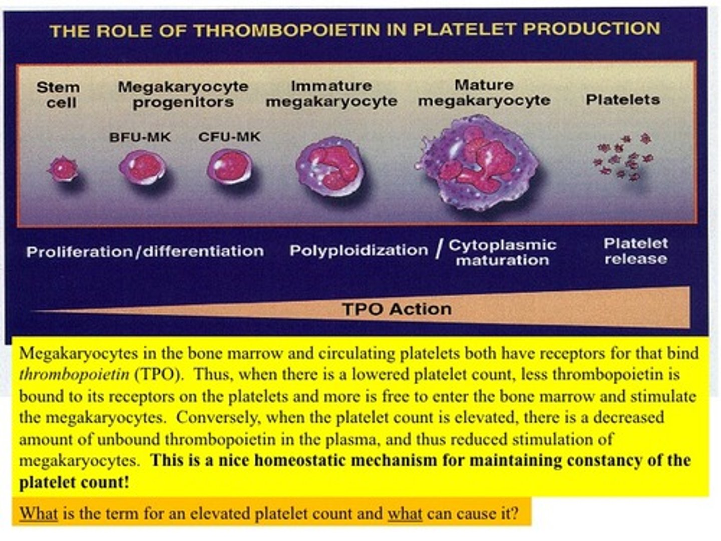
Megakaryocyte
A HUGE cell in red bone marrow that splits apart produces platelets
Thrombopoietin (TPO)
A hormone that stimulates platelet formation
How long do platelets live?
5-9 days
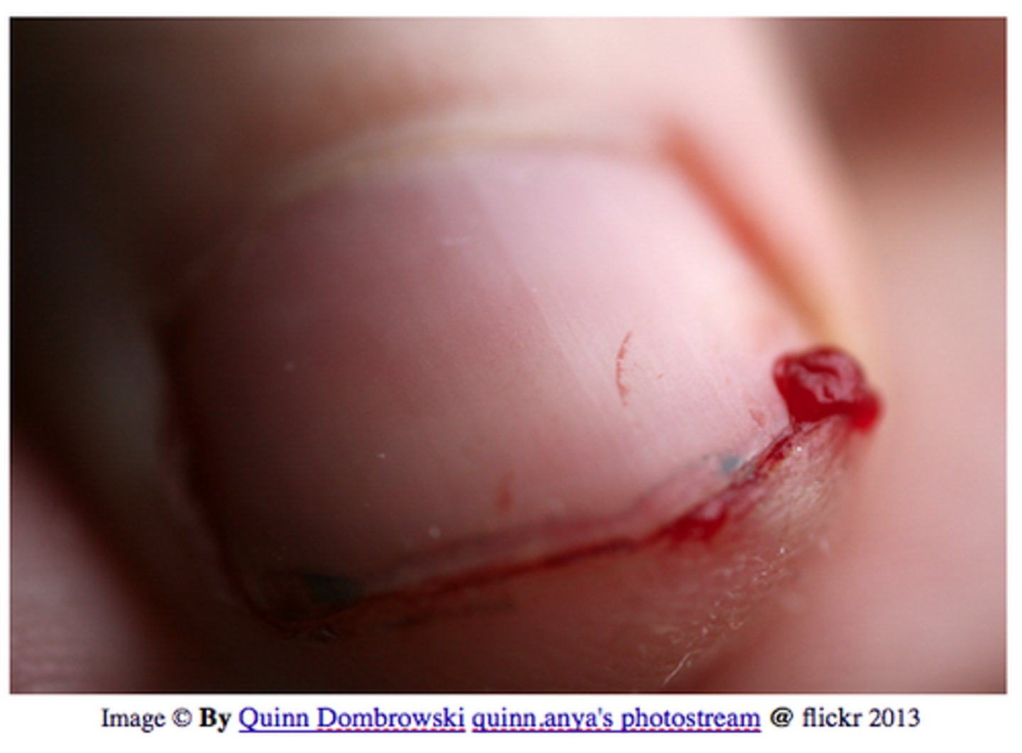
Hemostasis
To stop or control bleeding
3 Steps of hemostasis
1. Vascular spasm
2. Platelet plug formation
3. Coagulation
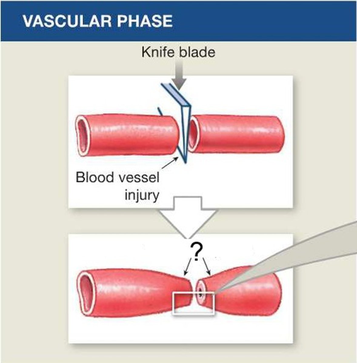
Vascular spasm
Contraction of the smooth muscle in the wall of a damaged blood vessel to prevent blood loss
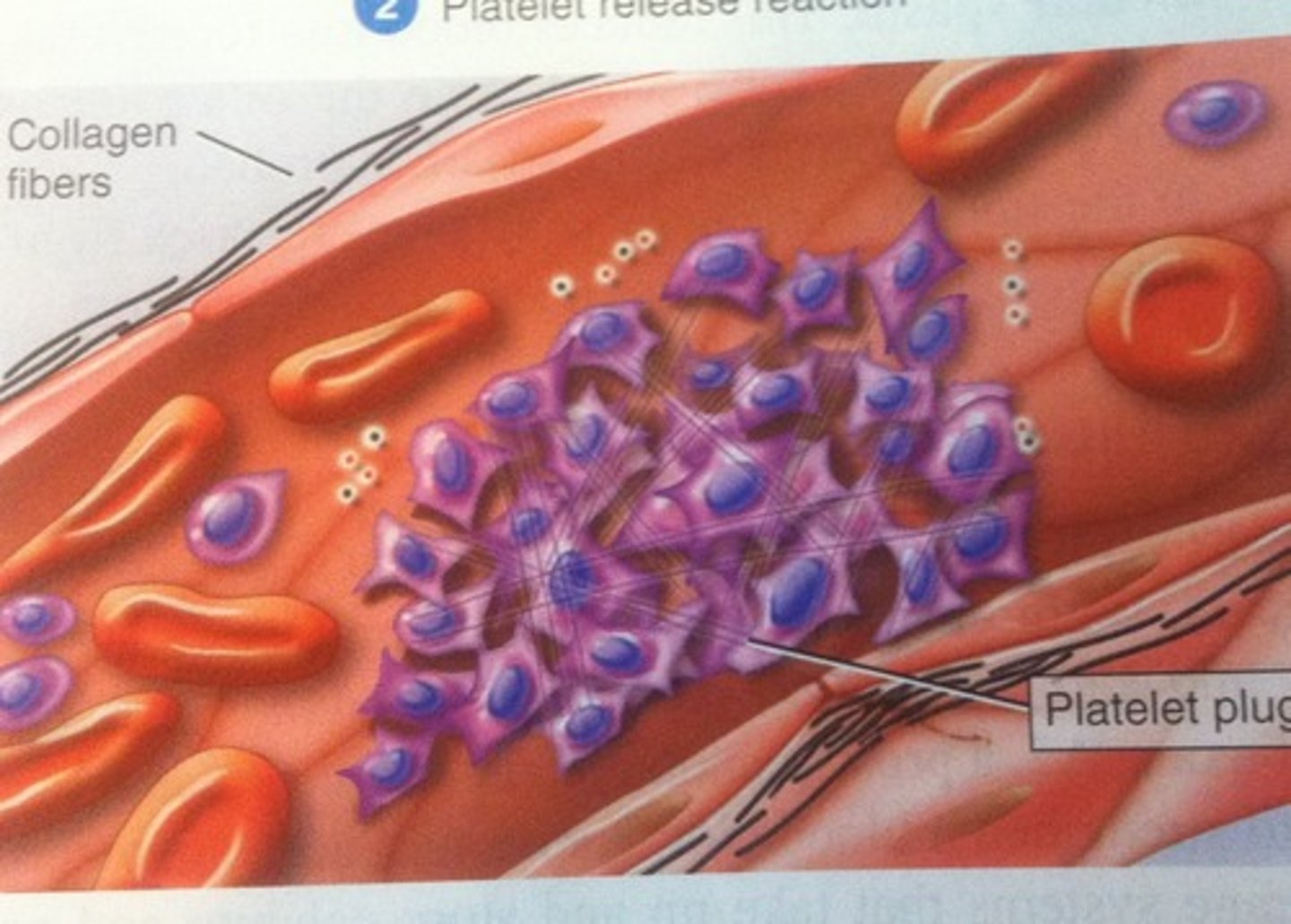
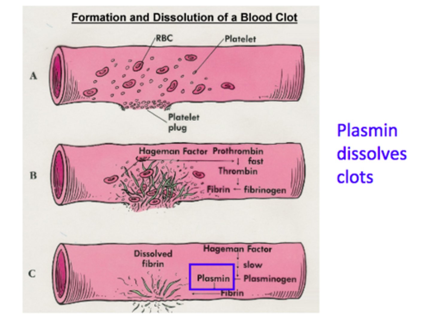
Platelet plug formation
When platelets are exposed to collagen after an injury to the blood vessel, platelets clump and form a temporary seal --> they release chemicals to call more platelets to the injury site
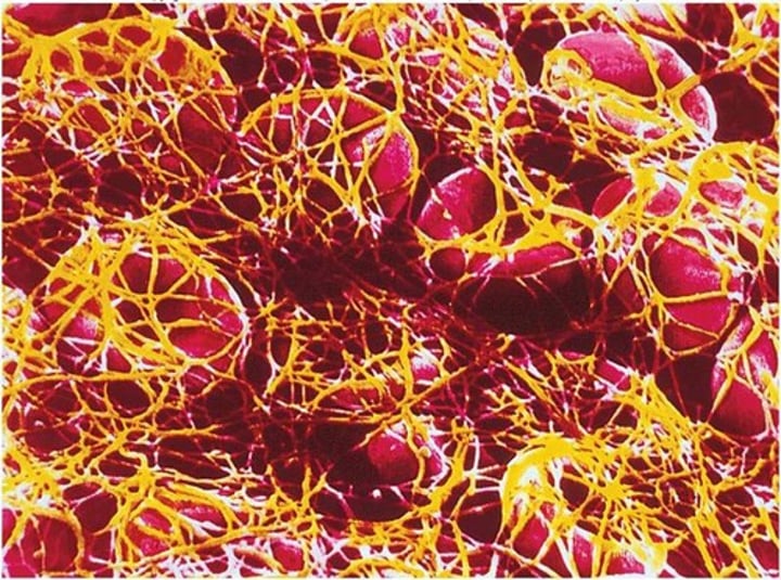
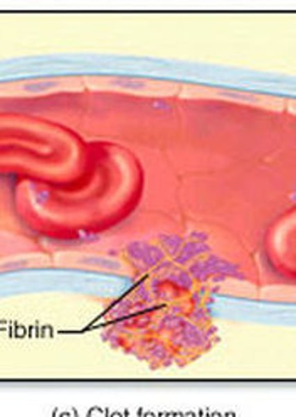
Coagulation
Blood clotting
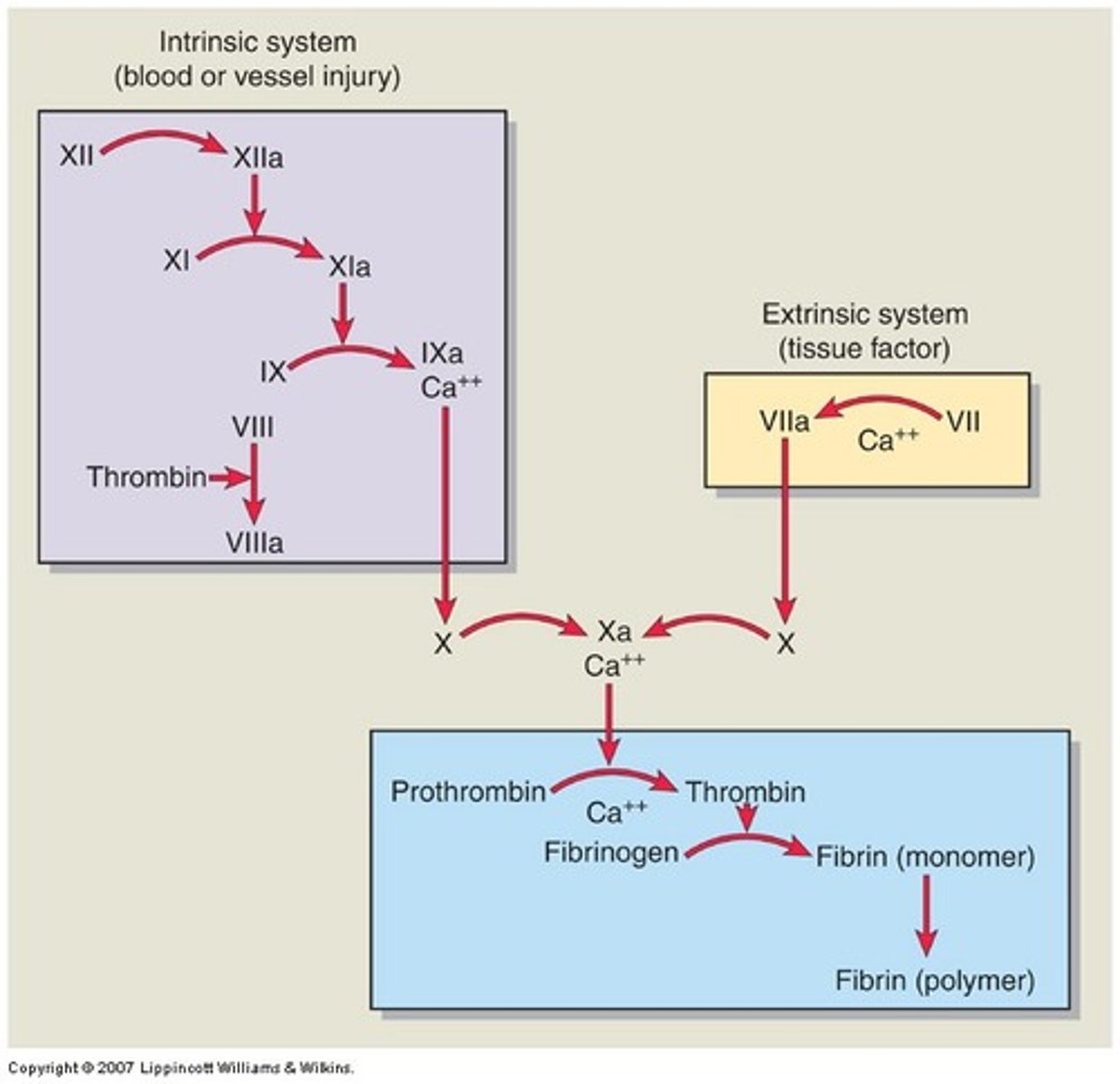
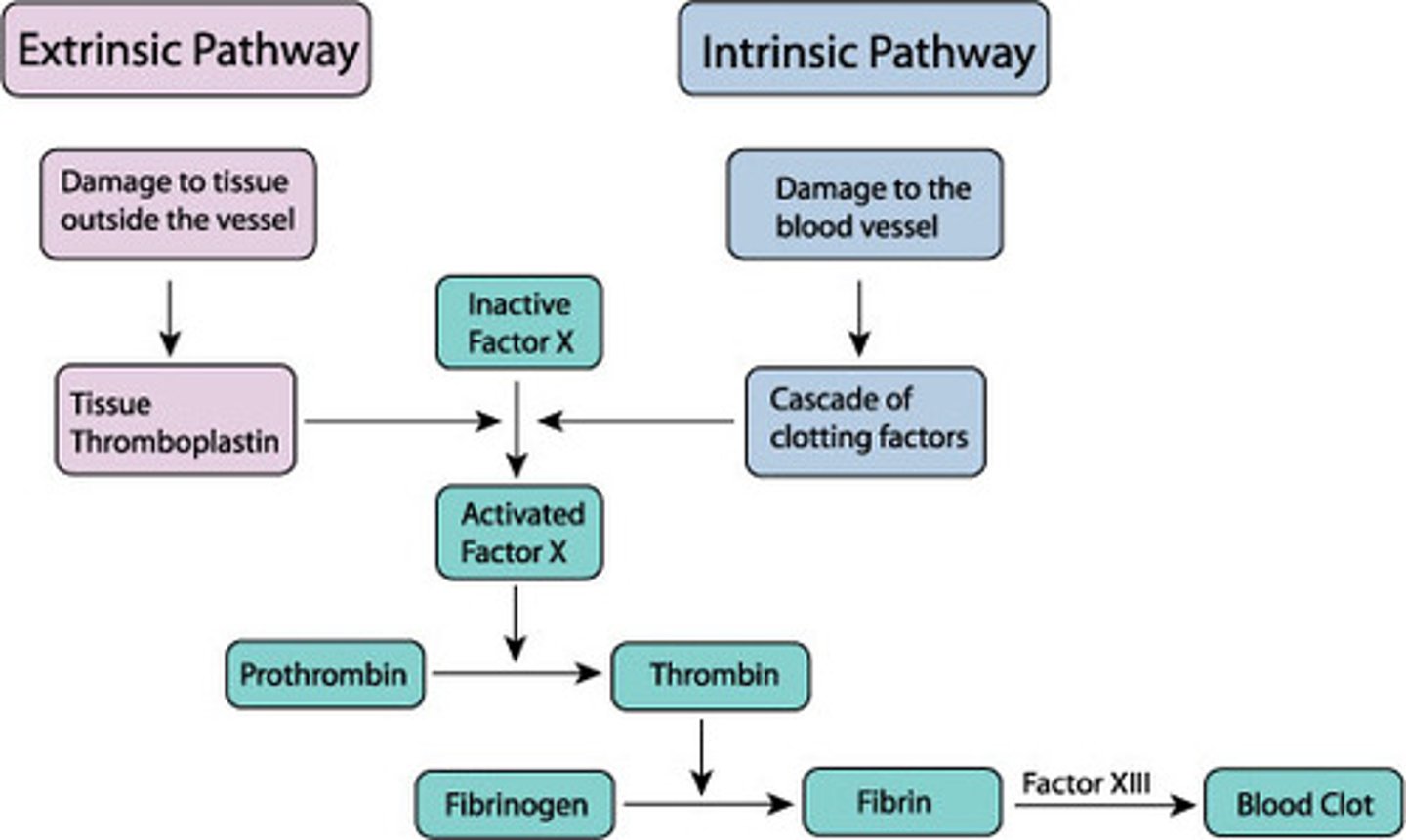
Intrinsic pathway of coagulation
Activated by internal injury; clotting factors are present within the blood and are activated by exposed collagen fibers
Extrinsic pathway of coagulation
Activated by external injury; clotting factors are located outside the blood and are activated by exposure to tissue factor
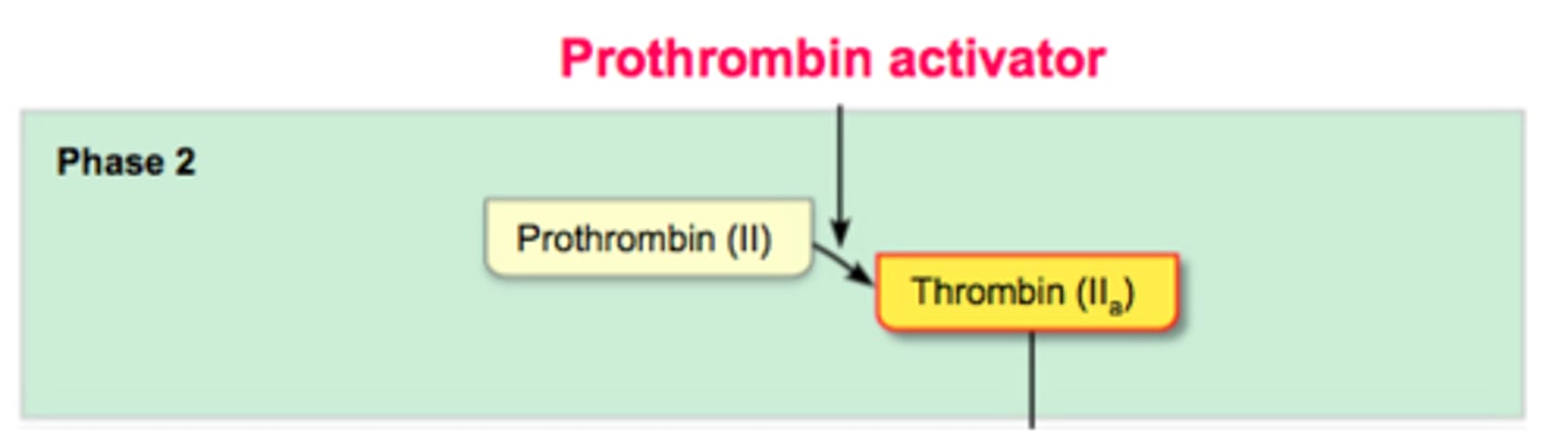
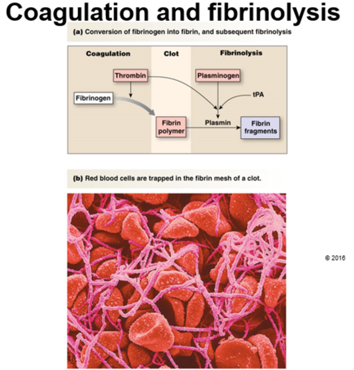
Phase 1 of coagulation
Prothrombinase is activated by either intrinsic or extrinsic pathway
Phase 2 of coagulation
Prothrombin is converted into the active enzyme thrombin
Phase 3 of coagulation
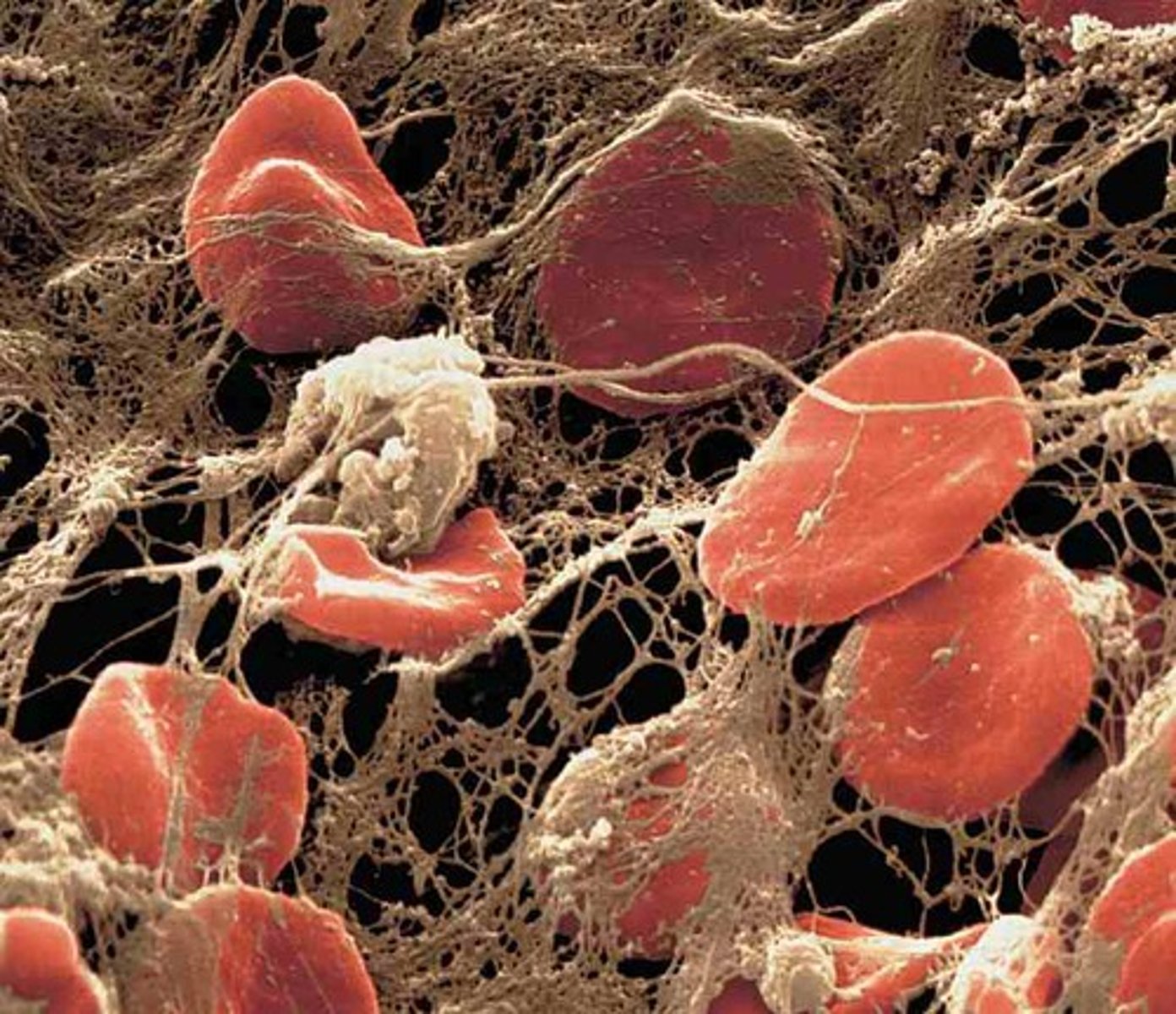
Thrombin converts fibrinogen to fibrin --> fibrin strands trap RBCs in a jelly substance
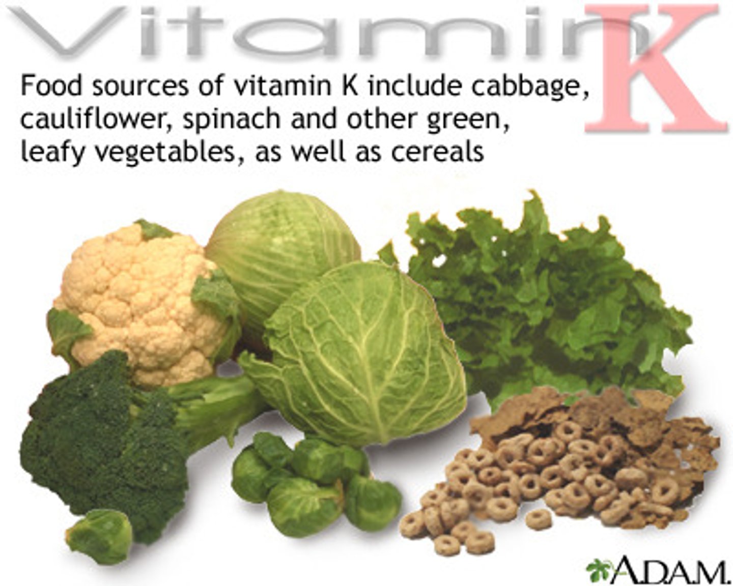
What role does vitamin K play in blot clotting?
Vitamin K is needed for the creation of 4 clotting factors
How are blood clots destroyed?
Blood clots are dissolved by the enzyme plasmin
Fibrinolysis
Breakdown and removal of a clot by plasmin ("splitting of fibrin")
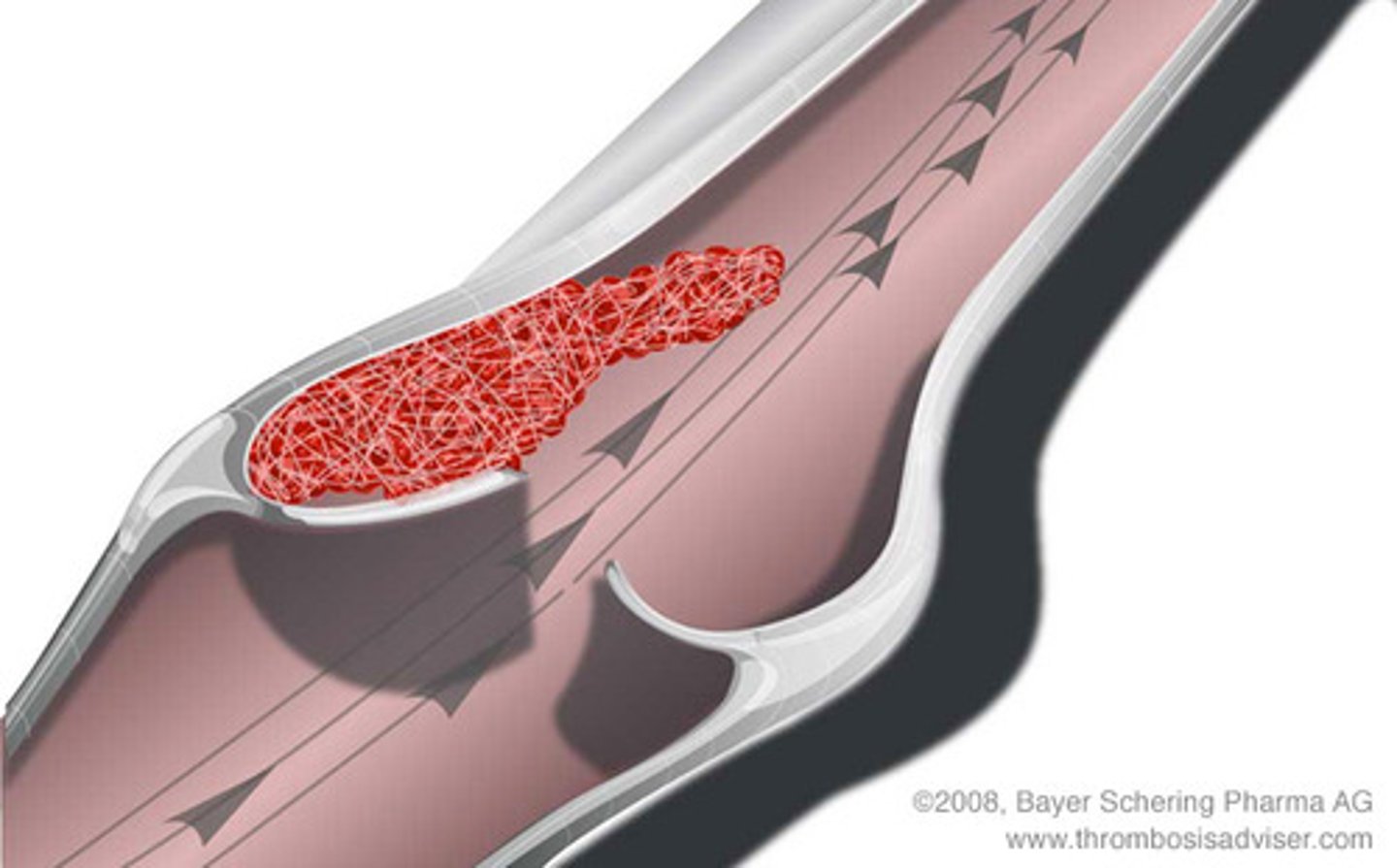
Thrombus
A blood clot that forms in an unbroken blood vessel; if the clot blocks blood circulation --> tissue death will occur
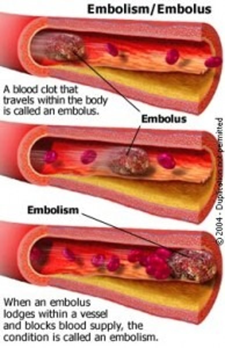
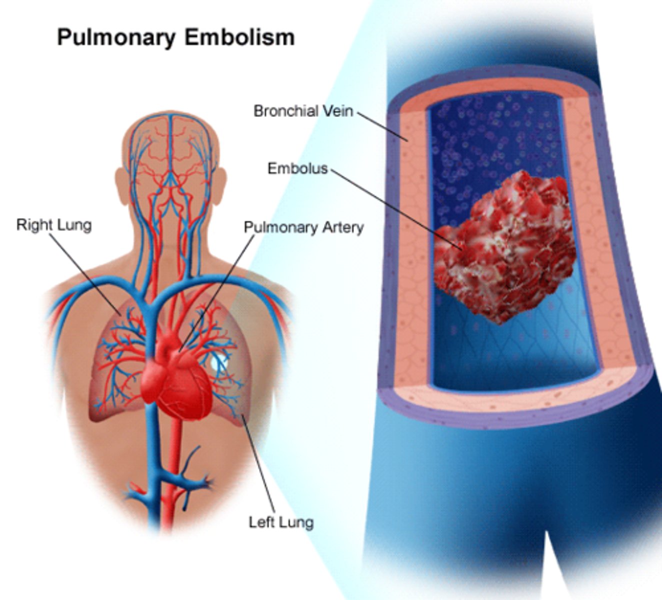
Embolus
A blood clot that breaks free and travels through the bloodstream
Embolism
When an embolus (a moving blood clot) lodges within a vessel and blocks the flow of blood
Anticoagulant drugs
Drugs that prevent the clotting of blood
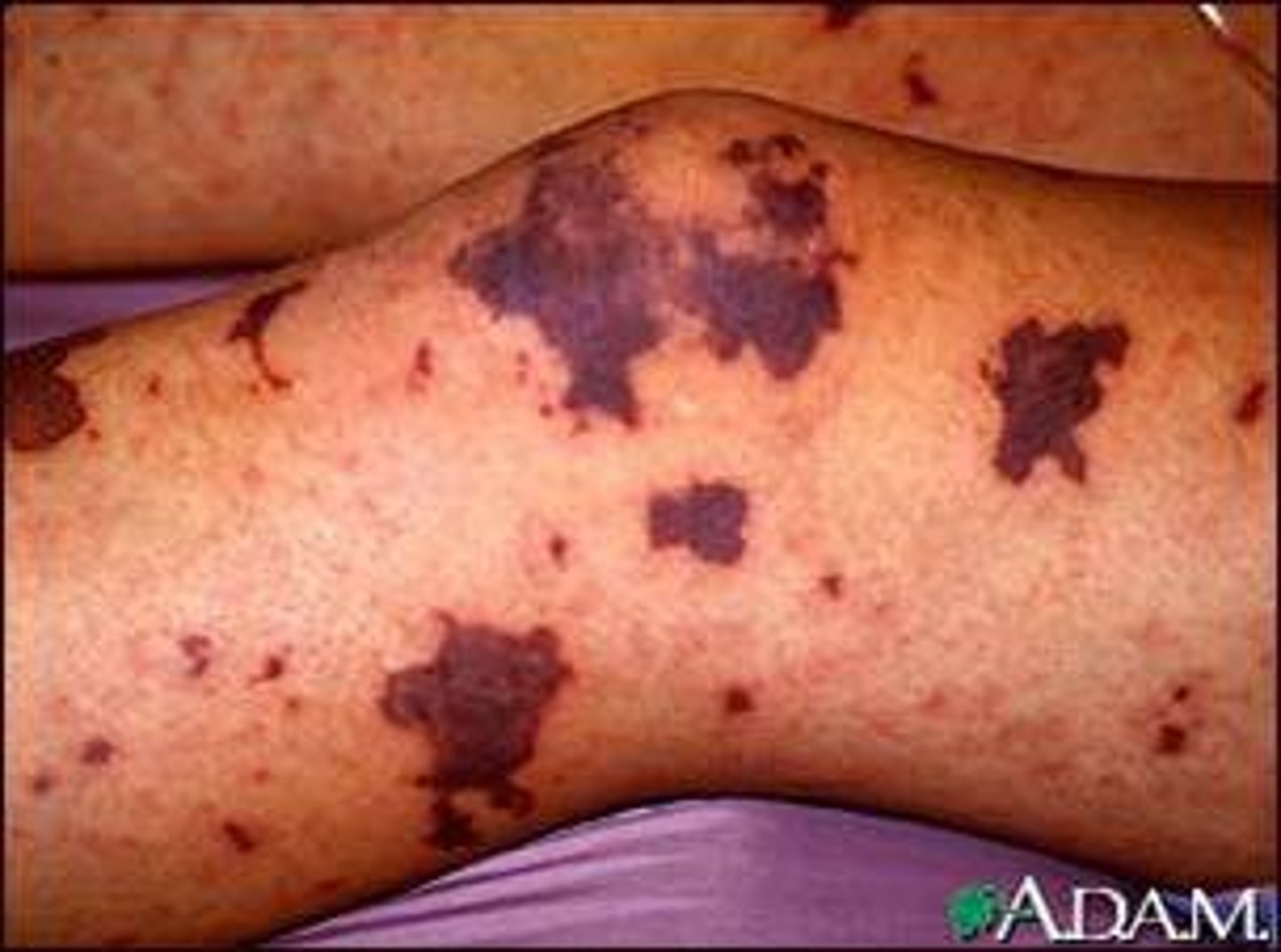
Thrombocytopenia
Low platelet count --> causes spontaneous bleeding and increased bleeding after an injury
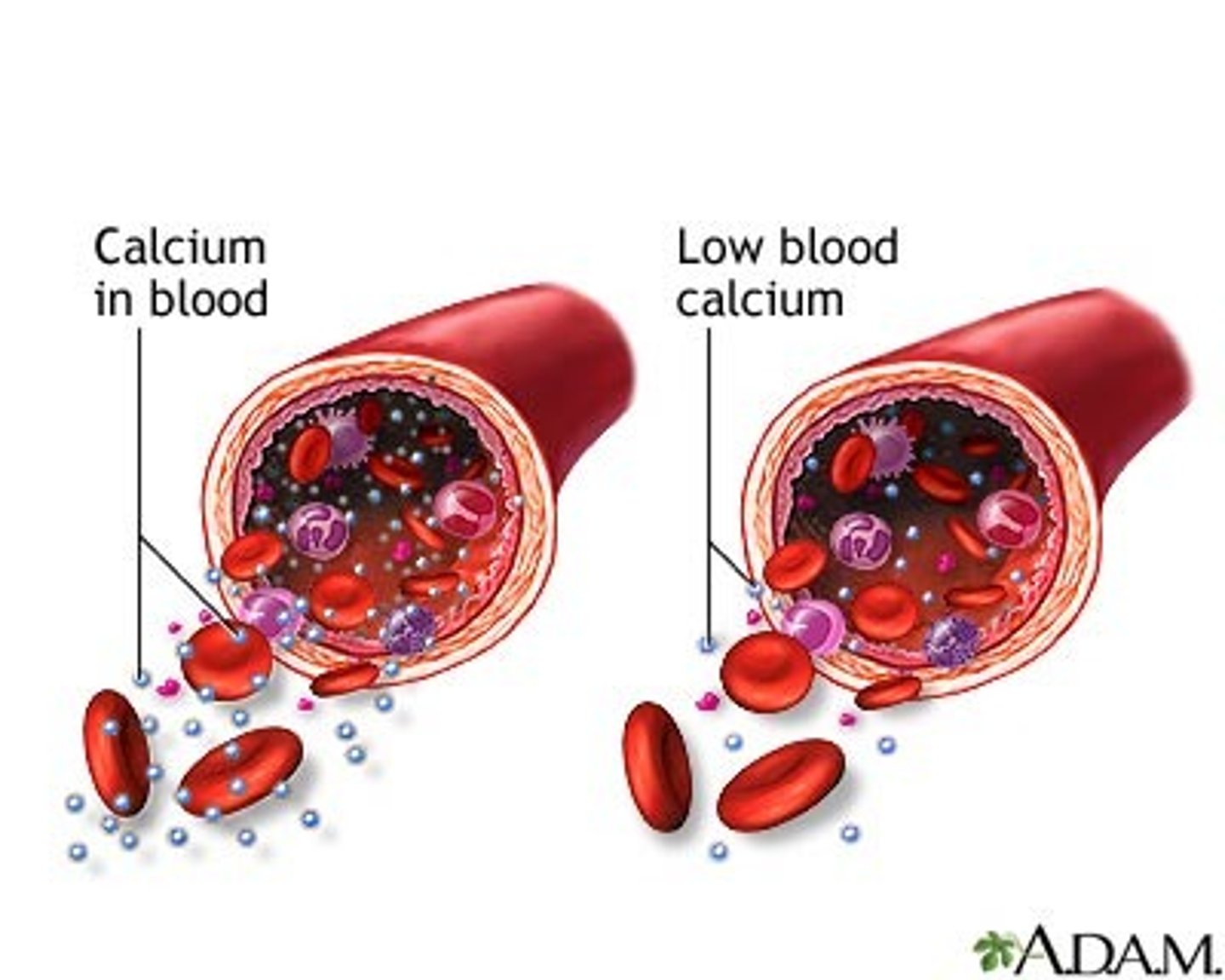
What role does calcium play in blood clotting?
Calcium minerals are needed for blood clotting
Blood groups (2)
ABO and Rh (positive or negative)
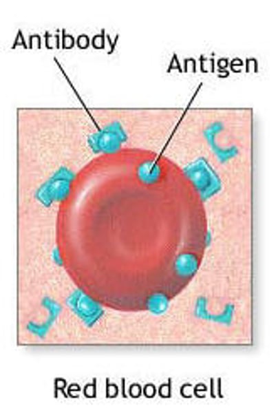
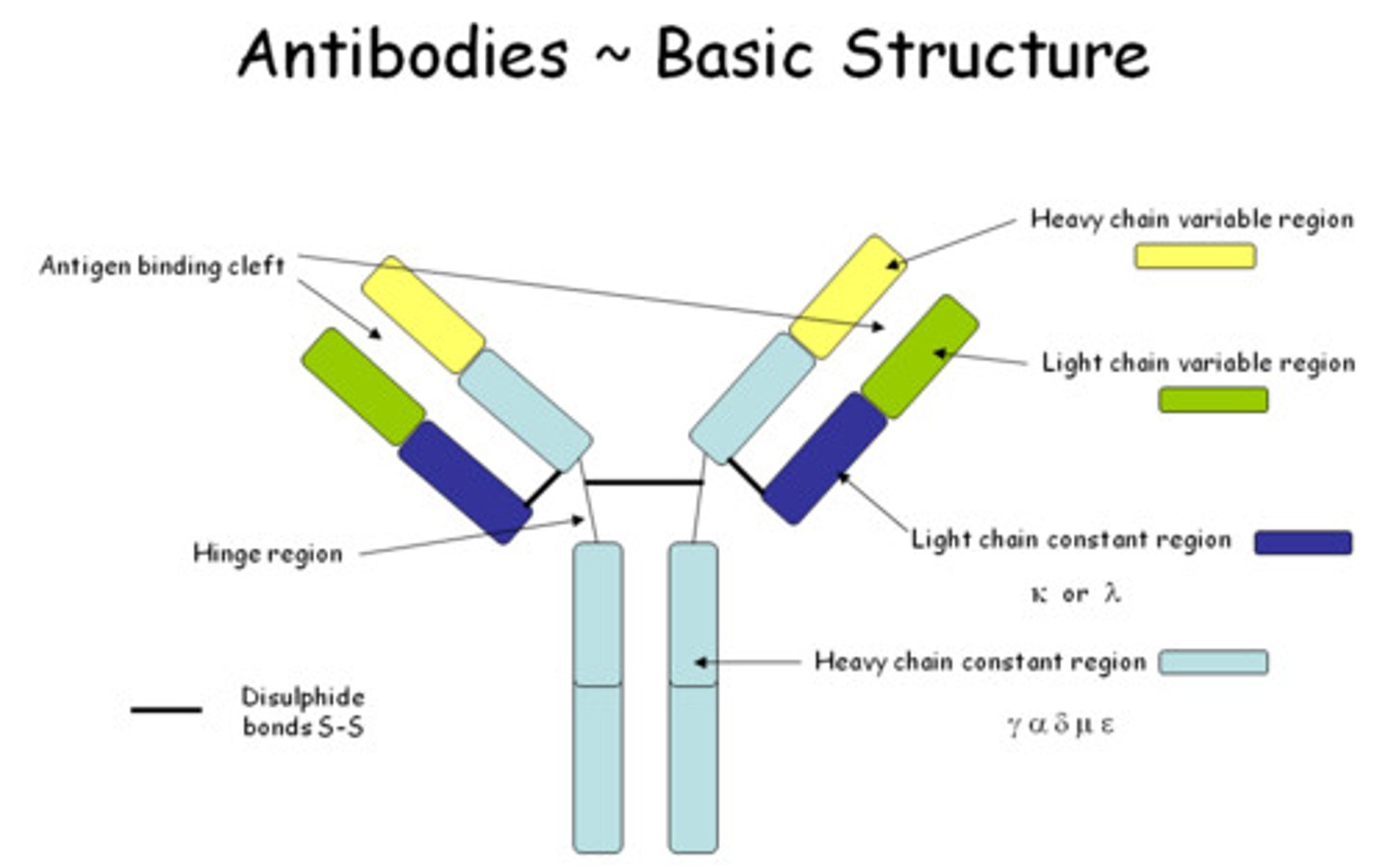
What are antigens?
Proteins present on the surface of RBCs that determine blood type
What are antibodies?
Proteins in blood plasma (globulins) that detect and attack antigen
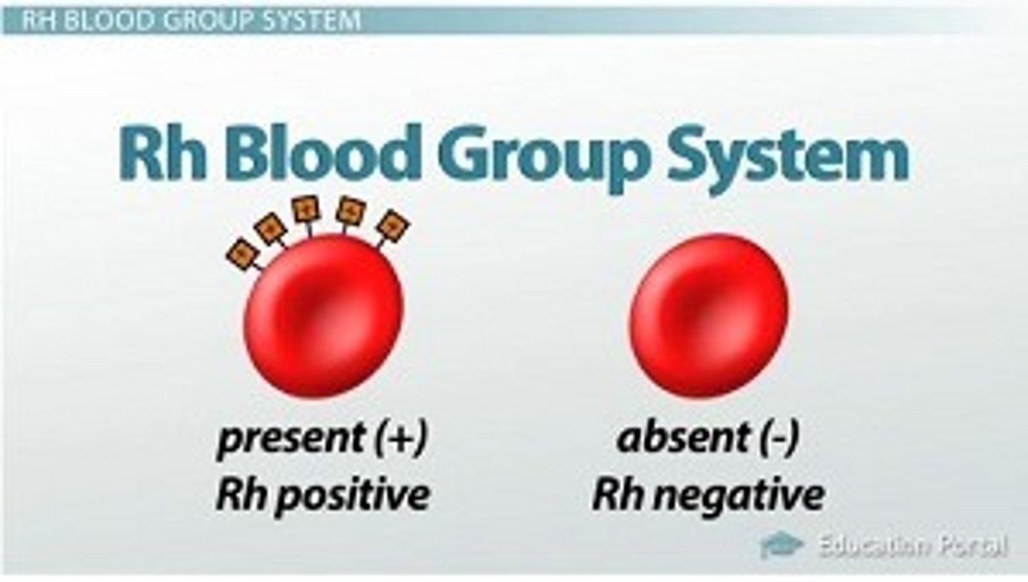
Positive blood type
The presence of Rh/D antigen --> positive
Negative blood type
Absence of Rh/D antigen --> negative
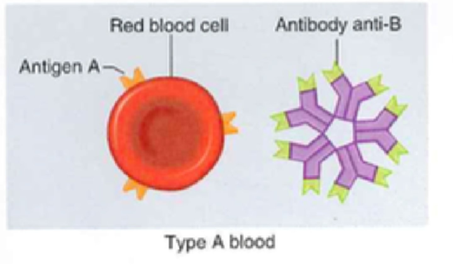
Type A blood
A antigens, B antibodies
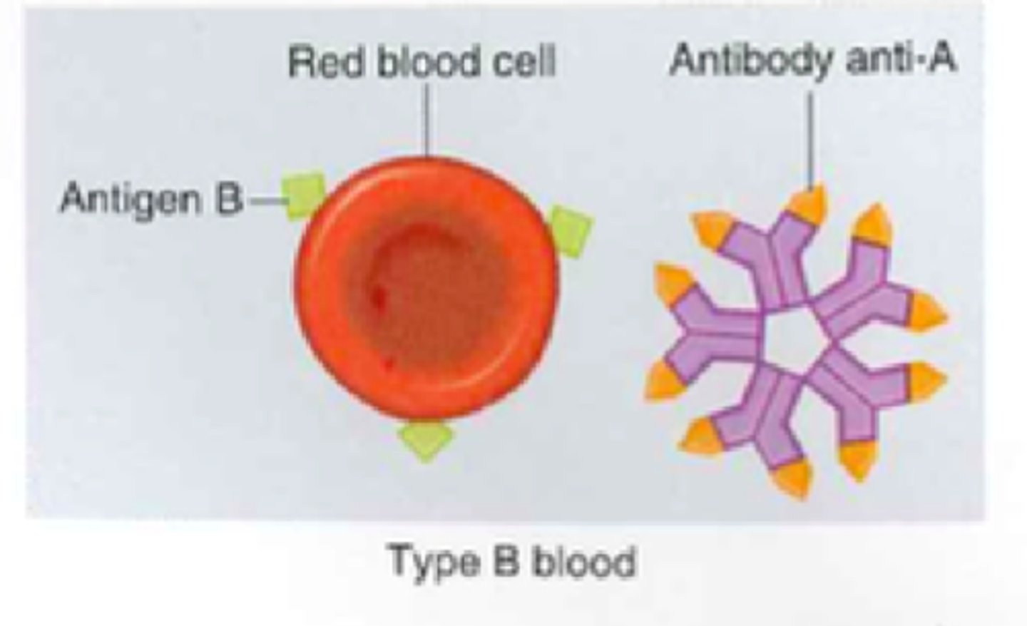
Type B blood
B antigens, A antibodies
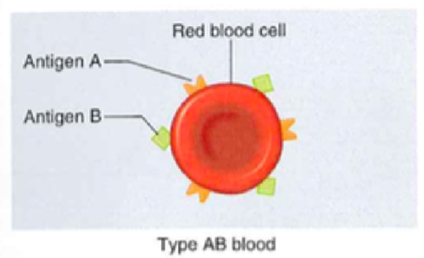
Type AB blood
A and B antigens, no antibodies (universal recipient)
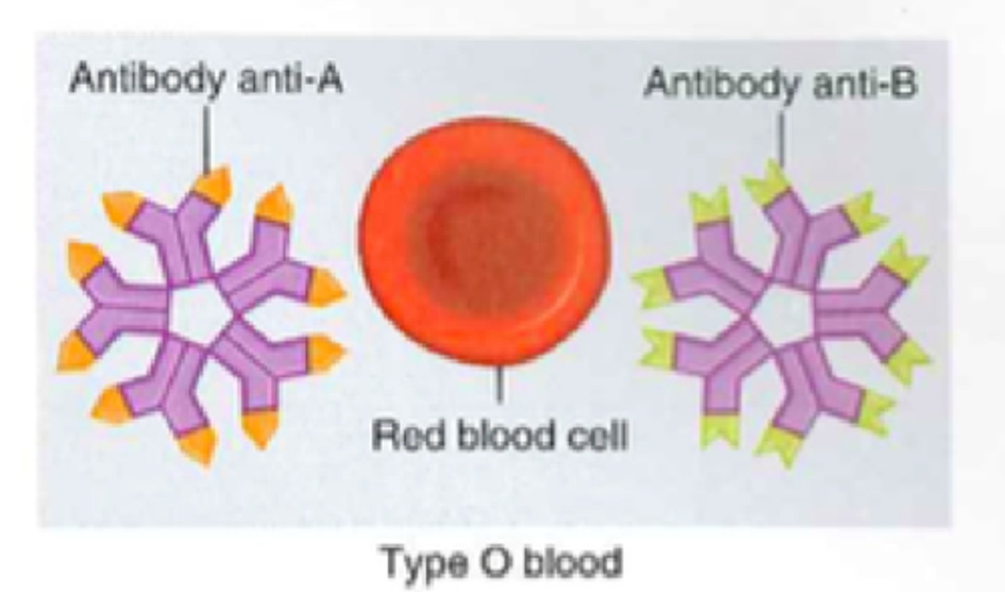
Type O blood
NO antigens, A and B antibodies (universal donor)
What happens when antibodies detect incompatible blood?
When antibodies detect an incompatible blood antigen, the RBCs will clump together (agglutination) and rupture (hemolysis)
Agglutination
The clumping of red blood cells; will result if blood types with different antigens are mixed
Rh incompatibility
Occurs when a woman who is Rh-negative becomes pregnant with a baby with Rh-positive blood --> the mother's antibodies will attack the baby's RBCs (because the mother's body views them as foreign objects)
Hemolysis
The destruction of red blood cells