Biomechanics Anatomy
1/86
There's no tags or description
Looks like no tags are added yet.
Name | Mastery | Learn | Test | Matching | Spaced | Call with Kai |
|---|
No study sessions yet.
87 Terms
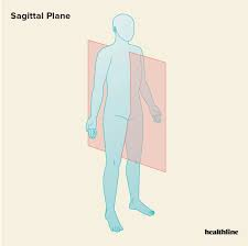
sagittal plane
a vertical plane that divides the body into right and left halves, primarily involving movements related to flexion and extension as well as forward and backward movements
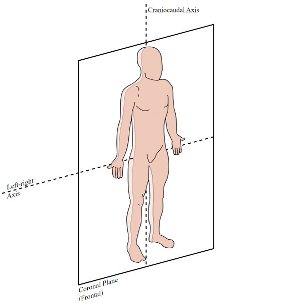
coronal plane
an anatomical plane that divides the body into dorsal and ventral sections (front and back, anterior and posterior). It is perpendicular to the sagittal and transverse planes.
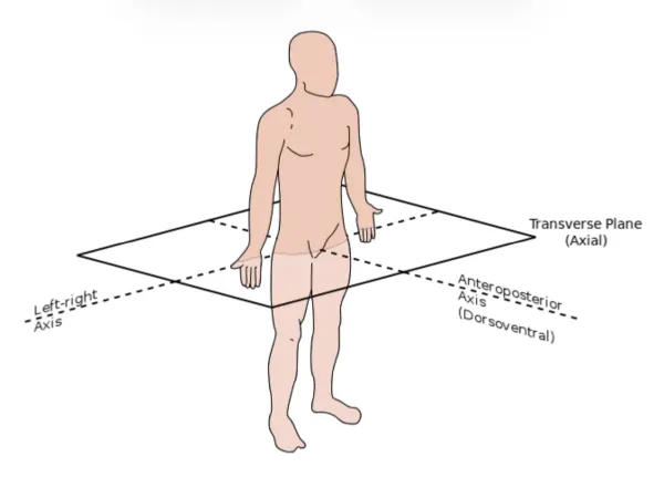
transverse plane
axial; a horizontal plane. It is perpendicular to both the sagittal and coronal planes, and parallel to the ground. It divides the body into an upper (superior) section and a lower (inferior) section
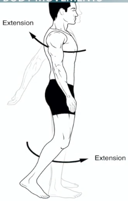
extension
a movement that increases the angle between two body parts. At the elbow: increasing the angle between the ulna and the humerus. At the knee: straightens the lower limb
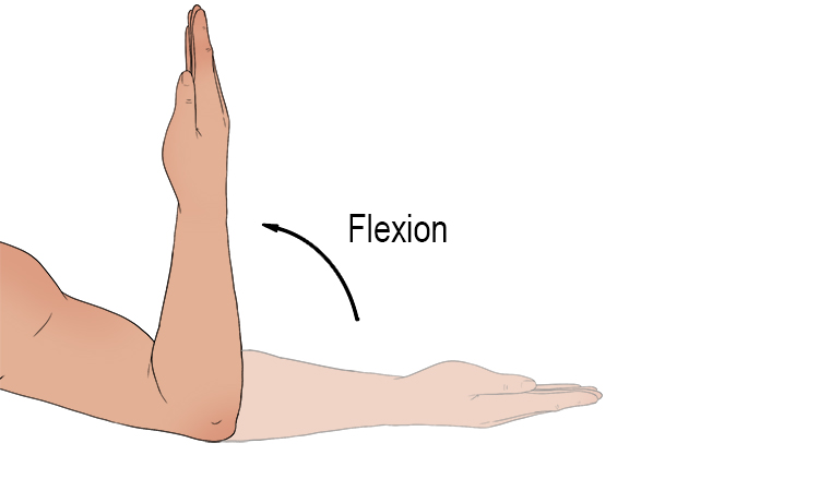
flexion
a bending movement that decreases the angle between a segment and its proximal segment. For example, bending the elbow, or clenching a hand into a fist
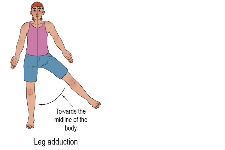
adduction
Movement towards the midline of the body, an imaginary line running down the middle of a body or any limb
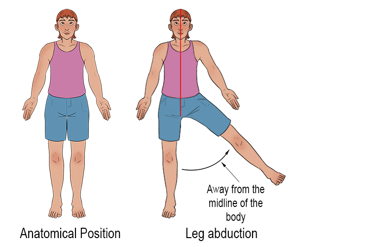
abduction
the motion of a limb or appendage away from the midline of the body. In the case of arms, it is the movement of the arms away from the body within the plane of the torso
pronation
medial rotation of the forearm so the palm faces posteriorly; palm facing downwards
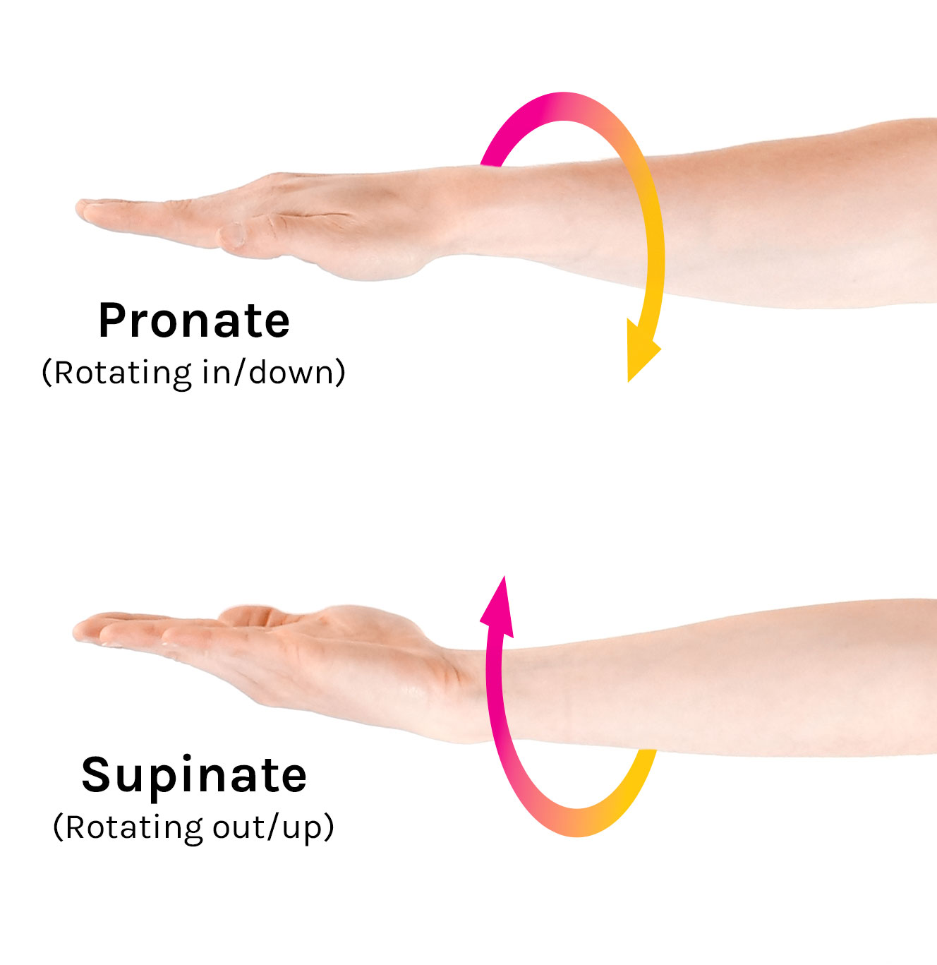
supination
lateral rotation of the forearm so the palm faces interiorly; palm facing upward

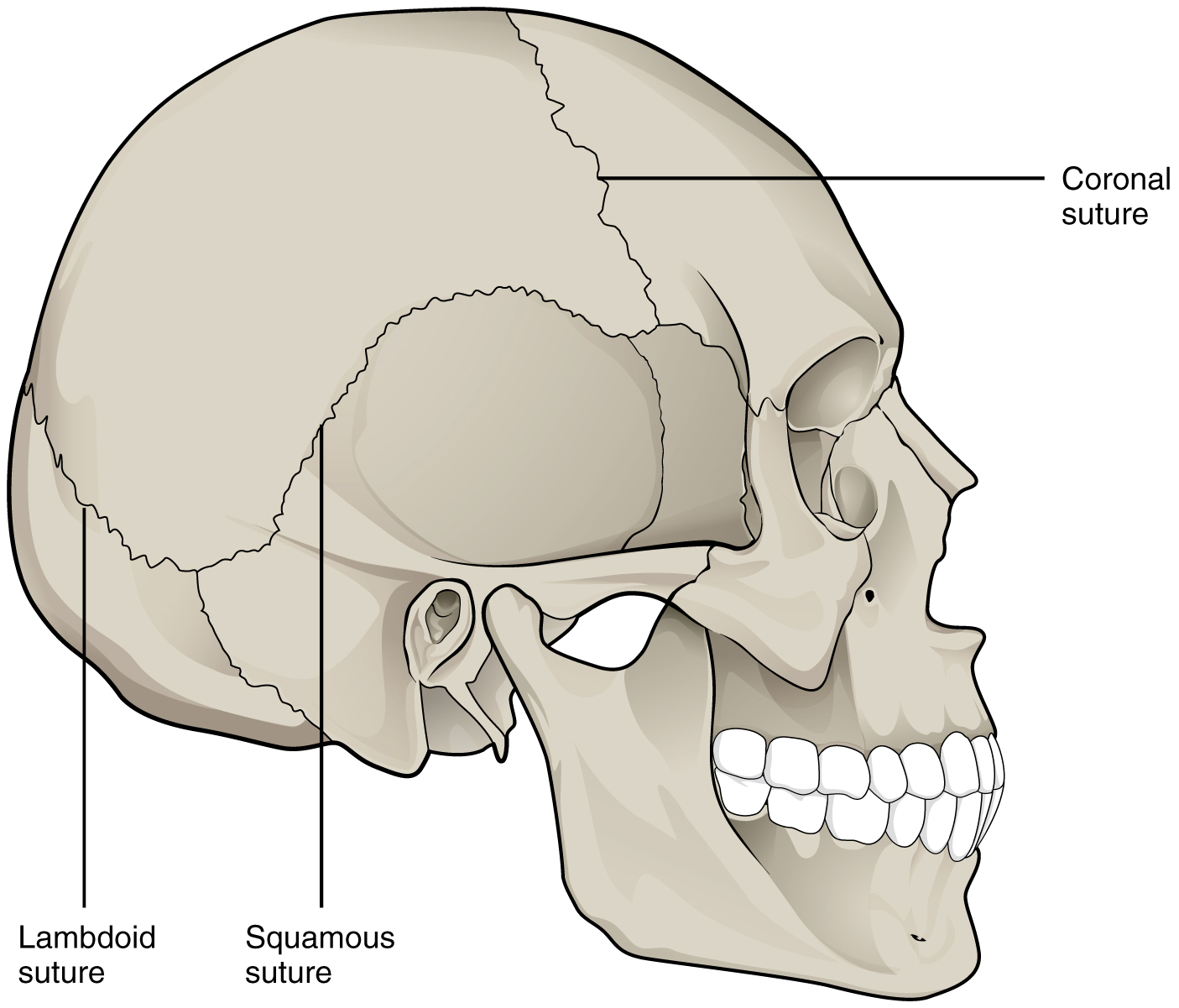
synarthrodial joints
In fibrous joints, the bones are joined by fibrous tissue, namely dense fibrous connective tissue, and no joint cavity is present. Formed by two tightly fitting bones, allows for no movement. Ex: skull
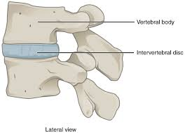
amphiarthrodial joints
Also known as a cartilaginous joint; a type of joint found in the human body that allows for some limited/relative movement. These joints are characterized by the presence of cartilage connecting the articulating bones, meaning there is no direct bone to bone contact. ex: vertebrae
diarthrodial joints
A synovial joint that joins bones or cartilage with a fibrous joint capsule that is continuous with the periosteum of the joined bones.
Allows for large degrees of varying motion. Have angular cavities, ligamentous capsules, synovial membranes and synovial fluid. Ex: knee
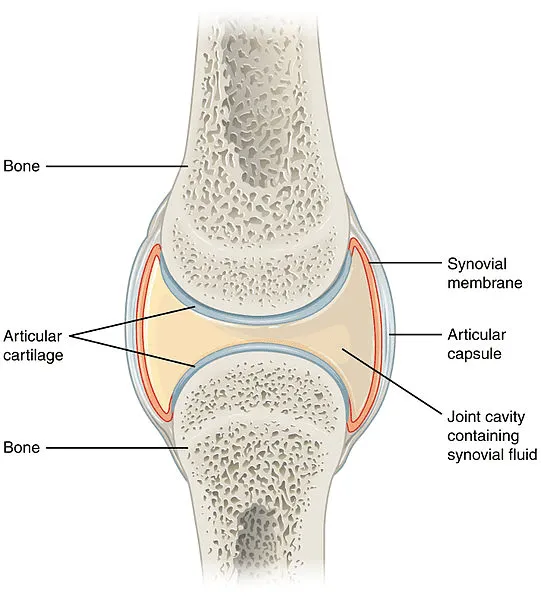
knee, shoulder, and hip
major joints in the body
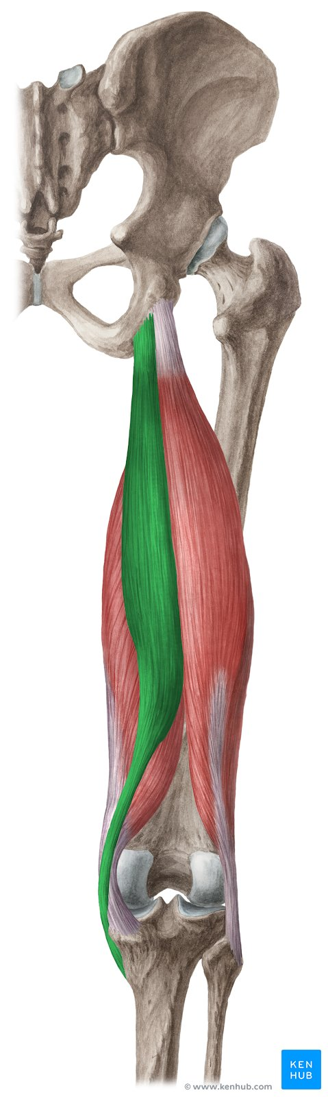
knee joint
major bones: femur (thigh bone) and tibia (shin bone). other bones: patella (knee cap) and fibula (lateral)
major ligaments in the knee
medial (tibia) collateral ligament (MCL), lateral (fibula) collateral ligament, anterior (front) cruciate ligament (ACL), posterior (back) cruciate ligament (PCL)
sartorius and quadriceps
major muscles within the knee joint
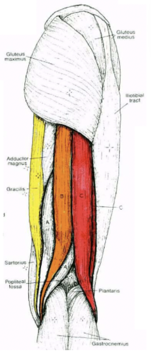
sartorius
a thin, long, superficial (strap-like) muscle in the anterior compartment of the thigh. Longest muscle in human body. Main flexor of the knee joint
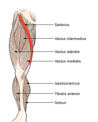
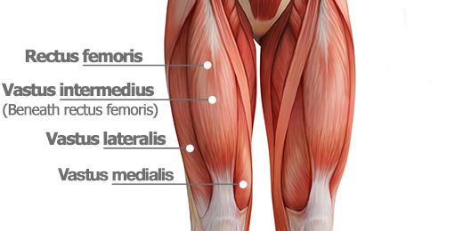
quadriceps
a large muscle group that includes the four prevailing muscles on the front of the thigh. Main extensor of the knee joint
composed of rectus femori, vastus intermedius (middle), vastus lateralis, vastus medialis
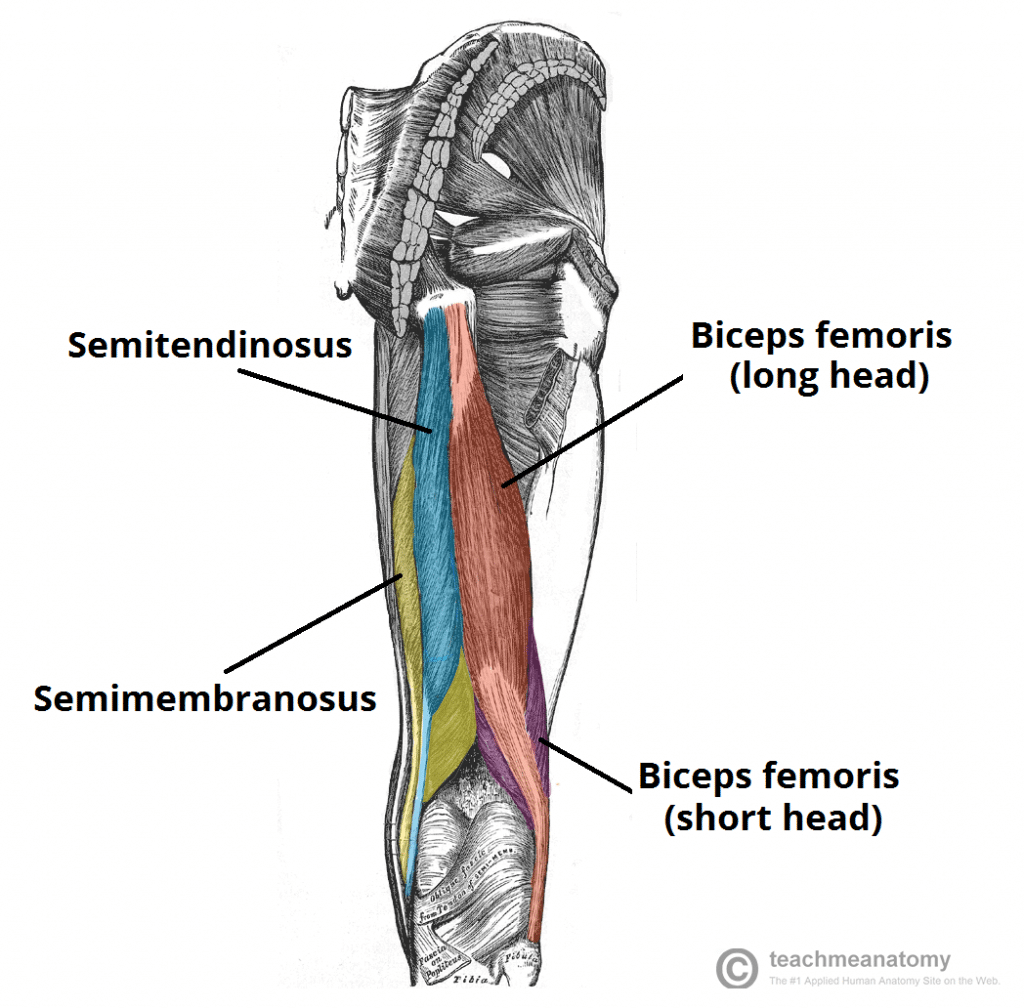
hamstrings
another muscle that works with the knee joint; any one of the three posterior thigh muscles in human anatomy between the hip and the knee: from medial to lateral, the gracilis, semitendinosus and biceps femoris. Also flexors of the knee joint
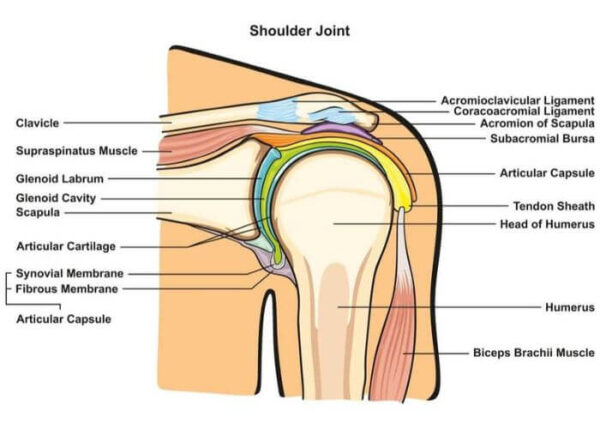
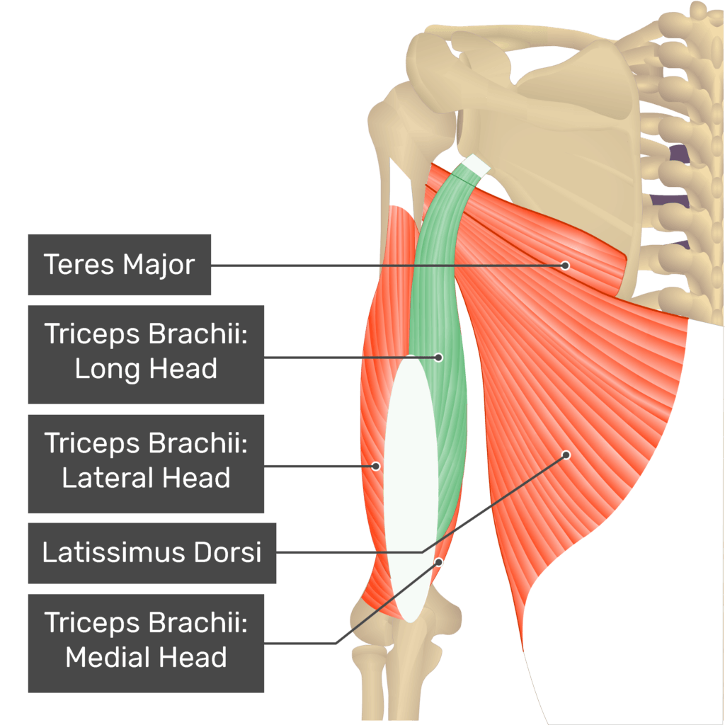
shoulder joint
major muscles responsible for movement: deltiod, pectoralis major, latissimus dorsi, teres major, biceps brachii, and triceps brachii
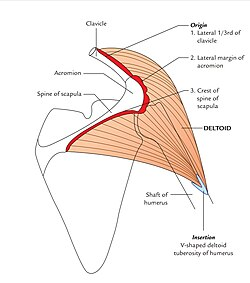
deltoid
the muscle forming the rounded contour of the human shoulder.
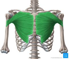
pectoralis major
the superior most and largest muscle of the anterior (front) chest wall. It is a thick, fan-shaped muscle that lies underneath the breast tissue and forms the anterior wall of the axilla.
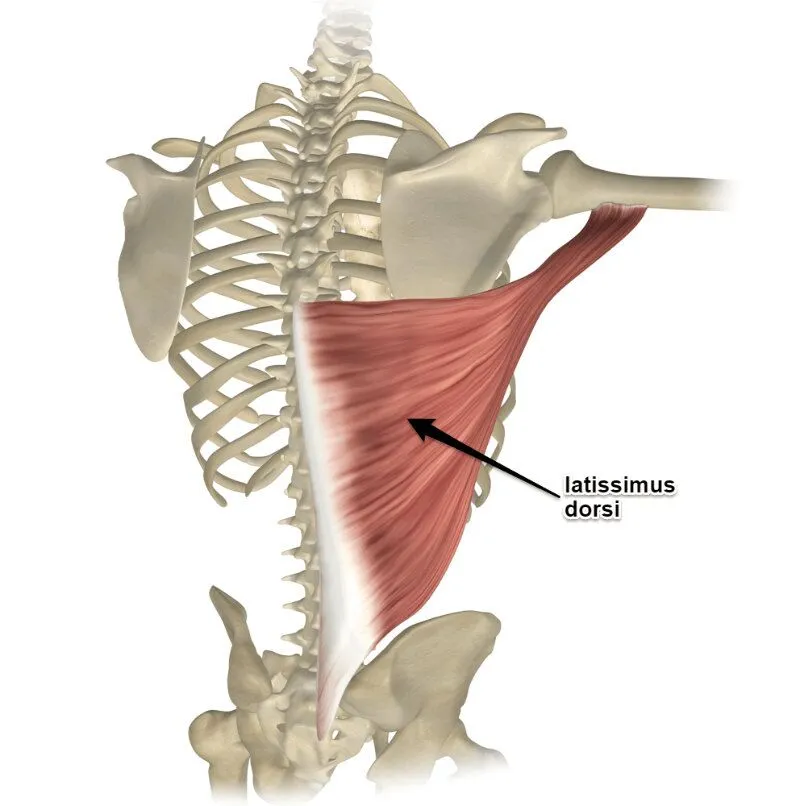
latissimus dorsi
one of the largest muscles of the back, stretching across the lower posterior thorax. Its primary function is in upper extremity movement.
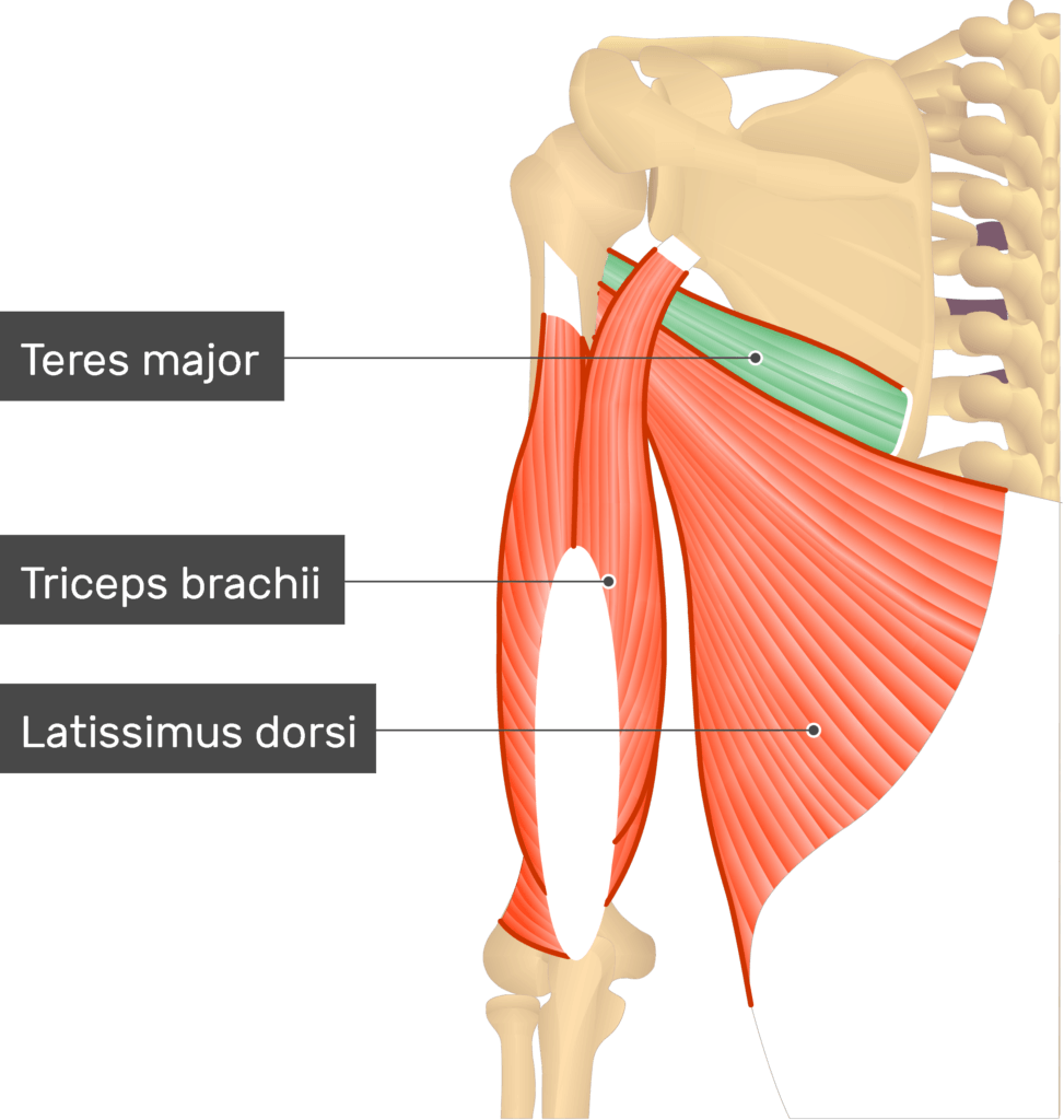
teres major
a thick but flattened, rectangular muscle that extends from the inferior posterior scapula to the medial lip of the intertubercular groove of the humerus. It functions synergistically with the latissimus dorsi to extend, adduct, and internally rotate the humerus
above and beneath the lats
biceps brachii
large muscle that lies on the front of the upper arm between the shoulder and the elbow

triceps brachii
large, thick muscle on the dorsal part of the upper arm. It often appears in the shape of a horseshoe on the posterior aspect of the arm. The primary function is the extension of the elbow joint.
muscles for adduction
pectoralis major, latissimus dorsi, and teres major that are involved in moving the shoulder towards the body's midline. And triceps brachii
muscles for abduction
deltoid works alone to move the shoulder away from the rest of the body
muscles for shoulder extension
pectoralis major, latissimus dorsi, teres major, triceps brachii
muscles for shoulder flexion
deltoid, pectoralis major, biceps brachii
muscles for medial rotation of shoulder
teres major, latissimus dorsi, deltoid, pectoralis major
muscles for lateral rotation of shoulder
deltoid
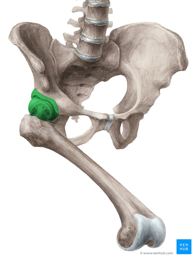
hip joint
connection points between your legs and your torso. Made up of your thigh bone (femur) and your hip bone (pelvis). Ball-and-socket joints that support your body weight and allow you to move your upper legs.
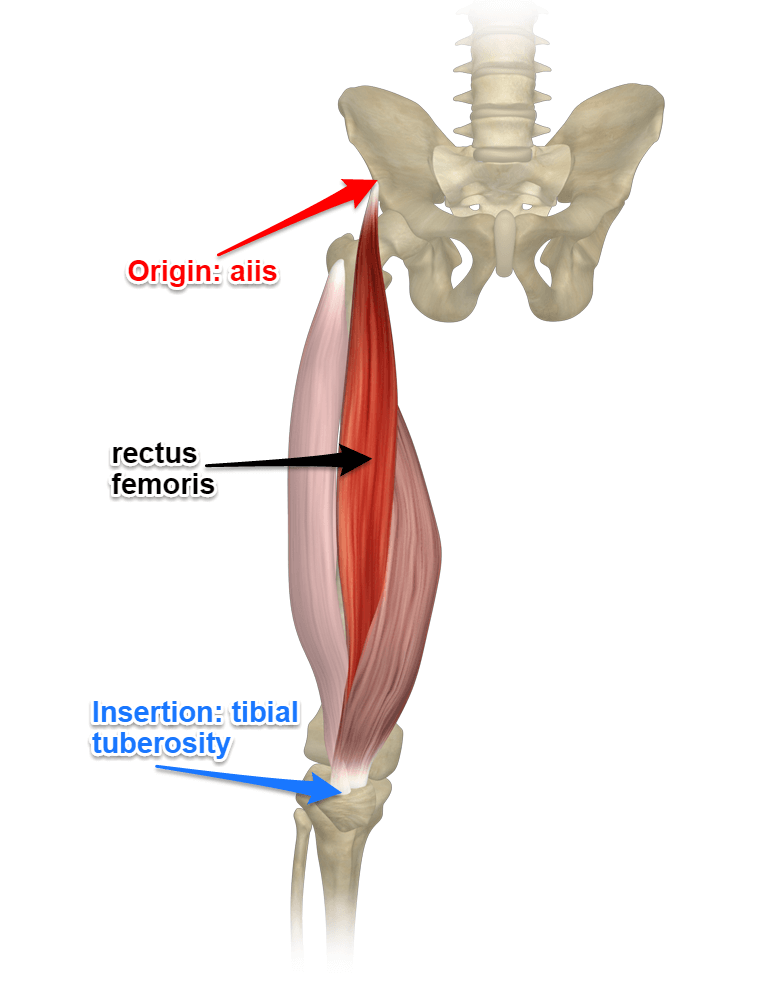
muscles for hip flexion
sartorius and rectus femoris
rectus femoris
one of the four quadriceps muscles of the human body; by acting on the hip joint, it helps with thigh flexion
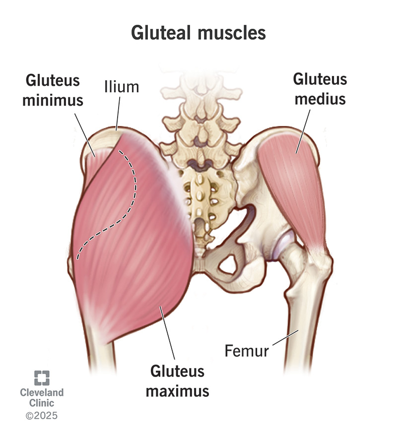
muscles for hip extension
gluteus maximus, semitendinosus, and biceps femoris
gluteus maximus
the most superficial and largest of the three muscles and makes up the bulk of the shape and form of the buttock and hip area. Biggest and strongest muscle in your body. Helps extend thigh bone out, away from the body, and helps keep your hips balanced and your trunk upright when you're sitting down.
semitendinosus
a long superficial muscle in the back of the thigh. Collectively with the other two muscles of the posterior compartment of the thigh, works to extend at the hip and flex at the knee
biceps femoris
a muscle of the posterior compartment of the thigh, and lies in the posterolateral aspect; part of the hamstrings and aids in extension of the hip joint
extensors of the knee joint
vastus lateralis, vastus medialis, vastus intermedius, rectus femoris
flexor of the knee joint
sartorius muscle movement
three types of muscle
skeletal, cardiac, and smooth
skeletal muscle
voluntary and striated; found throughout the body and functions to contract in response to a stimulus; contract to produce movement, sustain body posture and position, maintain body temperature, store nutrients, and stabilize joints
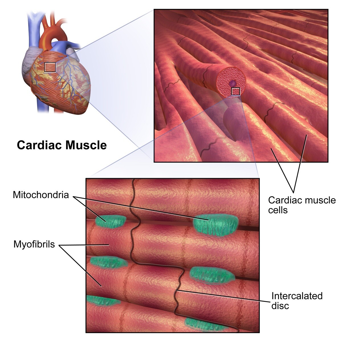
cardiac muscle
involuntary, striated, and auto-rhythmic; constitutes the main tissue of the wall of the heart; responsible for the contractility of the heart and, therefore, the pumping action
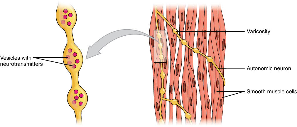
smooth muscle
involuntary and non-striated; muscle tissue in which the contractile fibrils are not highly ordered, occurring in the gut and other internal organs and not under voluntary control.
location of skeletal muscle
attached to bones (skeleton)
location of cardiac muscle
found only in the heart
location of smooth muscle
found in the walls of blood vessels and in the walls of organs of the digestive, respiratory, urinary, and reproductive tracts
function of skeletal muscle
movement of the body, and prevention of movement of the body
function of cardiac muscle
pumping of blood
function of smooth muscle
control of blood vessel diameter, movement of contents in hollow organs
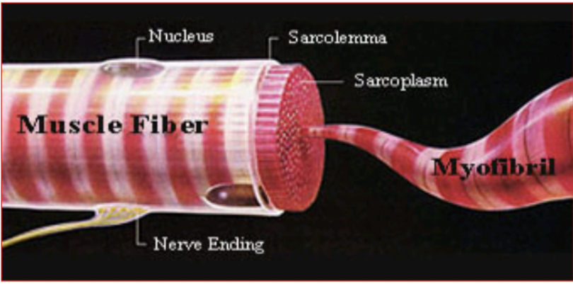
anatomical description of skeletal muscle
very large, cylindrical, multinucleated cells arranged in parallel bundles
anatomical description of cardiac muscle
short cells with blunt, branched ends. Cells joined to others by intercalated discsand gap junctions
anatomical description of smooth muscle
small, spindle-shaped cells joined to each other by gap junctions
initiation of contraction for skeletal muscle
only by a nerve cell
initiation of contraction for cardiac muscle
spontaneous (pacemaker cells), modifiable by nerves
initiation of contraction for smooth muscle
some contraction always maintained. Modifiable by nerves
voluntary
skeletal muscles are…. while cardiac and smooth are not
gap junctions
cardiac and smooth muscle contain these, while skeletal muscle does not; aggregates of intercellular channels that permit direct cell–cell transfer of ions and small molecules
functions of muscles
motion, movement of body fluids, regulation of body fluids (eg bladder), body stability, and heat production (through breakdown of ATP for energy for contraction)
characteristics of muscles
excitability - action potentials
contraction - generate force
extensibility - stretchable
elasticity
superficial fascia
connective and adipose tissue; storage, insulation, and protection; type of connective tissue
found directly under the skin and superficial adipose layers. It can show stratification both grossly and microscopically. Traditionally, it is described as being made up of membranous layers with loosely packed interwoven collagen and elastic fibers
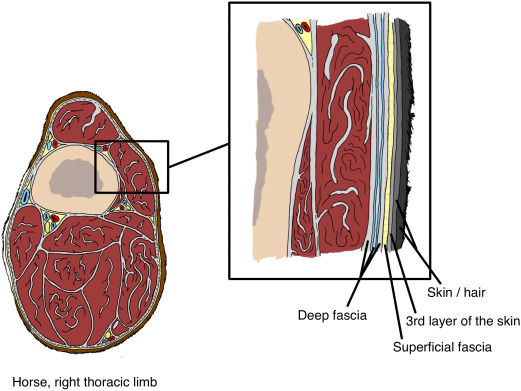
deep fascia
epimysium (whole muscle), perimysium (bundles of fibers), and endomysium (single muscle fiber)
surrounds your musculoskeletal system. It covers your muscles, bones, tendons, cartilage, nerves and blood vessels; layer of dense connective tissue that can surround individual muscles and groups of muscles to separate into fascial compartments.
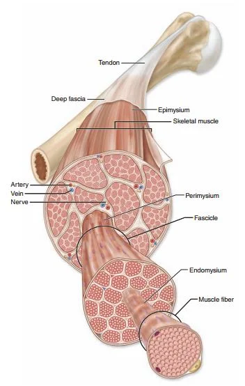
epimysium
a sheath of fibrous elastic tissue surrounding a muscle (fascia); It is a layer of dense irregular connective tissue which ensheaths the entire muscle and protects muscles from friction against other muscles and bones.
perimysium
the sheath of connective tissue surrounding a bundle of muscle fibers; the connective tissue that covers each single muscle fiber or myofiber or muscle cell.
endomysium
a wispy layer of areolar connective tissue that ensheaths each individual muscle fiber, or muscle cell. It also contains capillaries and nerves. It overlies the muscle fiber's cell membrane
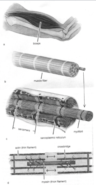
fascicles
muscle is composed of many bundles of these muscle fibers; they are covered by perimysium; a bundle of skeletal muscle fibers surrounded by perimysium, a type of connective tissue
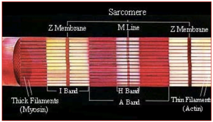
myofibrils
each muscle fiber is composed of numerous ________; the contractile elements of muscle; functional unit is the sarcomere; Function: produce muscle contraction and relaxation
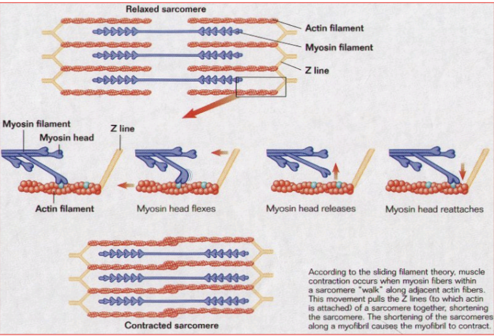
muscle contraction
occurs when myosin fibers within a sarcomere “walk” along adjacent actin fibers. This movement pulls the Z lines (to which actin is attached) of a sarcomere together, shortening the sarcomere. This shortening of the sarcomeres along a myofibril cause the myofibril to contract
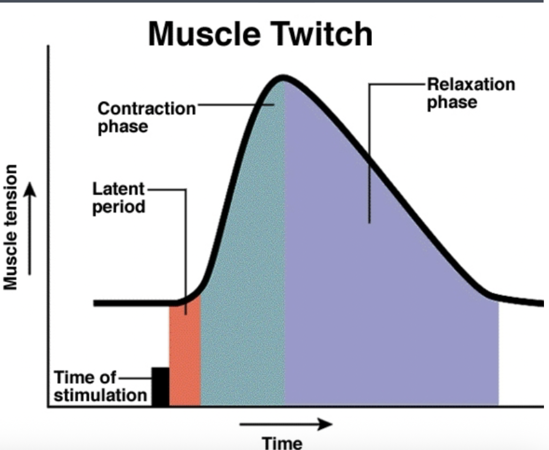
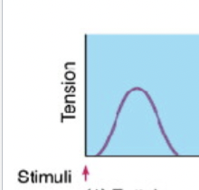
muscle twitch
muscle fibers that contract rapidly in response to a stimulus, producing a quick, brief contraction
stimulus for muscle contraction
???
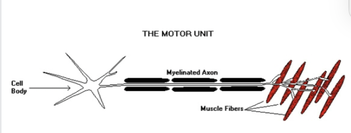
muscle fiber recruitment
describes a motor neuron (a nerve) and all the individual muscle fibers that it innovates. The activation of additional motor units to accomplish an increase in contractile strength in a muscle.
muscle tension vs length
a muscle is capable of being shortened to approximately 1/2 of its normal resting length & stretched about 2 times its normal resting length
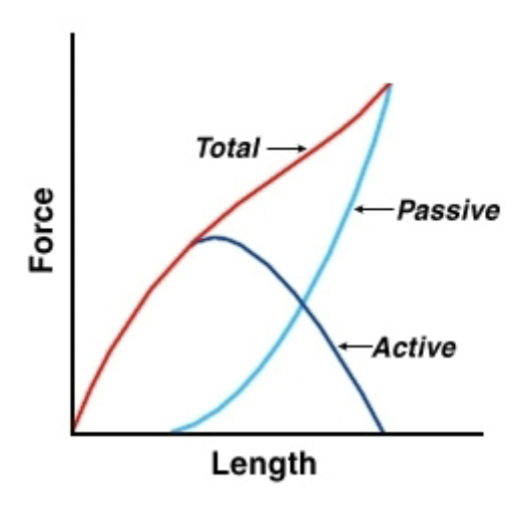
active tension
generated by the muscle in response to stimulus, and is the result of actin/myosin crossbridge cycling
passive tension
generated by stretch, occurs irrespective of stimulus, and is due to the elastic resistance by noncontractile proteins in the muscle (mainly titin). This increases (sometimes exponentially) at the upper limits of muscle length, whereas active tension peaks at the optimum sarcomere length and then declines towards zero
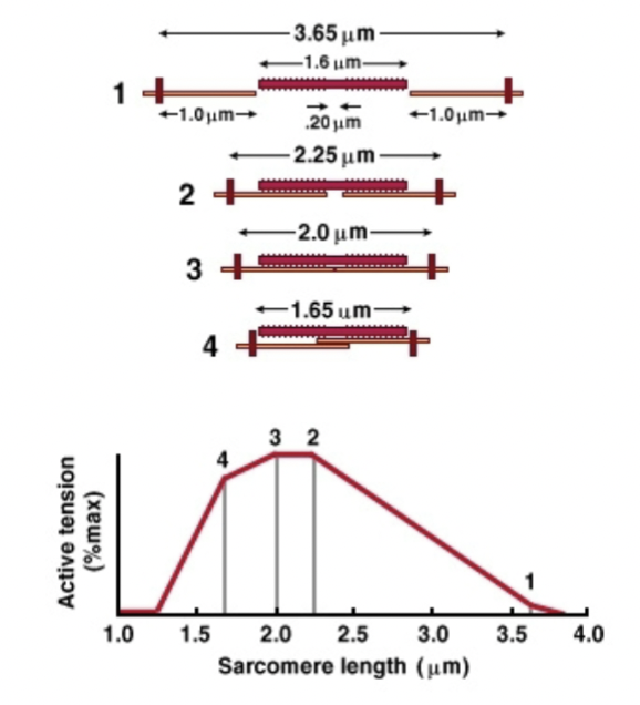
sarcomere tension vs length
The peak tension is produced when sarcomeres are at their resting length, as this provides the optimum alignment between the actin and myosin filaments. When the length of sarcomeres shortens, the actin filaments are pulled along the myosin filaments, which, in turn, pulls the z-lines closer to the myosin filaments.
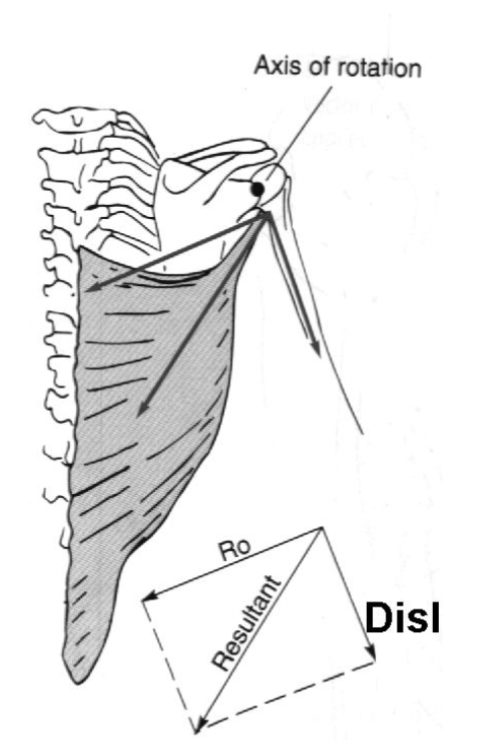
stabilizing components
a degree of parallel forces generated on the lever (bone and joint) when the muscles angle of pull is less than 90 degrees.
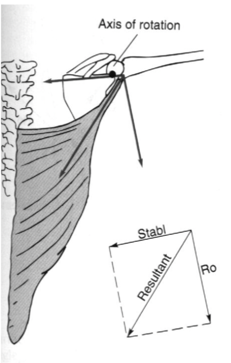
dislocating components
degree of parallel forces generated on the lever (bone and joint) when the muscle's angle of pull is greater than 90 degrees
muscle variables important for determining how much weight we can lift
Recruitment of fibers in a motor unit (nerve stimulus frequency)
Recruitment of motor units in a muscle
Recruitment of muscles in muscle group
Length of muscle
Orientation of muscle fibers
Mass of muscles (number of fibers)
motor unit recruitment
the process by which different motor units are activated to produce a given level and type of muscle contraction
muscle recruitment
describes a motor neuron (a nerve) and all the individual muscle fibers that it innovates
force generator
muscle substructure; quantifies the amount of force produced by muscle contractions. Muscle force generation is a fundamental part of the lower limb's locomotive strategies, stability, and energetics as it provides the active elements of internal controllable forces
Intercalated discs
structure in the heart composed of adherens junctions, desmosomes, and gap junctions, facilitating mechanical coupling and electrical communication between cardiac muscle cells.
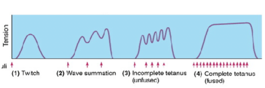
twitch
Single muscle contraction from one stimulus
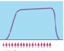
Wave summation
Several stimuli and series so quickly that twitches overlap and cause tension
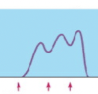
Incomplete tetanus
unfused; even more stimuli; lots of rapid interactions followed by very brief relaxation
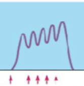
Complete tetanus
Fused; tons of stimuli; sustained contraction without relaxation