Pathomorph- Sem 6 L2 Esophagus, forestomach and stomach | Quizlet
1/44
There's no tags or description
Looks like no tags are added yet.
Name | Mastery | Learn | Test | Matching | Spaced | Call with Kai |
|---|
No analytics yet
Send a link to your students to track their progress
45 Terms
Esophageal Ecyasia is
Dilatation of the oesophagus & weakening of it's wall
mechanism and cause of Esophageal Ecyasia
• Mechanism= Achalasia- uncoordinated esophageal peristalsis w abnormal function of lower and upper sphincter
• Caused by
- denervation
- innervation disorders
- physical obstruction
-Idiopathic
Megaesophagus- 2 forms
1. Congenital
2.Acquired
1. Congenital Megaesophagus
=Partial Block of the lumen by persistent right aortic arch
• German Shepards, Irish setters, greyhounds
• Idiopathic denervation in other breeds eg. Great Danes and Labrador retrievers
2. Acquired Megaesophagus
• Dilation of oesophagus bc of failure of relaxation of the lower sphincter
• Caused by idiopathy, hypothyroidism, esophagitis, chronic gastric dilatation, polymyositis, myasthenia gravis
Esophagitis, Oesophageal erosions, ulcerations are caused by
• Reflux of gastric acid
• viral infections
• Iatrogenic
• irritant ingestion
Esophagitis, Oesophageal erosions, ulceration Pathomorph
• Hyperemia
• Exudate
• Inflammatory infiltrate
• Presence of erosions and ulcers
• Hyperplasia non-affected areas
Where are Foreign objects mostly found in the GIT
mostly in segments that can't fully expand eg. Dorsal to the larynx, thoracic inlet, base of cardia, diaphragmatic hiatus
Consequence of Foreign objects
1. Choke- clinical term of esophageal obstruction subsequent to stenosis or blockage
2. Necrosis of esophageal wall
- Pressure mucosal necrosis
- stricture formation after necrosis healing
- esophageal perforation/ inflammation of mediastinum
Clinical Manifestation of esophageal neoplasia
- weight loss
- painful swallowing - Regurgitation of undigested food
- Dysphagia
- 2ndary aspiration pneumonia
- rarely palpable intracervical mass
- sometimes complete esophageal obstruction
Esophageal Neoplasia is most common in, invades with what, in Equines can occur, what spread & tendency
• Tumour most common in older patients w/out clear breed & gender predisposition
• Invasion w Spirocerca Lupi is inovolved in pathogenesis of canine esophageal sarcomas in some geographic areas
• Squamous cell carcinoma after bracken fern consumption in Eq
• Most tumours locally inavasive w high spreading tendency
Histological types of Esophageal Neoplasia
-Leiomyoma
-Leiomyosacroma
-fibrosacroma
-osteosarcoma
Rumenitis is most commonly associated with
Lactic acidosis
Rumenitis Pathomorph
1. watery and acidic ruminal and intestinal contents
2. Often abdundant grain in rumen
3. Mucosa= brown and friable, detaches easily
4. Hydropic changes and coagulative necrosis of luminal epithelium - neutrophilic infiltration
5. Pale scars formed
Bloat or luminal tympany is
over-distention of rumen & reticulum by gases produced during fermentation
Primary bloat is
-dietary bloat
-Over production of stable foam
Secondary Bloat is
-Caused by physical or functional obstruction or stenosis of esophagus resulting in failure to eructate
- Tumors, foreign objects and innervation disorders
Mechanism of bloat
• Severe distension of rumen w fermentation gases
• Compression of Diaphragm, lungs, increased intrathoracic pressure
• Decrease in venous return to heart
• Generalised congestion cranial to the thorax inlet
• Death (50% of cases)
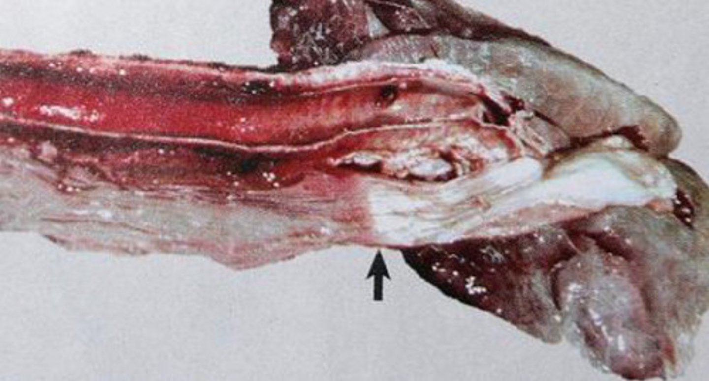
PM of Bloat
"Bloat Line”
- most reliable postmortem indicator of ante mortem bloat- sharp line of separation b/w the pale, bloodless distal oesophagus & congested proximal esophagus at the thoracic inlet
Gastric Dilatation and Volvulus is most common in
dogs esp large deep chested breed and pigs
Causes and predisposing factors of Gastric Dilatation and Volvulus
1. Distension of stomach w gas, fluid, food
2. Obstruction of cardia- prevents eructation and emesis
3. Obstruction of Pylorus- prevents passage of gastric contents into S.intestine
4. Relaxation of gastrohepatic lig
5. Postprandial exercise
6. Hereditary predisposition to gastric rotation
PM of Gastric Dilatation and Volvulus
• Rotation of the stomach clockwise on the vento-dorsal axis
• Rotation is 180-360 degrees
• Displacement of the spleen, torsion & congestion
• Twisted esophagus
Consequence of Gastric Dilatation and Volvulus
• Vascular compression, decreased venous drainage and hypoxia
• acid-base imbalances
• Antiperistaltic waves & atony
• Cardiovascular ischemia, arrhythmias and shock. Also cardiac collapse & death
Gastritis is?
term often applied to acute gastric injury w grossly visible hemorrhage or necrosis, when inflammatory processes, strictly speaking are almost absent or absent
what are the causes of Gastritis
• FBs eg. Bones
• Bacteria, Parasites, fungi, virus
• irritants and drugs
• Immune mediated mechanism
Chronic Giant Hypertrophic Gastropathy occurs in what? what are the clinical consequences?
Occurs in dogs
-weight loss
• vommitting, diarrhea
• Hypoproteinemia
Pathomorph of Chronic Giant Hypertrophic Gastrophy
• Folding of mucosal surface that form cerebriform mass
• Hypertrophy & hyperplasia of mucosal mem
• Variable inflammatory infiltrate
BRADSOT (BRAXXY) is
-acute abomatitis of sheep & rarely in calves due to clostridium septicum
-Occurs in cooler climates
BRADSOT PM
• Bloody abdominal fluid and congestion of abomasa serosa
• abomasal mucosa lesions= diffuse or involve demarcated foci
• Abomasal fold can be thickened, red, hemorrhagic or necrotic
-Hallmark= gelatinous Edema or emphysema of mucosa
Gastric Erosions/Ulcerations caused by
• Not proven, possible hereditary susceptibility
• Acquired abnormalities in protective barrier of mucosa
• Mast Cell Tumor - high level of histamine
• Helicobacter organism
Gastric Ulcerations in Dogs Caused by
• Often Idiopathic
• NSAIDS
• Uremia
• Mast cell tumours, gastrinoma
Gastric Ulcerations in dogs Clinically
• Vommiting
• Lack of appetite
- Abdominal pain
• Anemia- sudden sever or chronic haemorrhage
• Melana- bloody stool patchy black appearance
Gastric Ulcerations in dogs Pathomorph
- Solitary but often numerous lesions
• Localised mainly in antral mucosa or proximal duodenum
• Usually irregular to oval, from few mm to 5cm
• Often w actively bleeding floor or covered by clot
• All stomach can be filled w clotted black blood
Equine GAstric Ulcer Syndrome (EGUS) Causes
• Decrease in protective mechanisms
• Increase in aggressors action
• Non-glandular part: improper feeding, decreased peristalsis
• Glandular part: improper circulation
EGUS clinically
Poor performace
• Chronic Colic
• Loss of appetite
• Sometimes incidental finding w/out signs
EGUS Diagnosis
• Anamnesis, clinical signs
• gastroscopic exam
• response to treatment
Pathomorph of EGUS
• Small erosion, small to large ulcers
• Various number, size & intensity of lesion
• M often near margo plicatus
• Less common on lesser curvature, rare on greater curvature
• Possible severe bleeding
• Perforation of gastric wall in most sever cases
Gastric ulcer in Su caused by
• Feeding w finely ground grain in association w fermentative commensal bact
• Stressful husbandry: overcrowding, poor hygiene
• High dietary copper level, low protein, high level of unsaturated fatty acids
• Infection w Ascaris Suum or Helicobacter
Gastric ulcer in Su clinically
• Sudden Death
• Acute form: anaemia, weakness, inappetence, vomiting, melena
• Chronic form: anorexia w weight loss, intermittent melana
Gastric ulcer in Su PM
• Ulcers restricted to par esophagea, rarely elsewhere
• Lesions can be subtle or effect all par esophagus
• Hallmarks of paraketosis
• Floor of ulcers is covered necrotic debris & clot
• Inflammatory response
• Fatal bleeding of peritonitis= rare
GAstric parasites in horse
• Gastrophilus intestinal & nasalis
• Draschia Megastoma
GAstric parasites in Ru
- Hemonchus Contortus
• Ostertagia ostertagia & circumcincta
GAstric parasites in pigs
• Hyostrongylus rubidus
Gastric Neoplasia name types
1. Epithelial Tumours
2. Leiomyoma
3. Gastric Lymphoma
4. Carcinoma
Gastric Neoplasia rarity & clinically
- less than 1% of all malignancies mostly in smalls adult/older
• Progressive vomitting- hematemesis
• Anorexia
• w. loss