NPTE 2025: Muscle origin insertion, action, innervation
1/99
There's no tags or description
Looks like no tags are added yet.
Name | Mastery | Learn | Test | Matching | Spaced | Call with Kai |
|---|
No analytics yet
Send a link to your students to track their progress
100 Terms
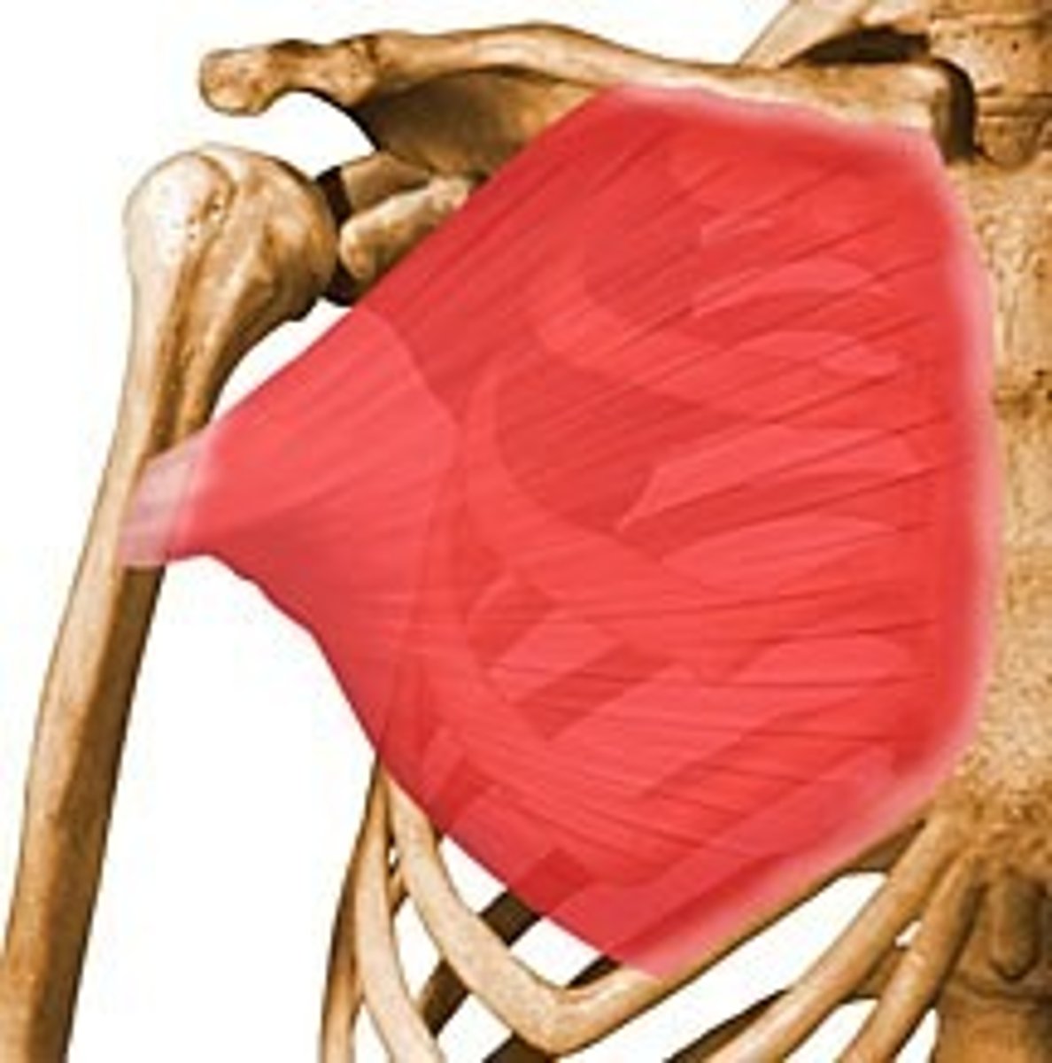
pectoralis major
O: Medial half of clavicle, Manubrium and body of sternum and cartilages of first 6-7 ribs
I: Crest of greater tubercle of humerus
A: Flexion, medial rotation and horizontal adduction of shoulder. Scapular protraction and depression
N: Lateral pectoral
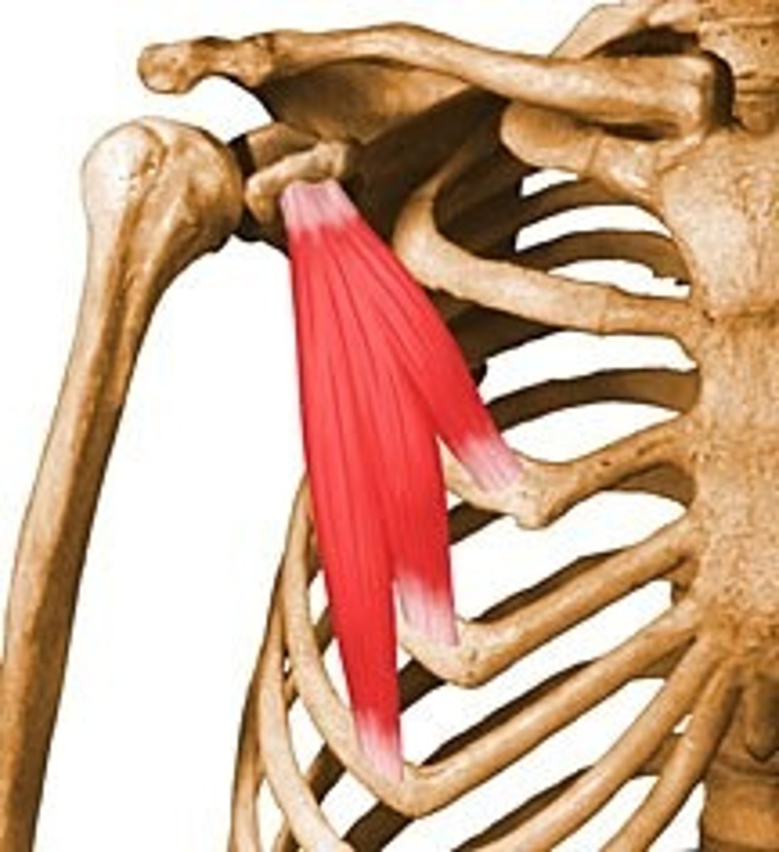
pectoralis minor
O: RIbs 2-5
I: Medial boarder of the coracoid process
A: Downward rotation of scapula, scapular protraction and depression
N: medial pectoral
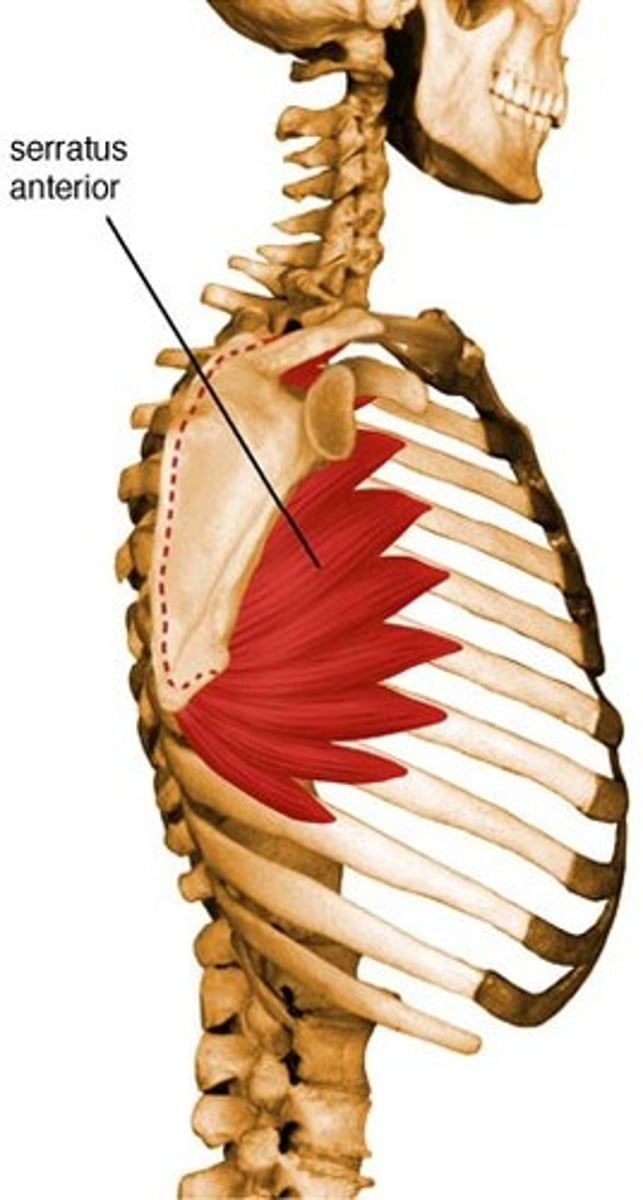
Serratus anterior
O: Ribs 1-8
I: Medial boarder, costal surface of scapula
A: Upward rotation of scapula, Scapular protraction and depression
N: Long thoracic
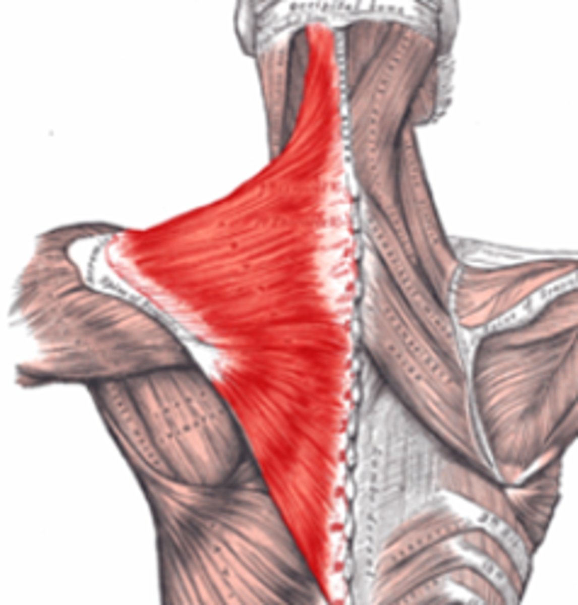
Trapezius
O: Superior nuchal line and external occipital protuberance, ligamentum nuchae, spinous processes and supraspinous ligaments of C7 and all thoracic vertebrae
I: Upper part: Lateral one third of the clavicle
Middle part: Acromion and upper lip of the spine of the scapula
Lower part: Spine of the scapula
A: Upper fibers- scapula elevation
Middle fibers- scapula retraction
Lower fibers- Scapula depression
Upper and lower fibers- Uperward rotation of scapula
N: CN XI
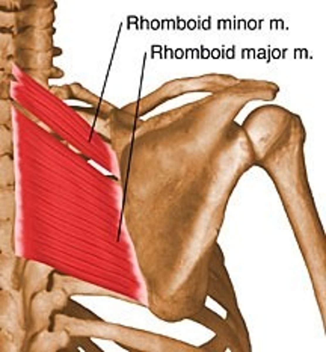
Rhomboid major and minor
O: Ligamentous nuchae and spinous processes of C7-T5
I: Medial border of scapula
A: Adduction, elevation and downward rotation of scapula
N: Dorsal scapular
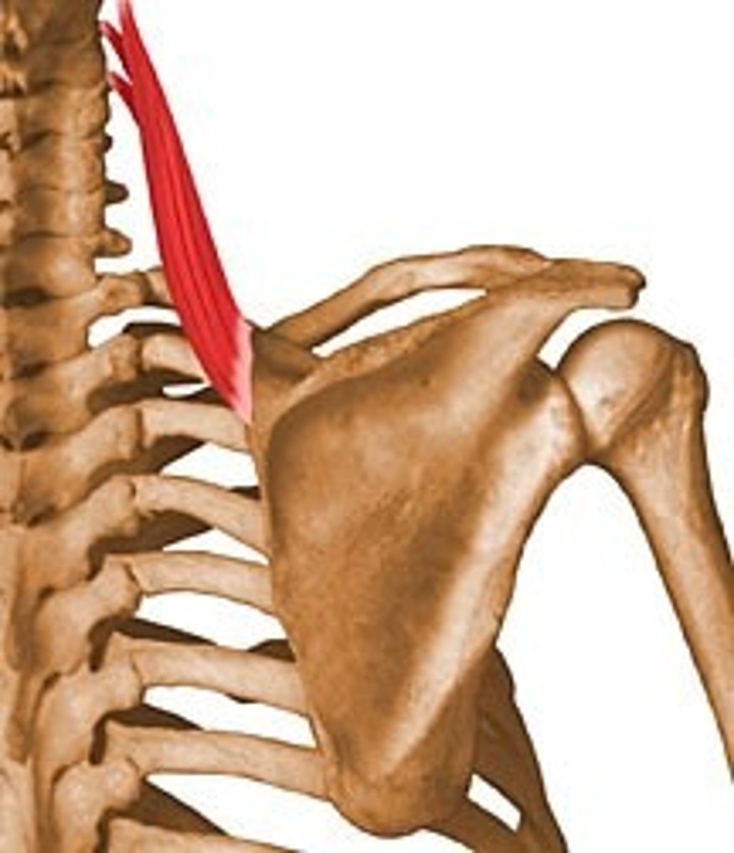
Levator scapulae
O: Transverse processes of C1-4
I: Medial boarder of scapula between superior angle and root of spine
A: Scapular Elevation and downward rotation
N: Ventral rami of SN C3-C4 and dorsal scapular nerve
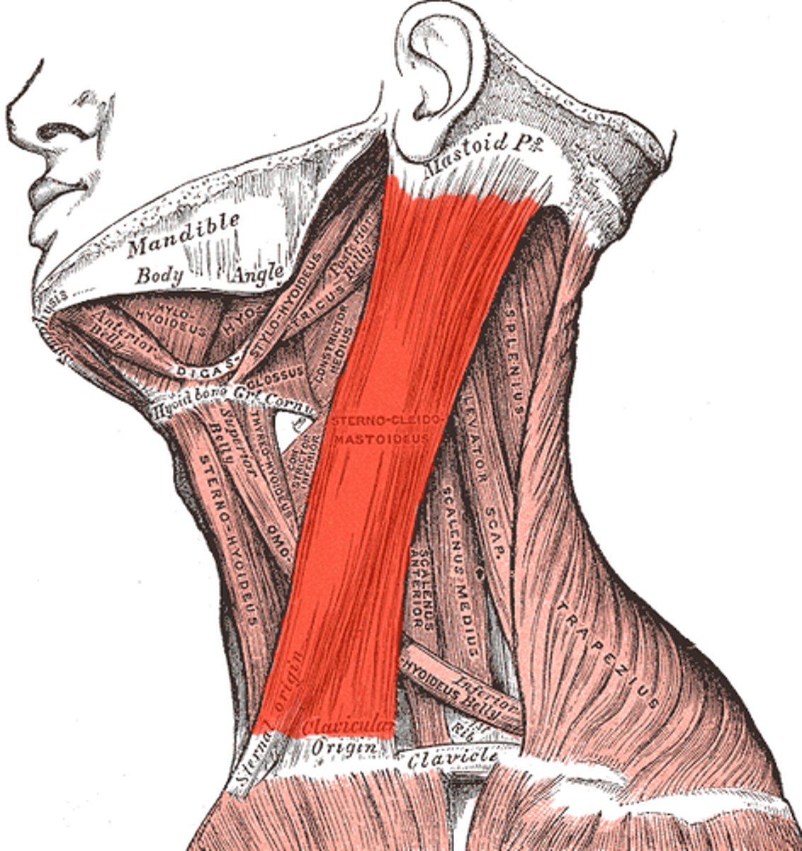
SCM
O: manubrium and medial portion of calvicle
I: mastoid process of temporal bone
A: Same side flexion and opposed side rotation of neck
N: CN XI
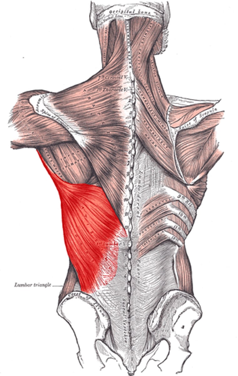
latissimus dorsi
O: Spinous process of 7th-12th thoracic vertebrae, through fascia from the lumbar and sacral vertebrae, last three or four ribs and slip from inferior angle of scapula
I: Intertubercular groove of humerus
A: Medial rotation, adduction and extension of shoulder joint
N: Thoracodorsal
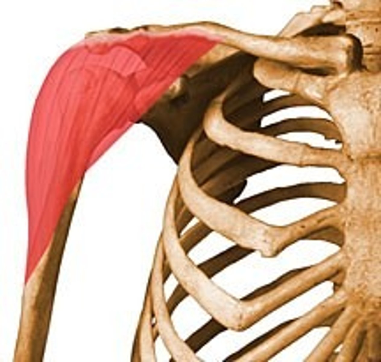
Deltoid
O: Anterior part: lateral end of clavicle
Middle part: acromion
Posterior part: spine of the scapula
I: deltoid tuberosity of humerus
A: abduction at shoulder
Anterior fibers: flex the shoulder
Posterior fibers: extend the shoulder
N: axillary
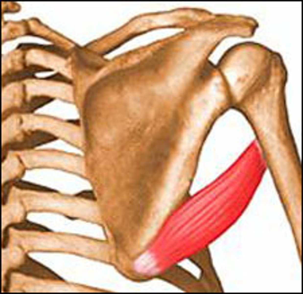
Teres major
O: Inferior angle of scapula
I: crest of lesser tubercle of Humerus
A: Shoulder medial rotation, extension, adduction
N: Lower subscapular
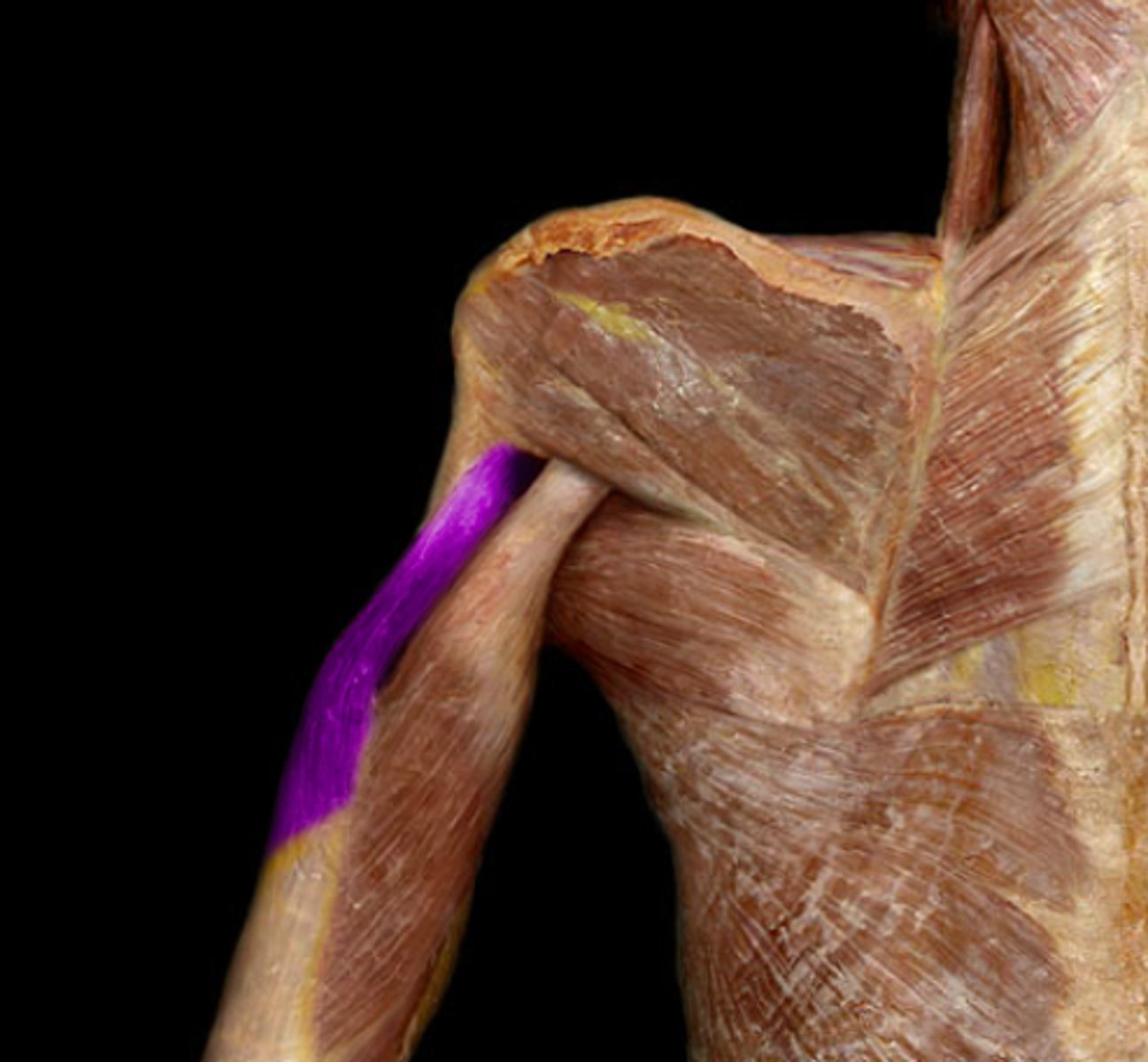
Coracobrachialis
O: Coracoid process of scapula
I: Medial aspect of middle shaft of the humerus
A: flexion of arm
N: musculocutaneous
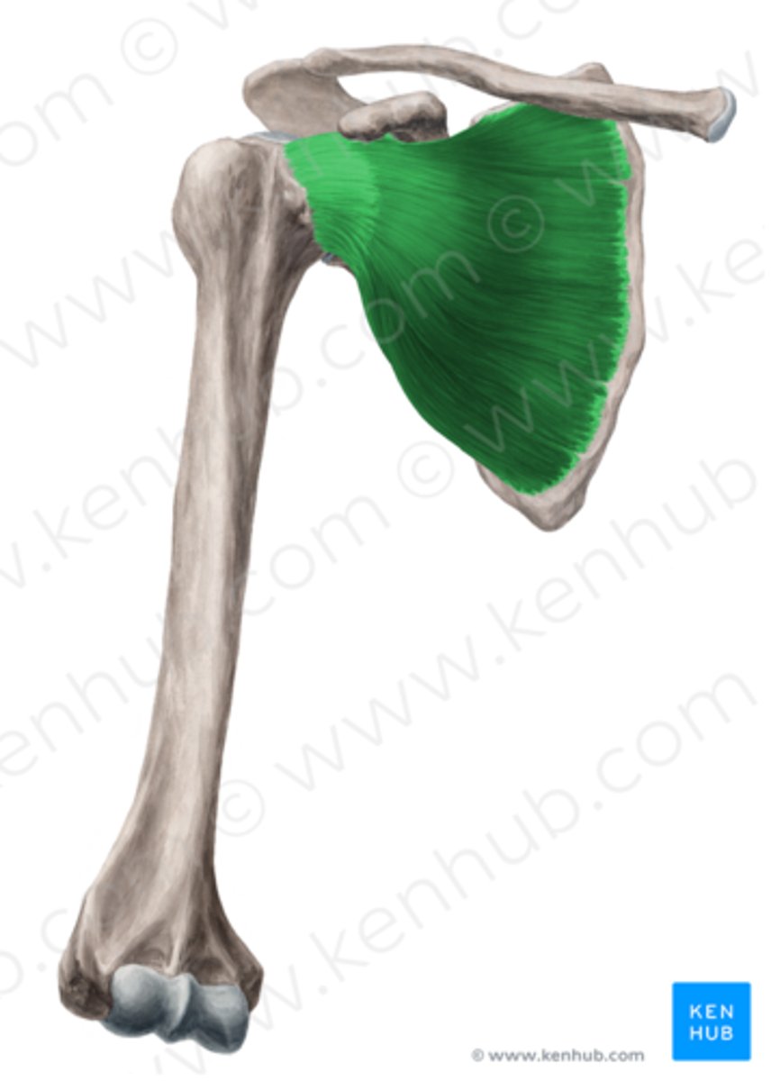
Subcapularis
O: Subscapular fossa of scapula
I: lesser tubercle of humerus
A: Medially rotate shoulder, adduct at shoulder
N: upper and lower subscapular nerves
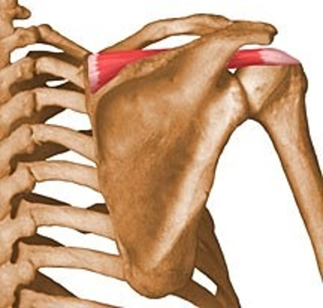
Supraspinatus
O: Supraspinatus fossa of scapula
I: Upper facet of greater tubercle of humerus
A: shoulder abduction up to 15 degrees
N: Suprascapular
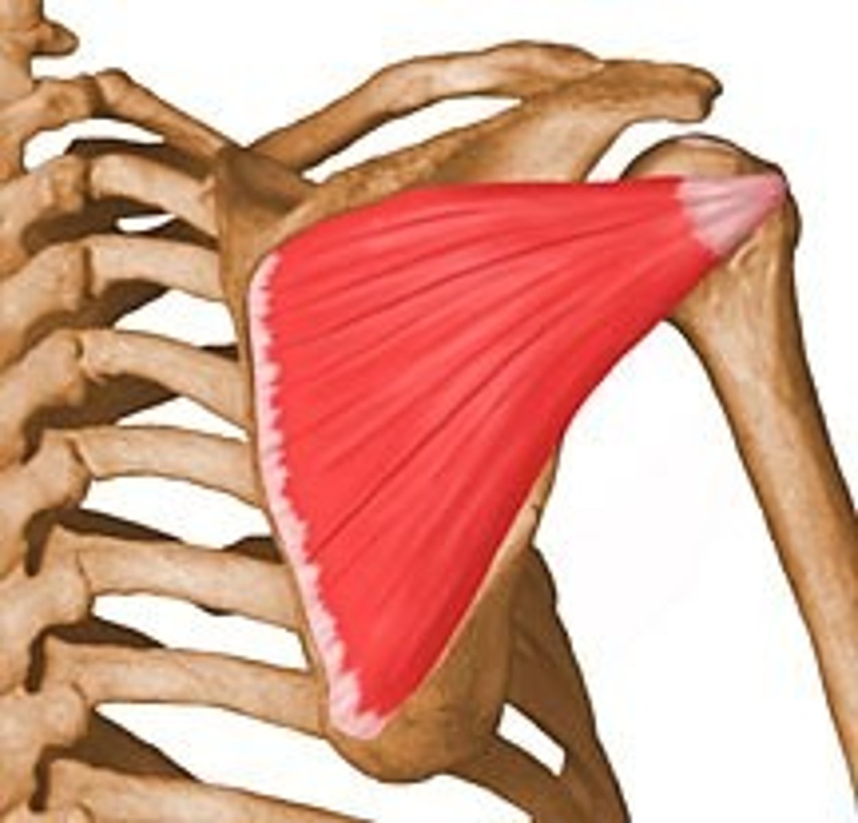
Infraspinatus
O: infraspinatus fossa of scapula
I: Middle facet of greater tubercle of humerus
A: lateral rotation at shoulder
N: Suprascapular
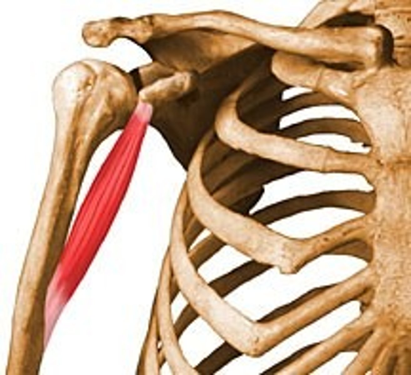
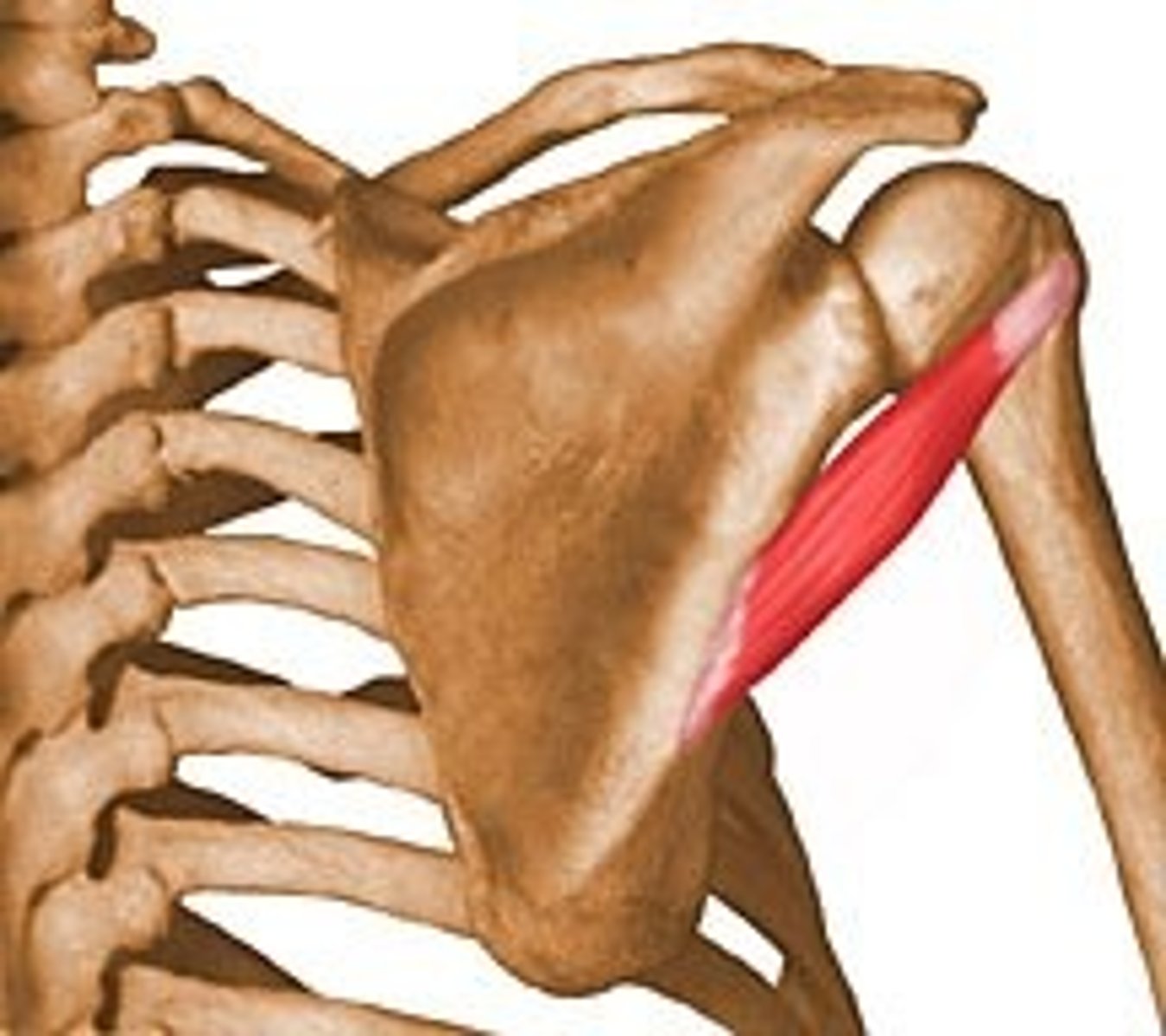
Teres minor
O: lateral border of scapula
I: lower facet of greater tubercle of humerus
A: lateral rotation at shoulder
N: axillary
Long head of biceps
O: supraglenoid tubercle of scapula
I: Bicipital tuberostiy of radius
A: flexion of elbow, supination of forearm, flexion of shoulder
N: musculocutaneous
Short head biceps
O: coracoid process of scapula
I: bicipital tuberosity of radius
A: Flexion of elbow, supination of forearm, flexion of shoulder
N: Musculocutaneous
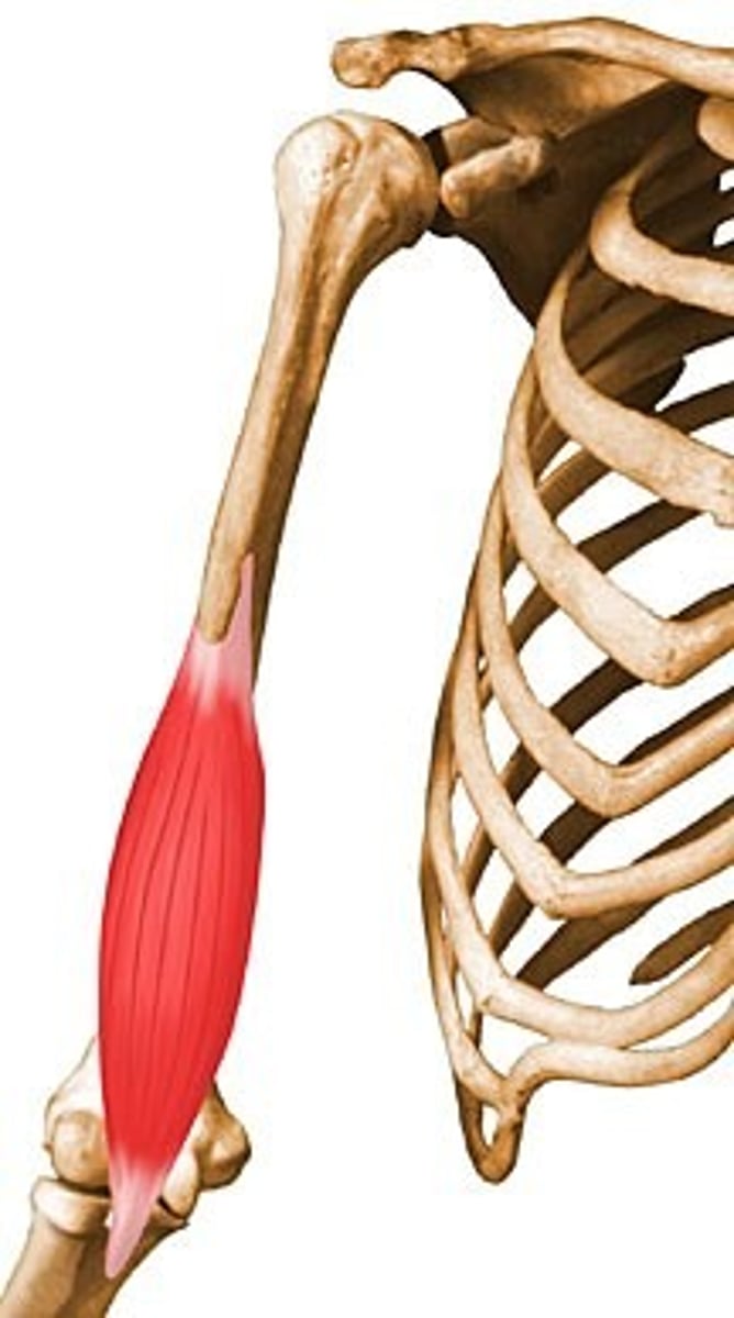
Brachialis
O: Distal aspect of the anterior surface of the humerus
I: Coronoid process and tuberosity on the proximal ulna
A: flex elbow
N: musculocutaneous
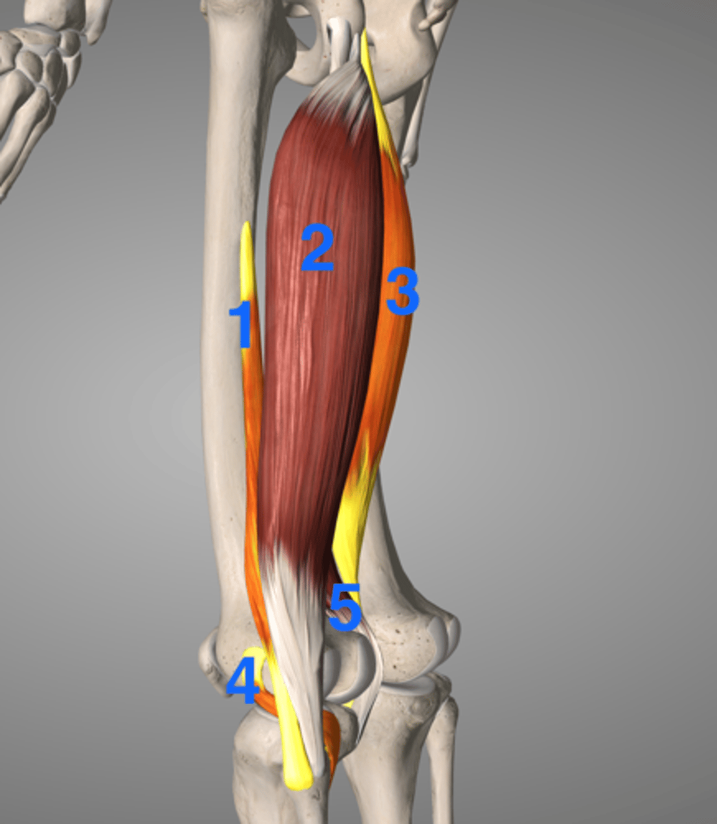
Lateral head of triceps
O: Posterior humerus, superior and lateral to the radial groove
I: olecranon process of ulna
A: extend elbow and shoulder
N: radial
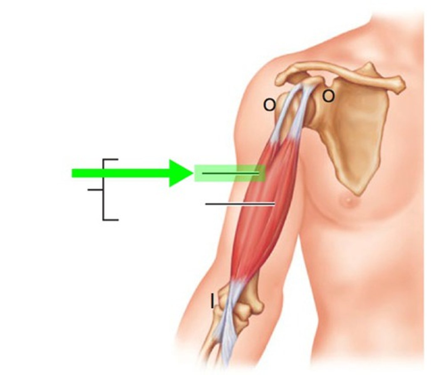
Long head of triceps
O: infraglenoid tubercle of scapula
I: olecranon process of ulna
A: extend elbow and soulder
N: radial
Medial head of triceps
O: Posterior humerus, inferior and medial to the radial groove
I: Olecranon process of ulna
A: extend elbow and shoulder
N: radial
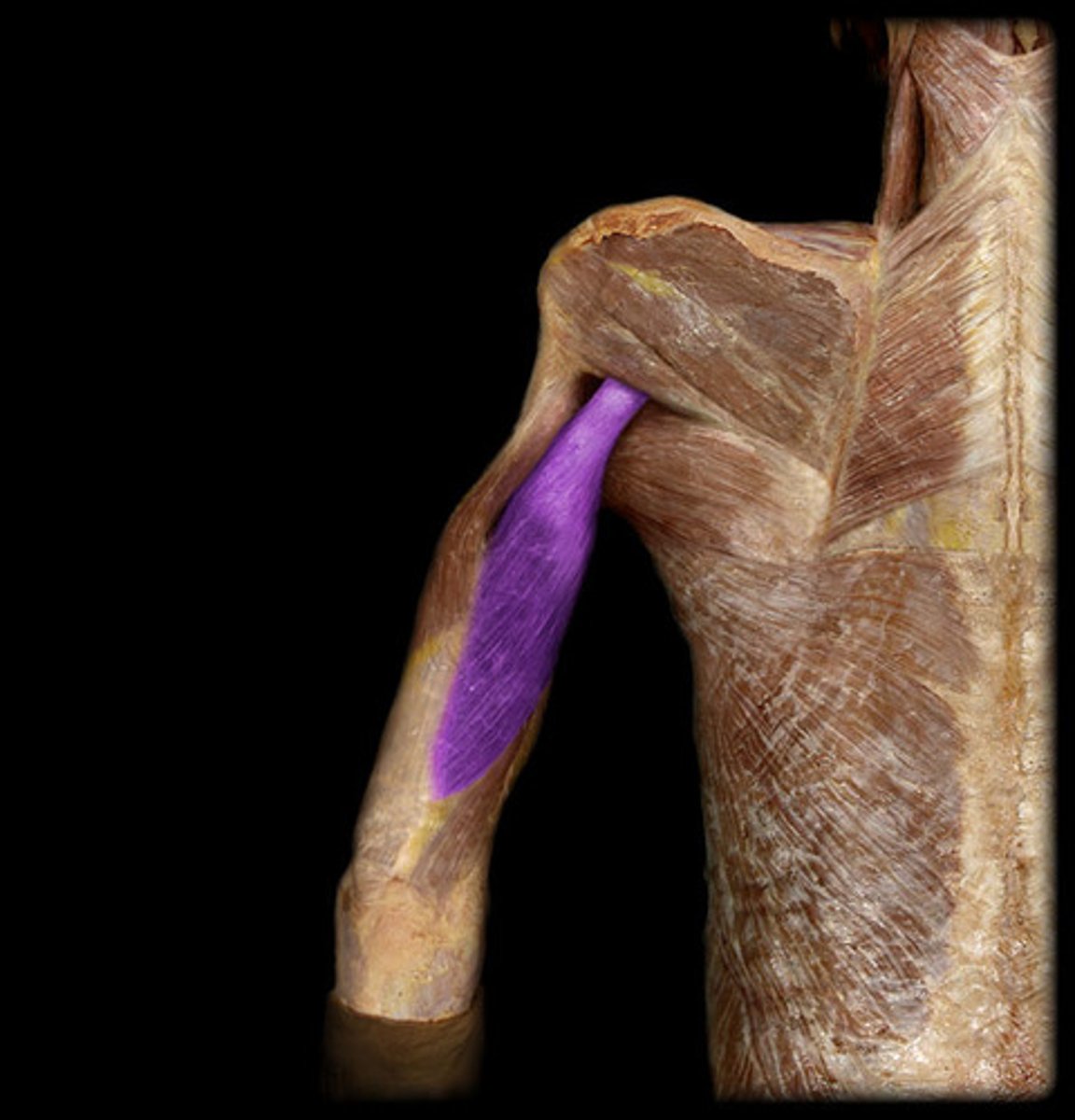
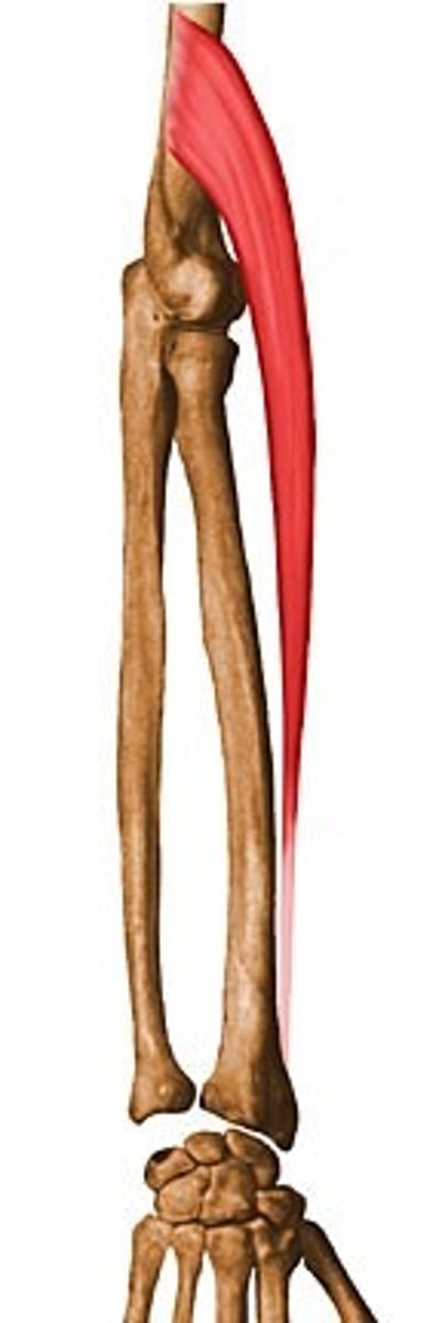
Brachioradialis
O: upper two third of the lateral supracondylar ridge of the humerus
I: Near styloid process at the distal radius
A: Elbow flexion especially in mid-prone position
N: Radial
Anconeus
O: Posterior side of the lateral epicondyle of the humerus
I: Between the olecranon process and proximal surface of the posterior side of the ulna
A: Weak elbow extensor
N: Radial
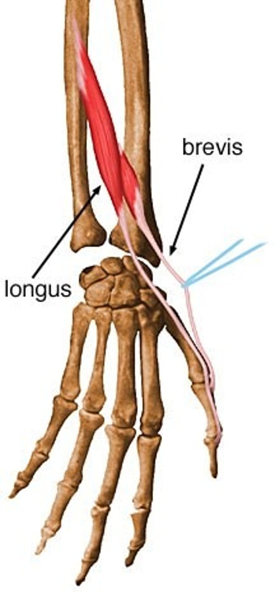
Extensor carpi radials longus and brevis
O: common extensor origin lateral epicondyle of the humerus
I: Base of 2nd metacarpal (ECRL)
Base of 3rd metacarpal (ECRB)
A: Wrist extension and radial deviation
N: radial
Extensor digitorum
O: Common extensor origin lateral epicondyle of humerus
I: By four tendons, each to the base of the extensor mechanism and to the dorsal base of the proximal phalanx of the fingers
A: Extends the MCP joints, extends the IP joints of the second through fifth digits
N: radial
Extensor digiti minimi
O: ulnar side of the belly of the extensor digitorum
I: Tendon usually divides, joining the ulnar side of the tendon of the extensor digitorum
A: Extends the MCP joints, extends the IP joints of the little finger
N: radial
Extensor carpi ulnaris
O: Common extensor origin-lateral epicondyle of the humerus
I: base of the 5th metacarpal
A: Extension of wrist and ulnar deviation
N: radial
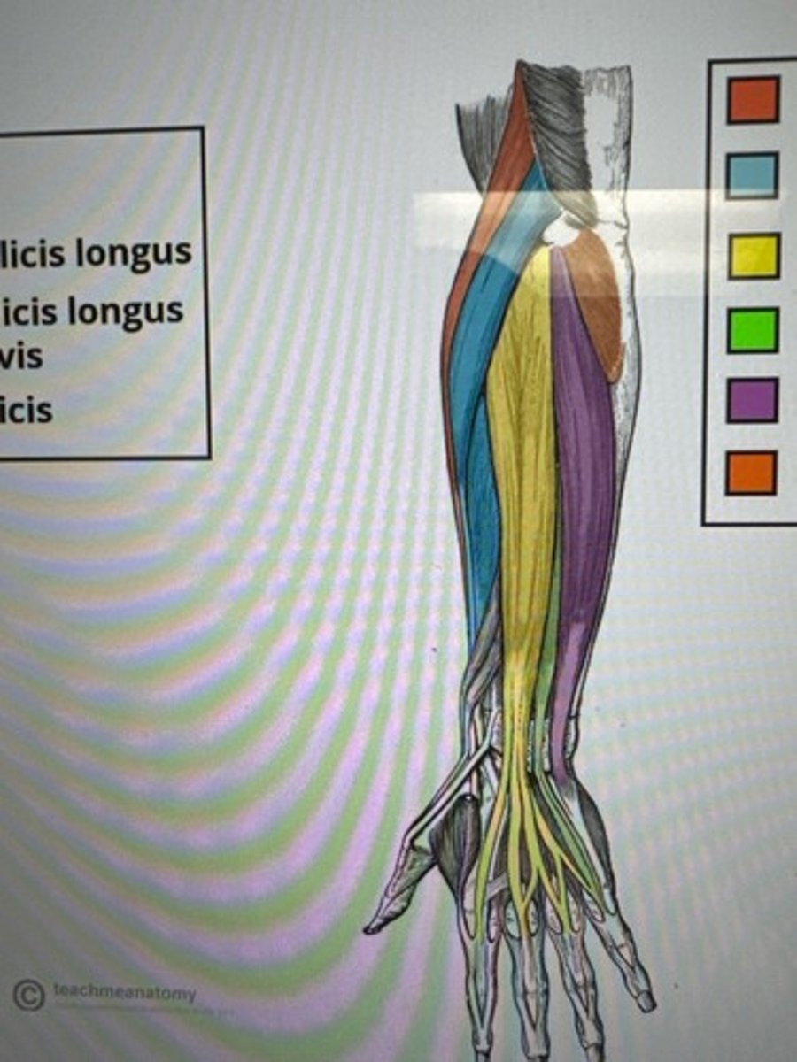
Abductor pollicis longus
O: posterior surface of the middle part of the radius and ulna, and adjacent interosseous membrane
I: Base of 1st metacarpal
A: 1st digit ABD and extension at CMC joint
N: radial
Extensor pollicis brevis
O: Posterior surface of the middle to distal parts of the radius and adjacent interosseous membrane
I: proximal phalanx of thumb
A: 1st digit extension at MCP, CMC
N: radius
Extensor pollicis longus
O: Posterior surface of the middle part of the ulna and adjacent interosseous membrane
I: Distal phalanx of thumb
A: 1st digit extension at MCP IP joints, DIP
N: radial
Extensor indicis
O: posterior surface of the middle to distal part of the ulna and adjacent interosseous membrane
I: Tendon blends with the ulnar side of the index tendon of the extensor digitorum
A: 2nd digit extension and wrist extension
N: radius
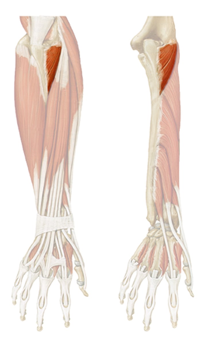
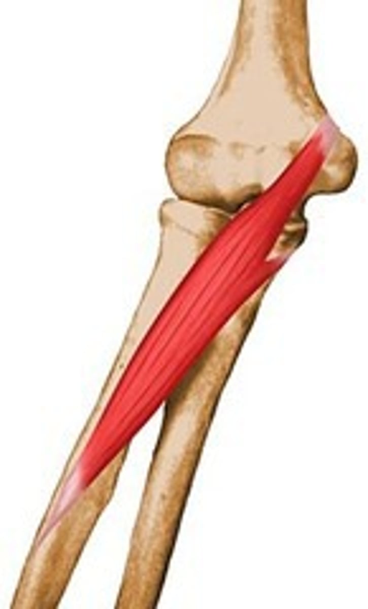
Supinator
O: lateral epicondyle of the humerus and supinator crest of the ulna
I: Lateral surface of the proximal radius
A: supination forearm
N: radial
Pronator teres
O: humeral head: medial epicondyle
Ulnar head: medial to the tuberosity of ulna
I: Lateral surface of the middle radius
A: pronation of forearm, elbow flexion
N: Median
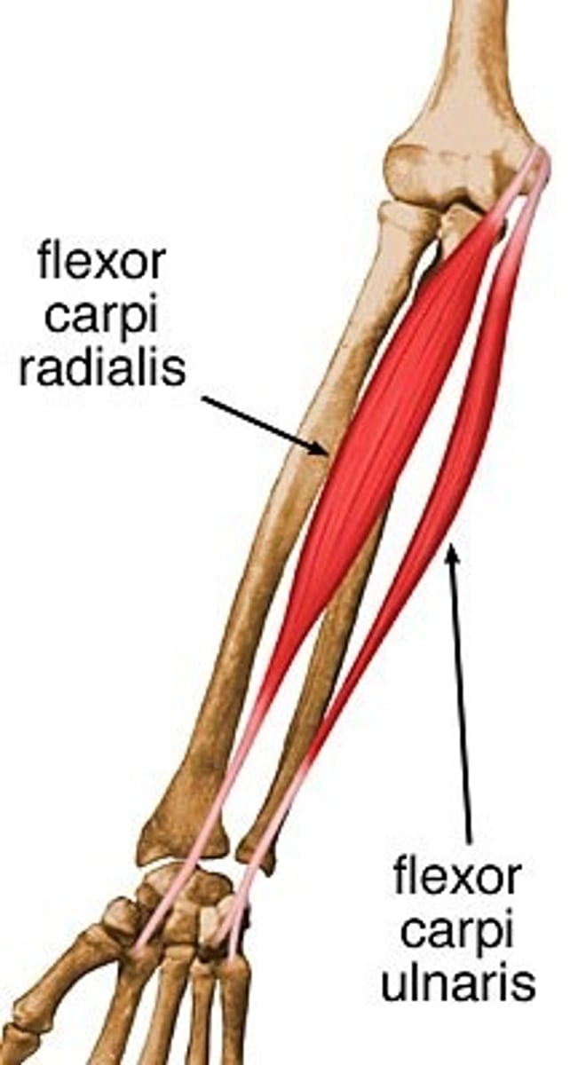
Flexor carpi radialis
O: Common flexor origin medial epicondyle of the humerus
I: 2nd and 3rd metacarpals
A: Wrist flexion, radial deviation
N: median
Palmaris Longus
O: Common flexor origin medial epicondyle of the humerus
I: carpal ligament and palmar aponeurosis of the hand
A: Flexion of wrist
N: median
Flexor carpi ulnaris
O: Humeral head: common flexor origin- medial epicondyle of the humerus
Ulnar head: posterior border of the middle one third of ulna
I: Pisiform bone, pisohamate and pisometacarpal ligaments, and palmar base of 5th metacarpal bone
A: wrist flexion, ulnar deviation
N: ulnar
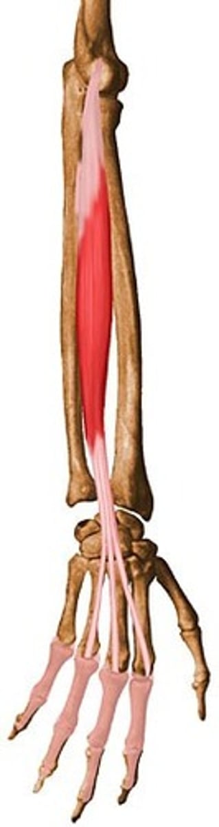
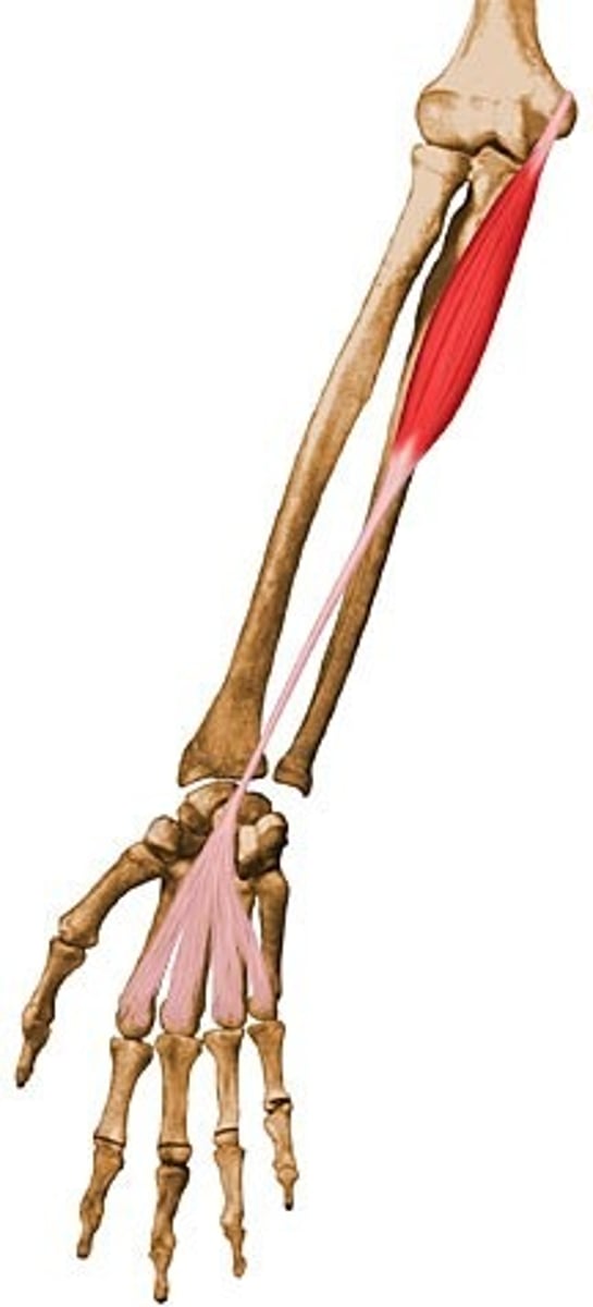
Flexor digitorum superficialis
O: Humeroulnar head: common flexor origin, and the medial side of coronoid process of ulna
Radial head: oblique line just distal and lateral to the bicipital tuberosity
I: by four tendons, each to the sides of the middle phalanges of the fingers
A: flexion of PIP joint of fingers
N: Median
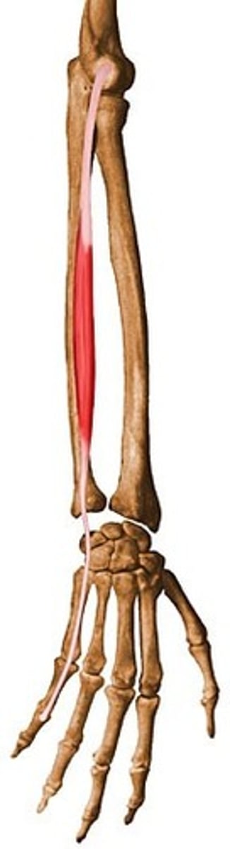
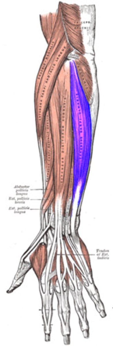
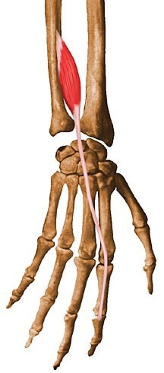
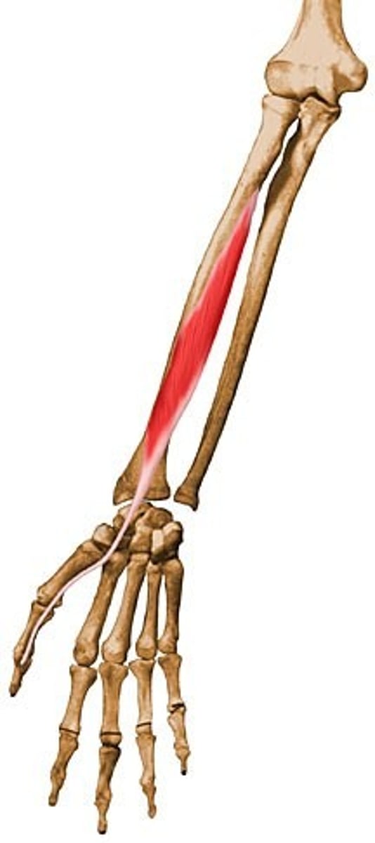
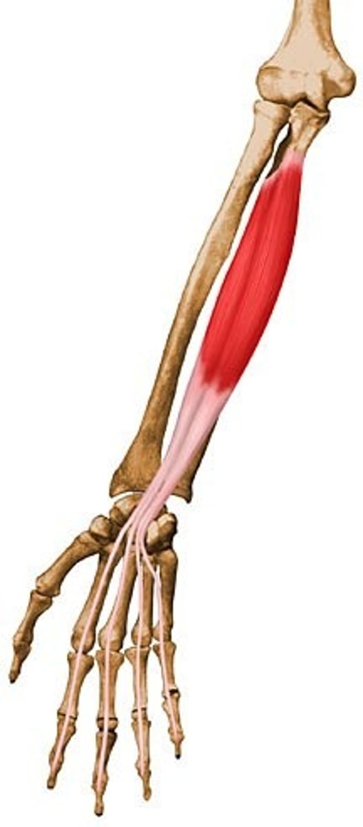
Flexor pollicis longus
O: Middle part of the anterior surface of the radius and adjacent interosseous membrane
I: Distal phalanx of thumb
A: Flexion of distal phalanx of thumb
N: Median
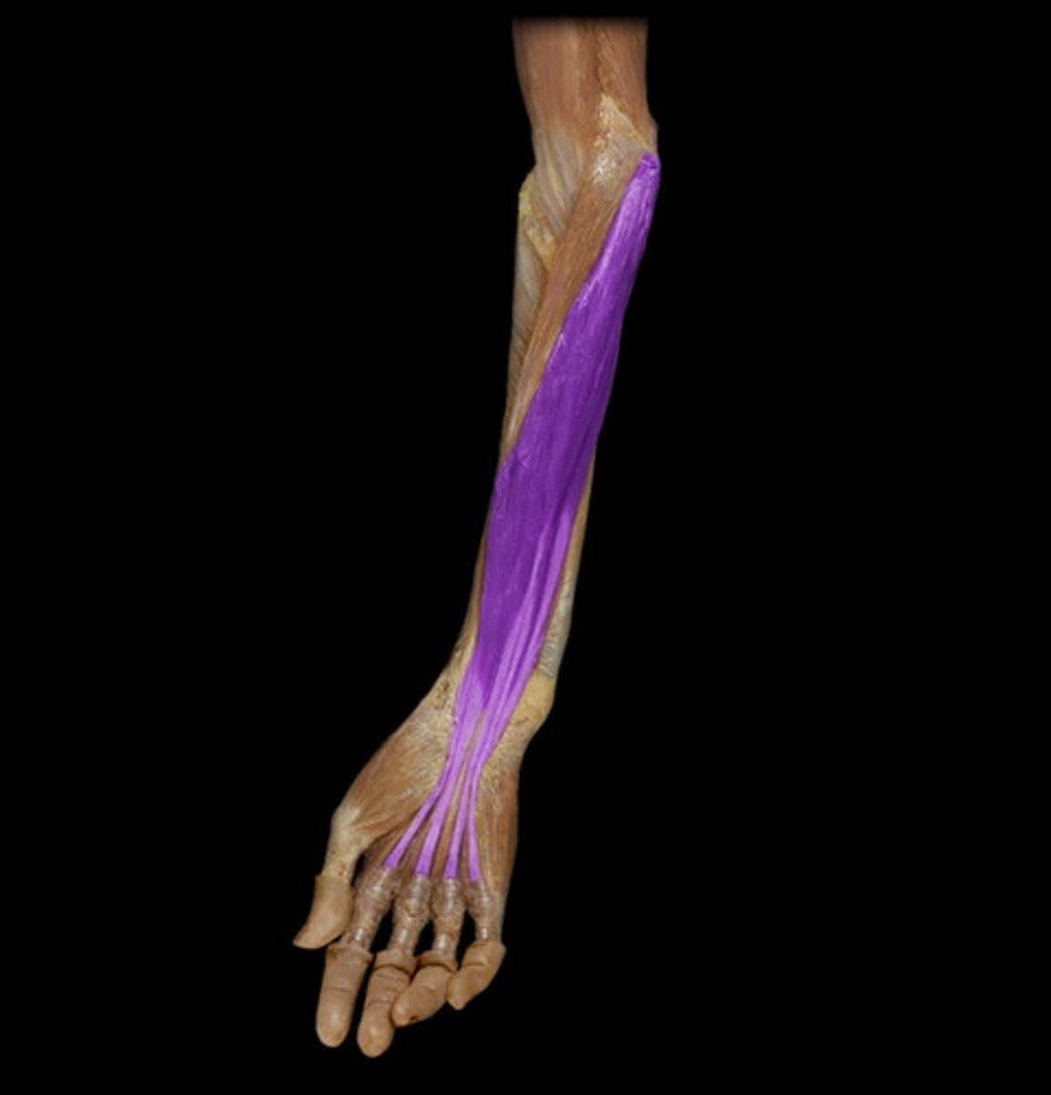
Flexor digitorum profundus
O: Proximal 3/4 of the medial side of the ulna and adjacent interosseous membrane
I: By four tendons, each to the palmar base of the distal phalanges of the fingers
A: FLex wrist, flex all joints to DIP in digits 2-5
N: medial half ulnar
lateral half medial
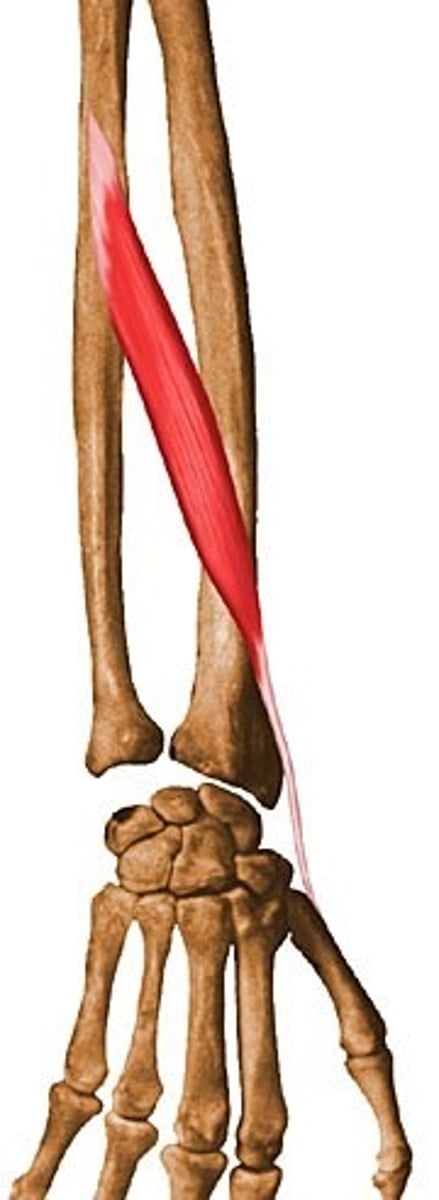
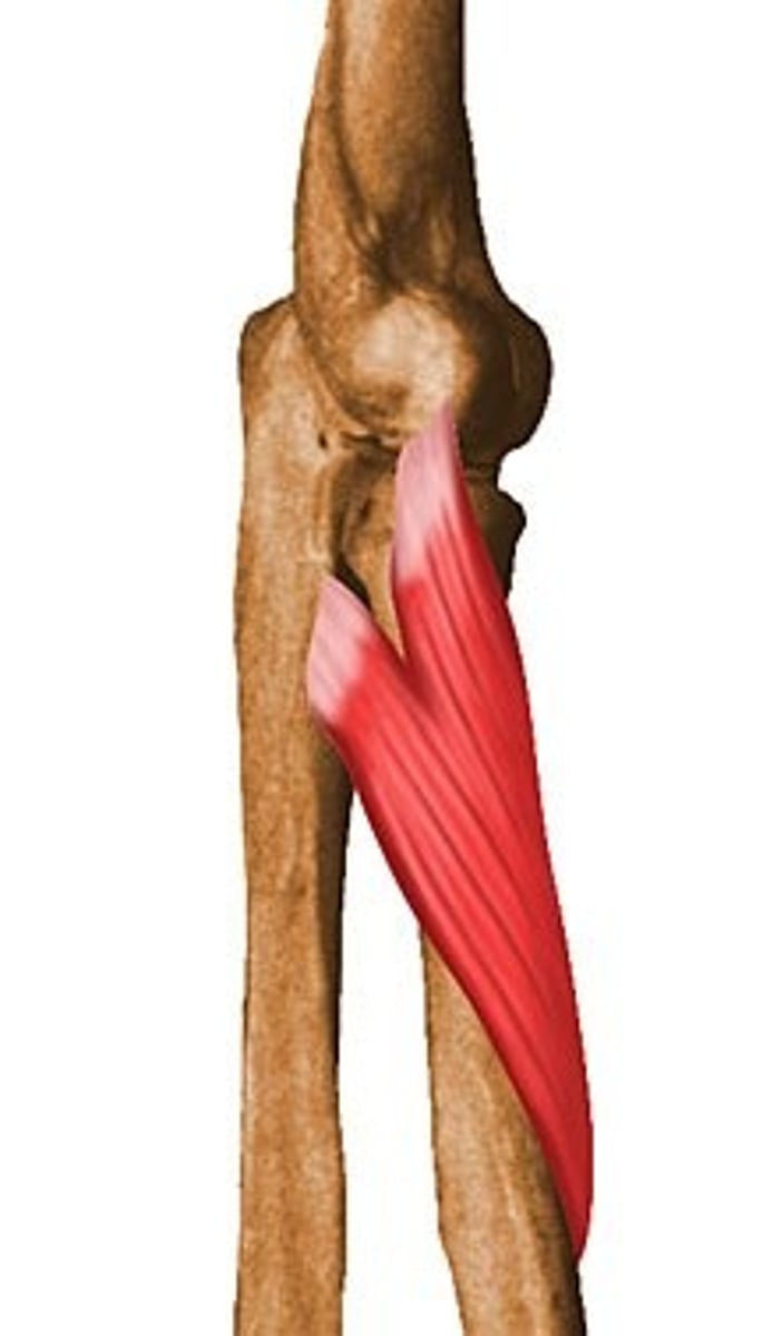
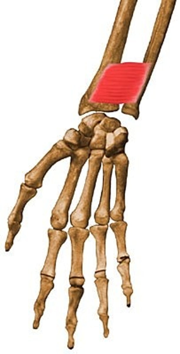
Pronator quadratus
O: Anterior surface of distal ulna
I: Anteiror surface of distal radius
A: pronates forearm
N: Median
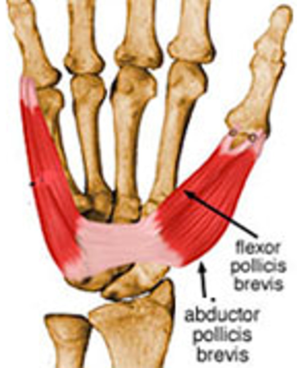
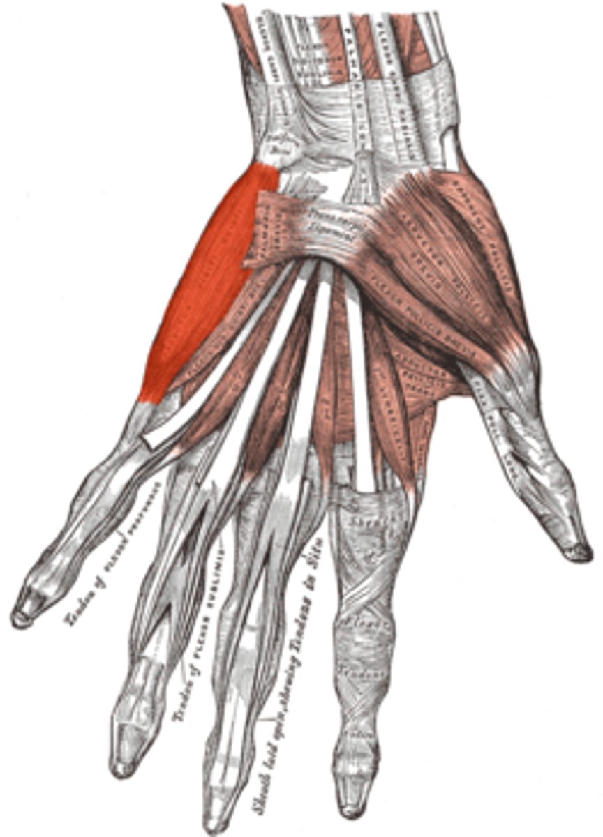
Abductor pollicis brevis
O: palmar tubercles of the trapezium and scaphoid
I: base of proximal phalanx of thumb
A: abduction of thumb at MCP, CMC, joint
N: median
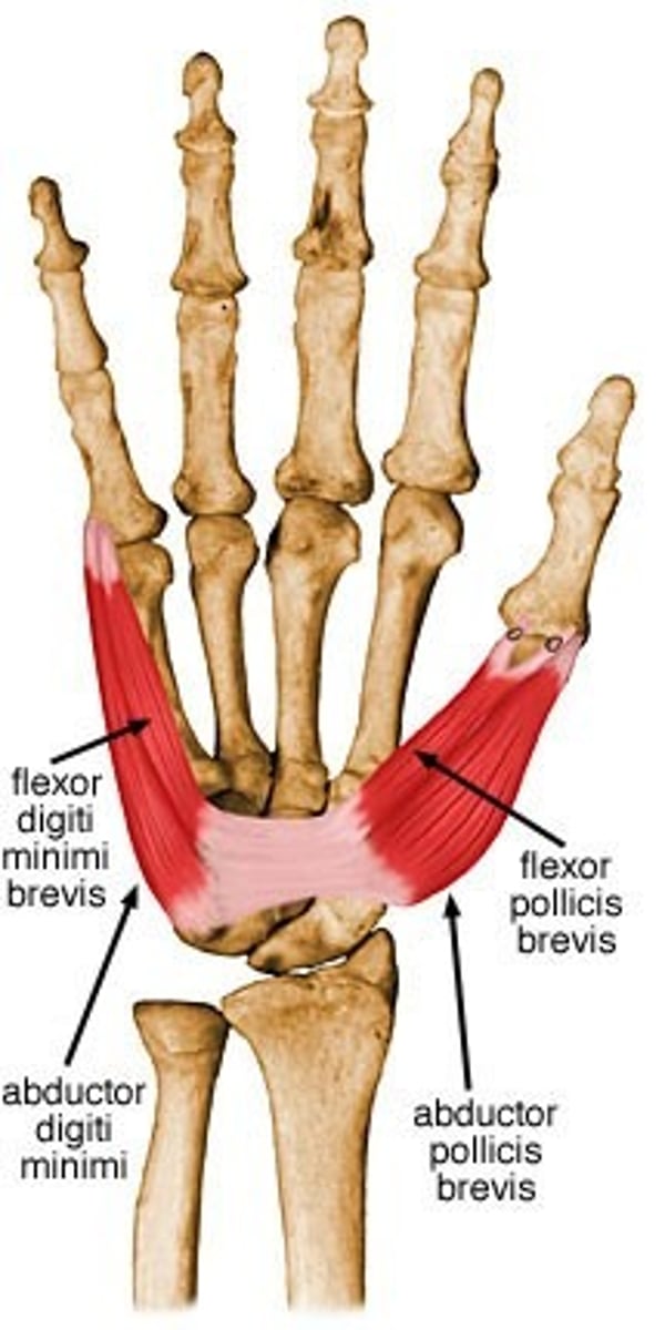
Flexor pollicis brevis
O:Transverse carpal ligament and palmar tubercle of trapezium
I: Radial side of proximal phalanx of thumb
A: flexion of thumb
N: median
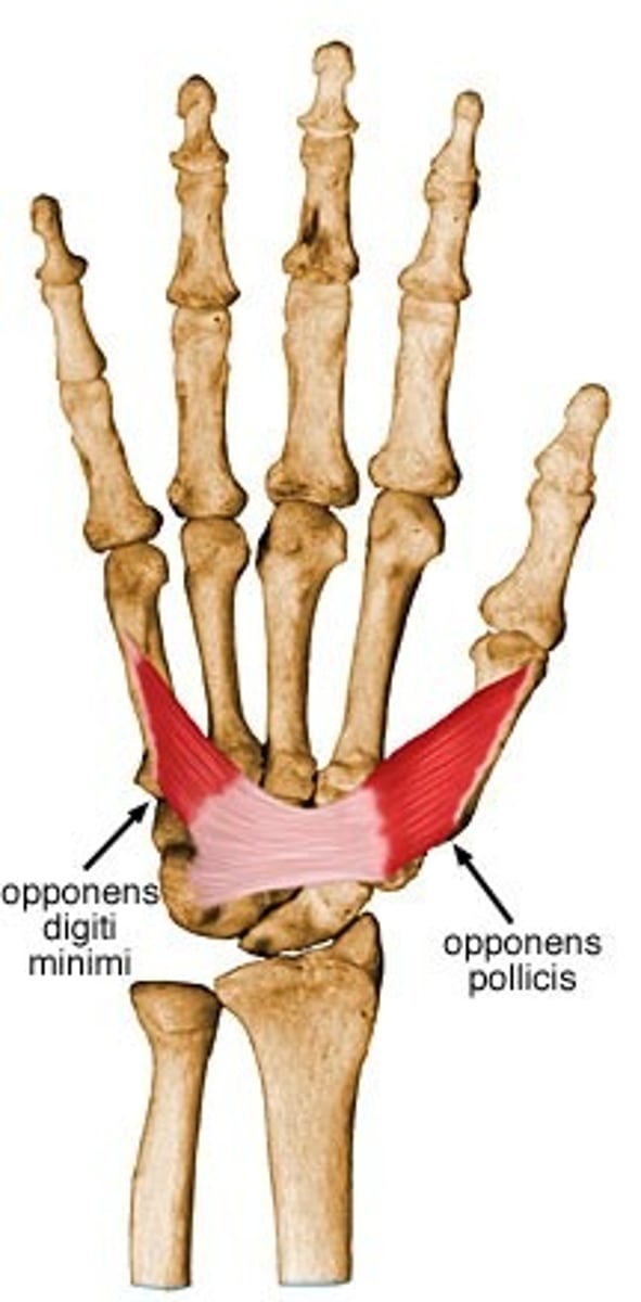
Opponens pollicis
O: Transverse carpal ligament and palmar tubercle of trapezium
I: shaft of the thumb metacarpal
A: opposition of thumb
N: median
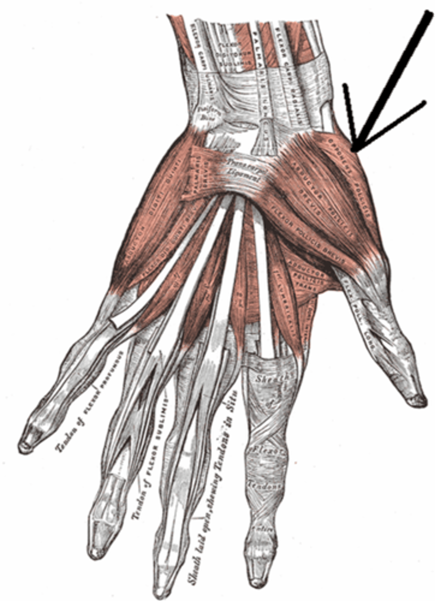
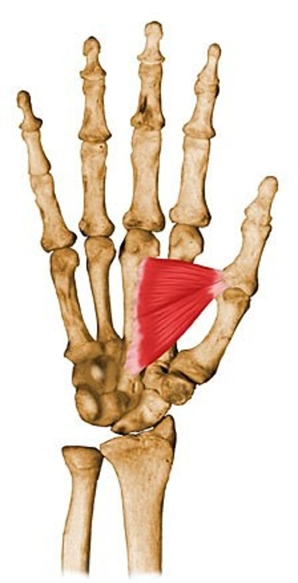
Adductor pollicis
O: oblique head: capitate bone and base of second and third metacarpal
Transverse head: palmar surface of 3rd metacarpal
I: Ulnar side of the base of the proximal phalanx of the thumb
A: adduction of thumb
N: ulnar
Abductor digiti minimi of the hand
O: Pisiform bone, and tendon of the flexor carpi ulnaris
I: base of the proximal phalanx of the little finger
A: abduction of the little finger at MCP joint
N: ulnar
Flexor digiti minimi of the hand
O: Hook of the hamate
I: base of the proximal phalanx of the little finger
A:Flexion of little finger
N: ulnar
Opponens digiti minimi
O: Transverse carpal ligament and hook of the hamate
I: shaft of the fifth metacarpal
A: Flexion of 5th metacarpal, lateral rotation producing opposition
N: ulnar
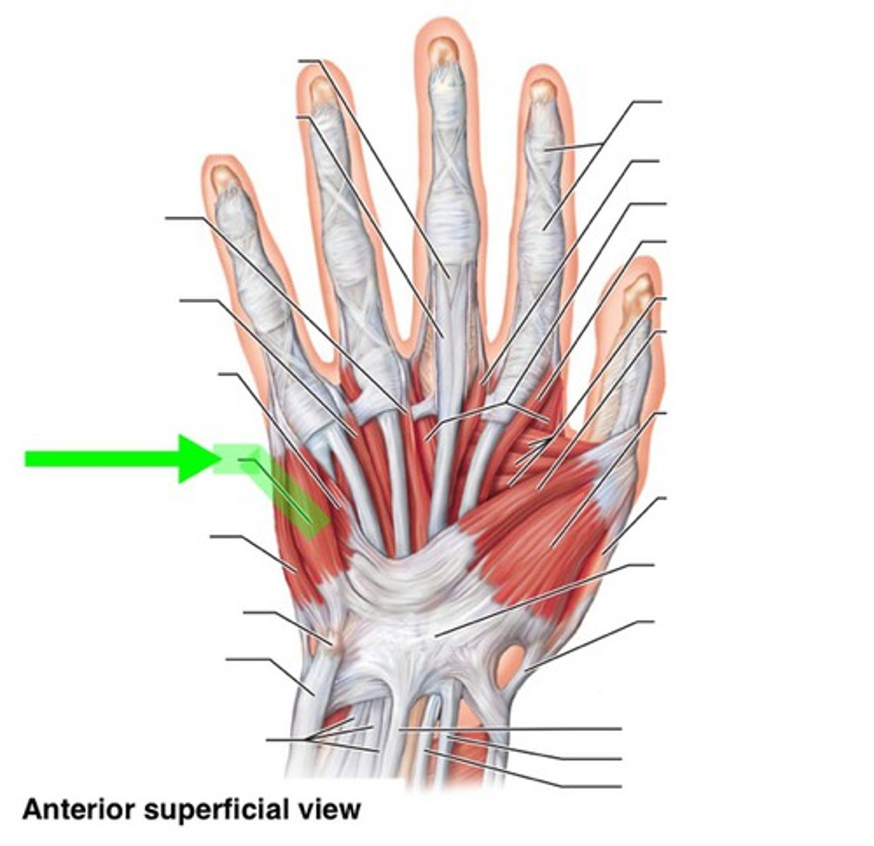
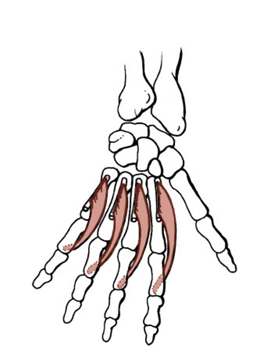
Lumbricals of the hand
O: First and second: radial surface of the flexor profundus tendons of the index and middle fingers, respectively
Third: adjacent sides of the flexor profundus tendons of the middle and ring fingers
Fourth: adjacent sides of the flexor profundus tendons of the ring and little fingers
I:Into the radial border of the extensor expansion on the dorsum of the respective digits
A: Flexion at MCP, Extension at IP
N: Medial two: ulnar nerve
Lateral two: median nerve
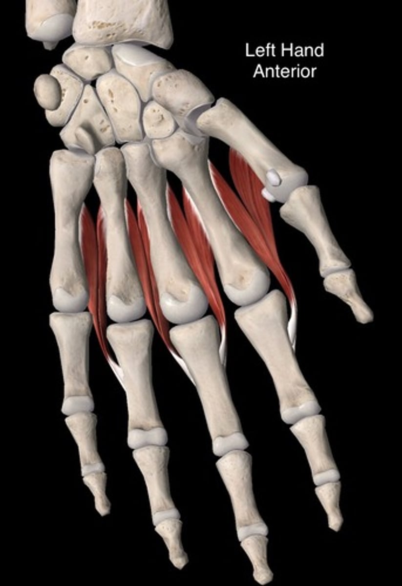
Dorsal interossei of the hand
O: First: adjacent sides of the first and second metacarpal
Second: adjacent sides of the second and third metacarpal
Third: adjacent sides of the third and fourth metacarpal
Fourth: adjacent sides of the fourth and fifth metacarpal
I: First: radial side of the index finger, chiefly to the base of the proximal phalanx
Second: radial side of the middle finger
Third: Ulnar side of the middle finger, chiefly into extensor expansion
Fourth: ulnar side of the ring finger
A: Abduction 2nd, 3rd, 4th digits away from middle finger
N: ulnar
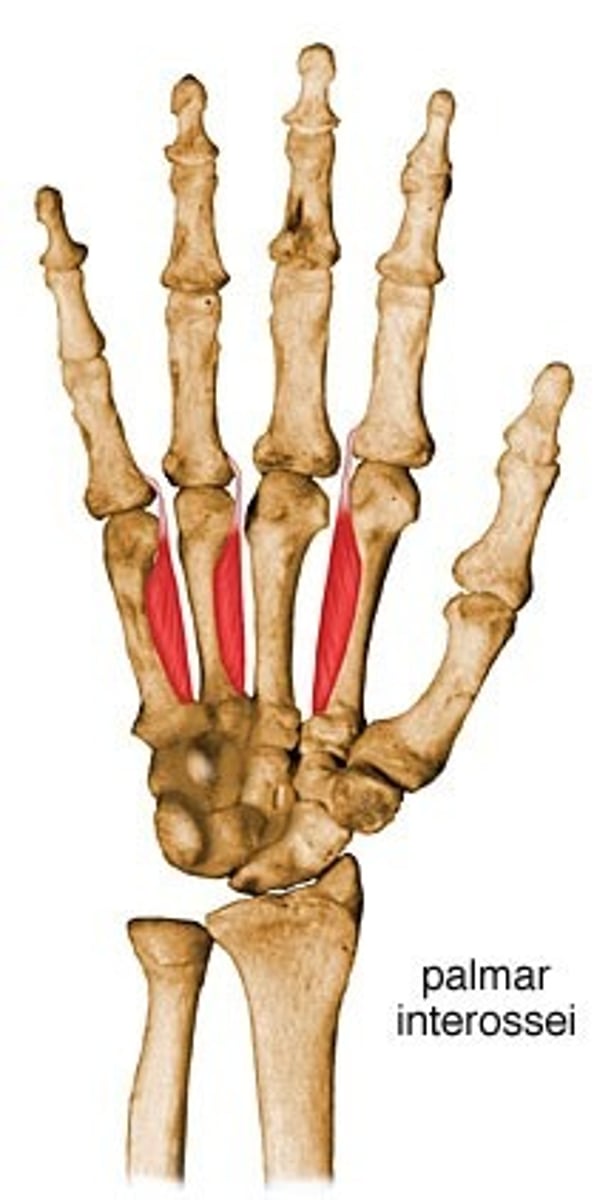
Palmar interossei
O: First: ulnar side of the thumb metacarpal
Second: ulnar side of the second metacarpal
Third: radial side of the fourth metacarpal
Fourth: radial side of the fifth metacarpal
I: First: Ulnar side of the thumb
Second: ulnar side of the index finger
Third: Radial side of the ring finger
Fourth: Radial side of the little finger
A: Adduction of 2nd, 4th, 5th digits
towards middle finger, flexion of digit
at MCP, extension at IP
N: Ulnar
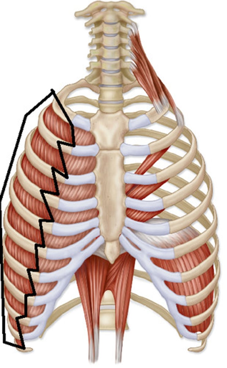
External intercostals
O: lower boarder of a rib
I: Upper border of rib below
A: elevate ribs during inspiration
N:Intercostal nerves
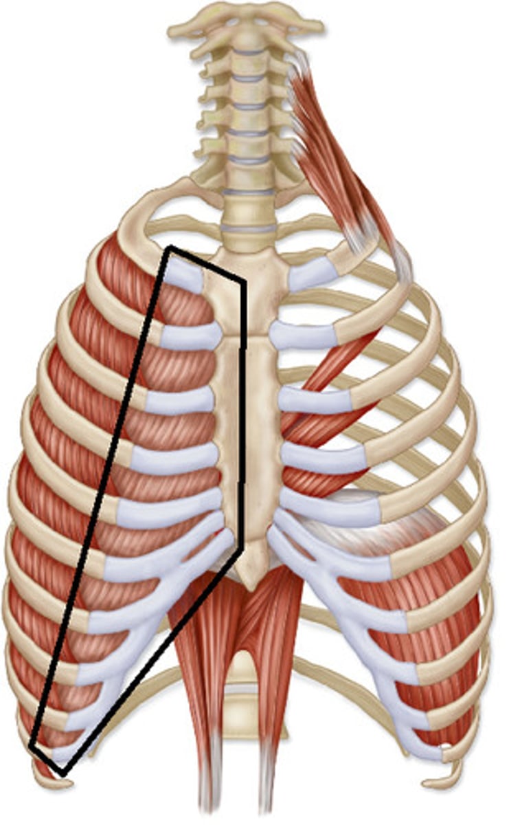
Internal intercostals
O: inner surface of a rib and costal cartilages
I: Upper boarders of the adjacent ribs below
A: draws ribs downward during forced expiration
N: Intercostal nerves
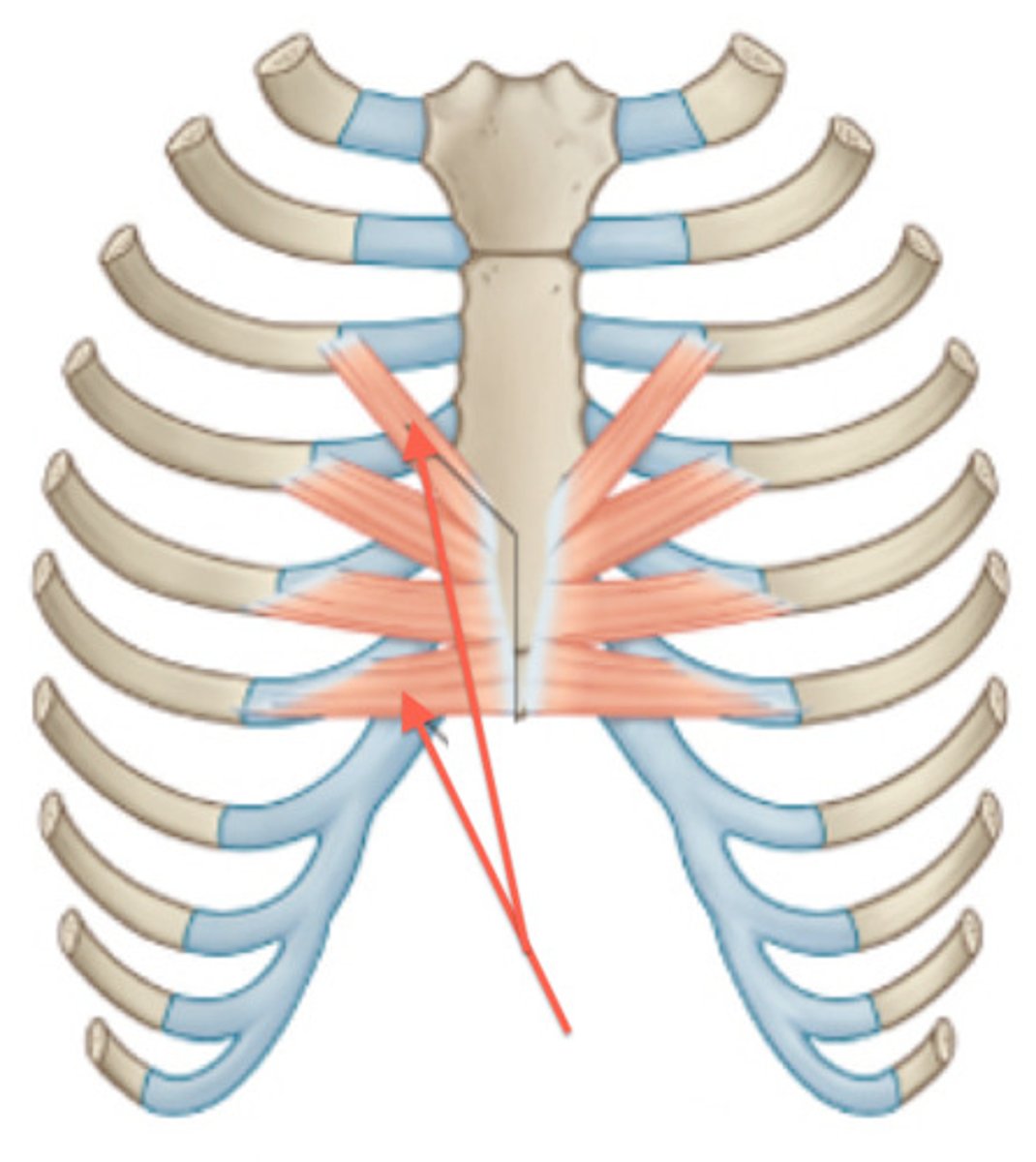
Transversus thoracis
O: inner surfaces of the lower third of the body of the sternum and adjacent surfaces of the xiphoid process
I: Internal surfaces of the sternocostal joints associated with the second (or third) through the sixth ribs
A: depresses the ribs
N: intercostal nerve
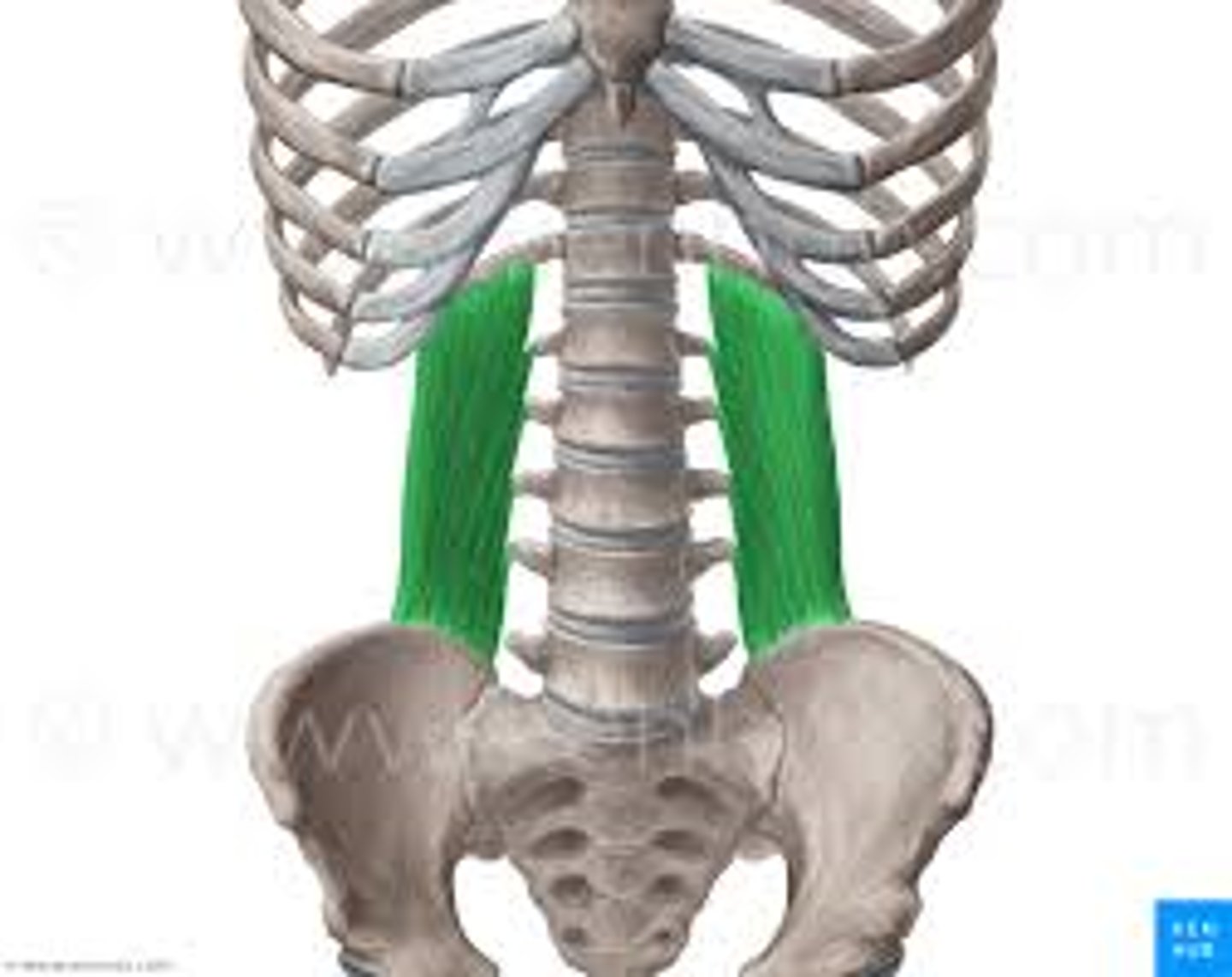
Quadratus lumborum
O: illiolumbar ligament and crest of the ilium
I: Rib 12 and tips of the transverse processes of the L1 to L4
A: Extension and side flexion of lumbar spine, expiration
N:Ventral ramus of spinal nerve roots T12-L3
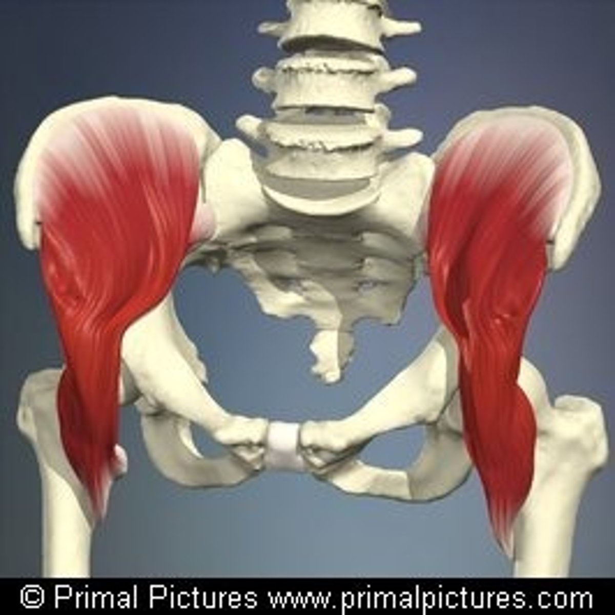
Psoas major
O: transverse processes and lateral bodies of the last thoracic and all lumbar vertebrae including the intervertebral discs
I: Lesser trochlear of femur
A: Forward flexion and side flexion of lumbar spine, FLexion of the hip joint
N: Branches of ventral rami L1-L3
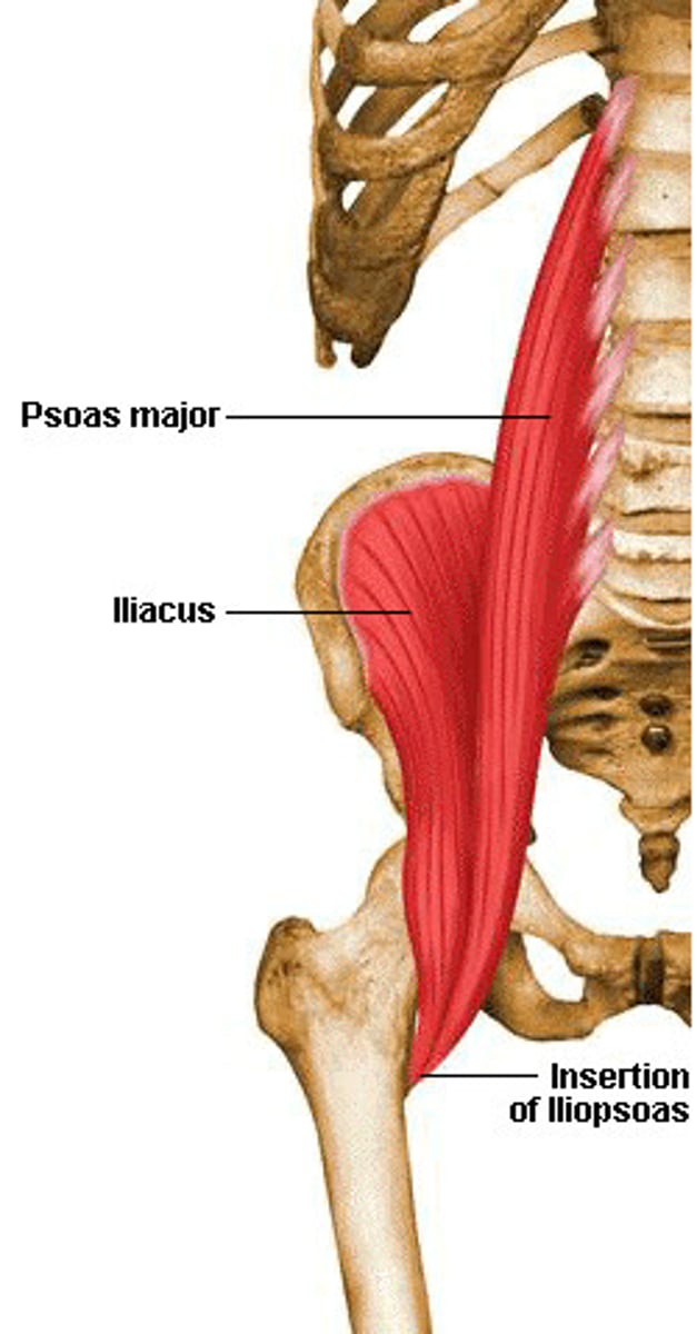
Iliacus
O: supeiror two thirds of the iliac fossa, inner lip of the iliac crest
I:Lesser trochanter of femur
A: Flexion of hip joint
N: femoral nerve
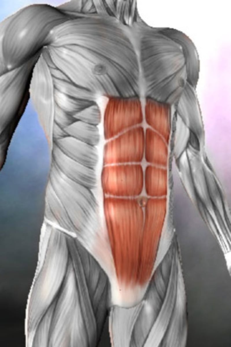
Rectus abdominis
O: Pubic crest and anterior pubic ligament
I: On the front of the wall of the thorax, along a horizontal line passing laterally from the xiphoid process, and cutting in that order, the 7th 6th, and 5th costal cartilages
A: Expiration, flexion of the spine
N: Lower 6 or 7 thoracic nerves
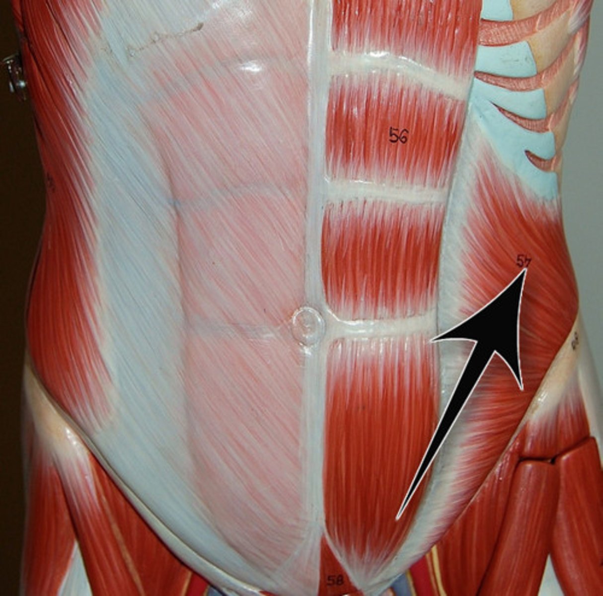
External oblique
O: Lateral side of ribs 4-12
I: Iliac crest, linea alba, and contralateral rectus sheaths
A: Bilaterally: flexion of the trunk and posterior tilt of the pelvis
Unilaterally: lateral flexion and contralateral rotation of the trunk
N: Lower 6 thoracic nerves
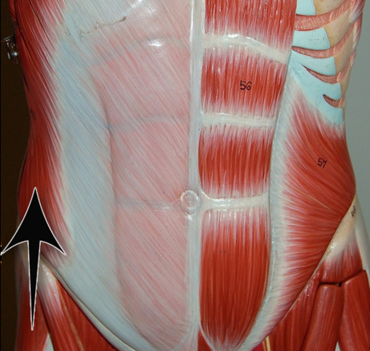
Internal oblique
O: iliac crest, inguinal ligament, and thoracolumbar fascia
I: ribs 9-12, linea alba, and contralateral rectus sheaths
A: Unilaterally: lateral flexion and ipsilateral rotation of the trunk
Bilaterally: flexion of the trunk and posterior tilt of the pelvis
N: Lower six thoracic nerves and the first lumbar nerve
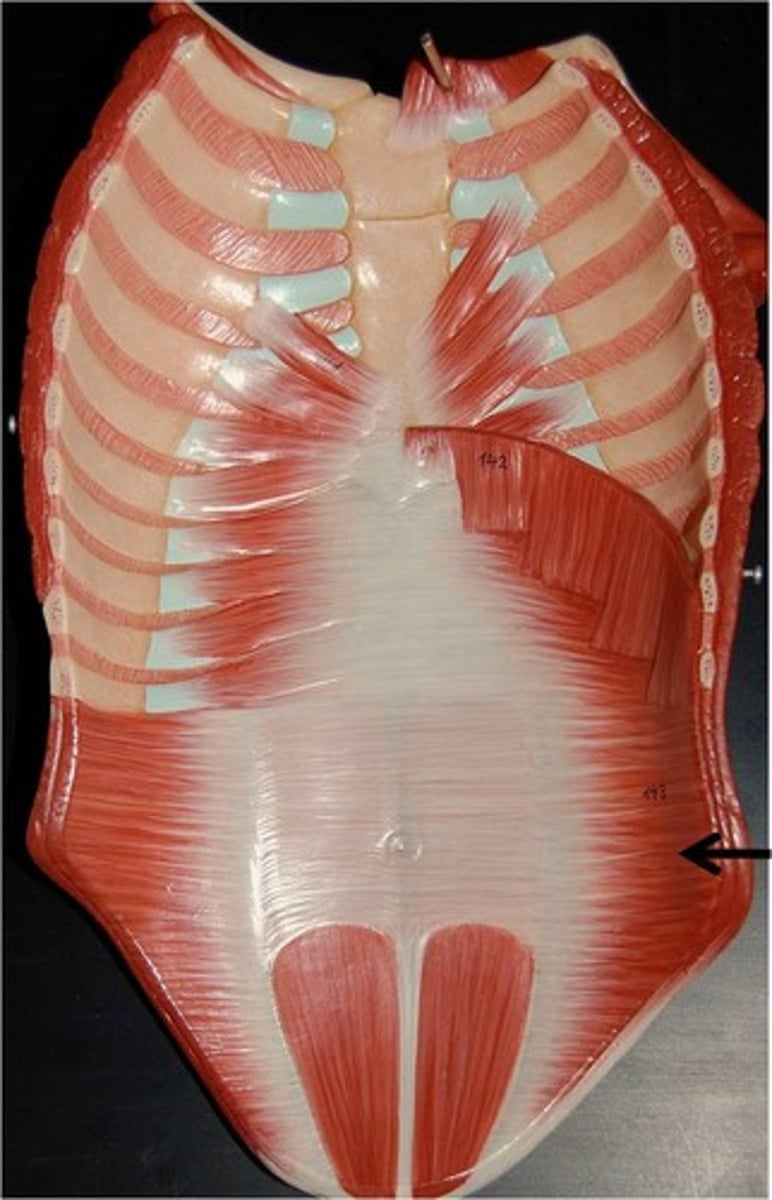
Transversus abdominis
O: Iliac crest, thoracolumbar fascia, inner surface of the cartilages of ribs 6-12 and the inguinal ligament
I: linea alba
A: Bilaterally: stabilization of attachment sites for other abdominal muscles, compression of the abdominal cavity
N: Lower six thoracic nerves and the first lumbar nerve
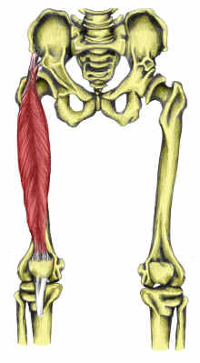
Rectus femoris
O: anterior inferior iliac spine
I: Base of the patella and, via the patellar tendon, the tibial tuberosity
A: extends knee and flexes hip
N: femoral
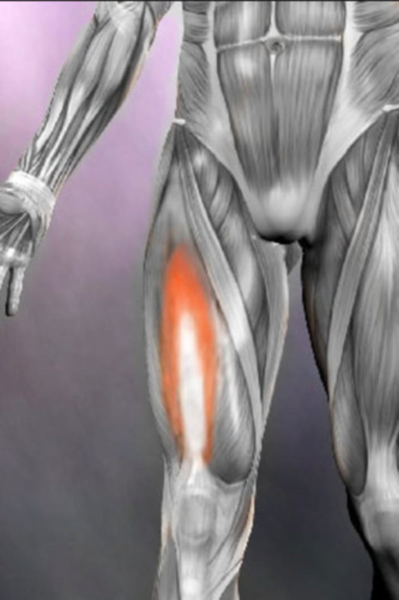
Vastus lateralis
O: upper region of intertrochanteric line, greater trochanter, lateral region of the gluteal tuberosity, lateral lip of the linea aspera
I: Lateral capsule of the knee, base of the patella, and, via the patellar tendon, the tibial tuberosity
A: extends knee
N: femoral
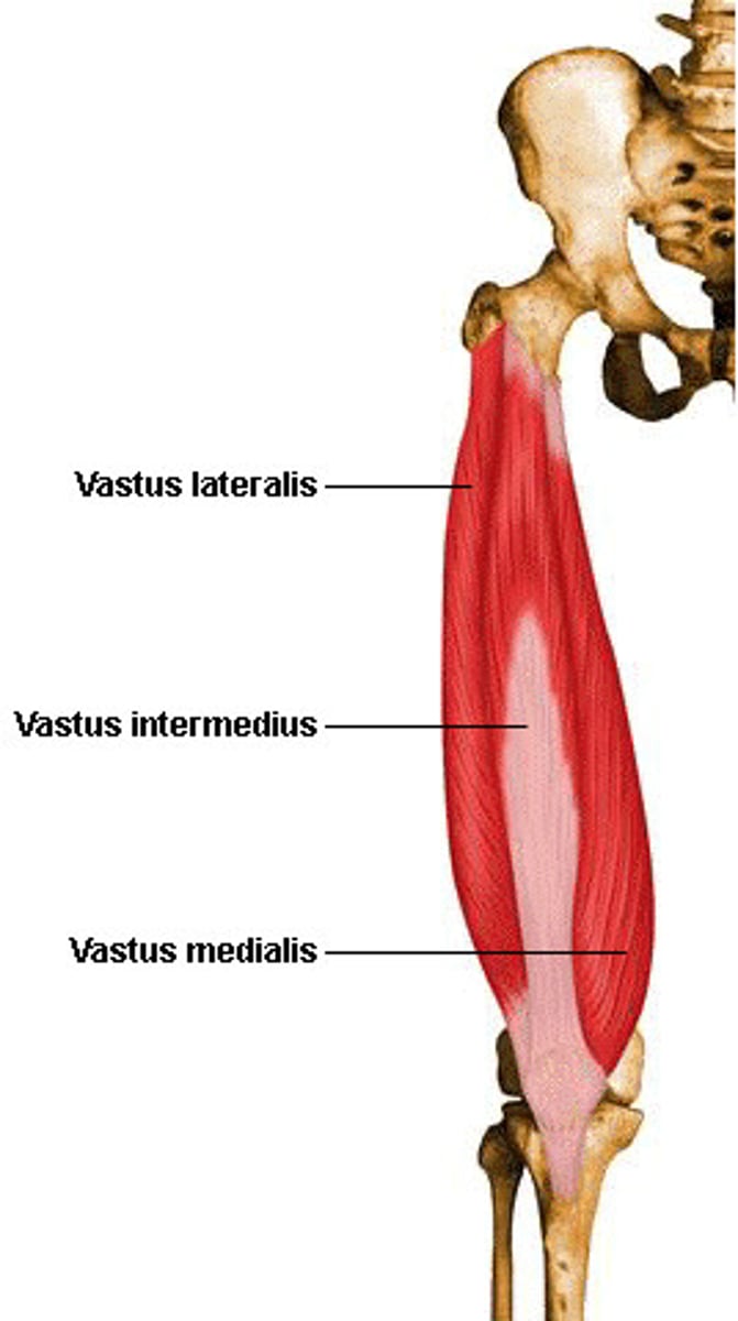
Vastus medialis
O: lower region of intertrochanteric line, medial lip of linea aspera, proximal medial supracondylar line
I: Medial capsule of the knee, base of the patella, and, via the patellar tendon, the tibial tuberosity
A: Extends knee
N: femoral
Vastus intermedius
O: Anterior-lateral regions of the upper two thirds of the femoral shaft
I: Lateral base of the patella, and, via the patellar tendon, the tibial tuberosity
A: extends knee
N: femoral
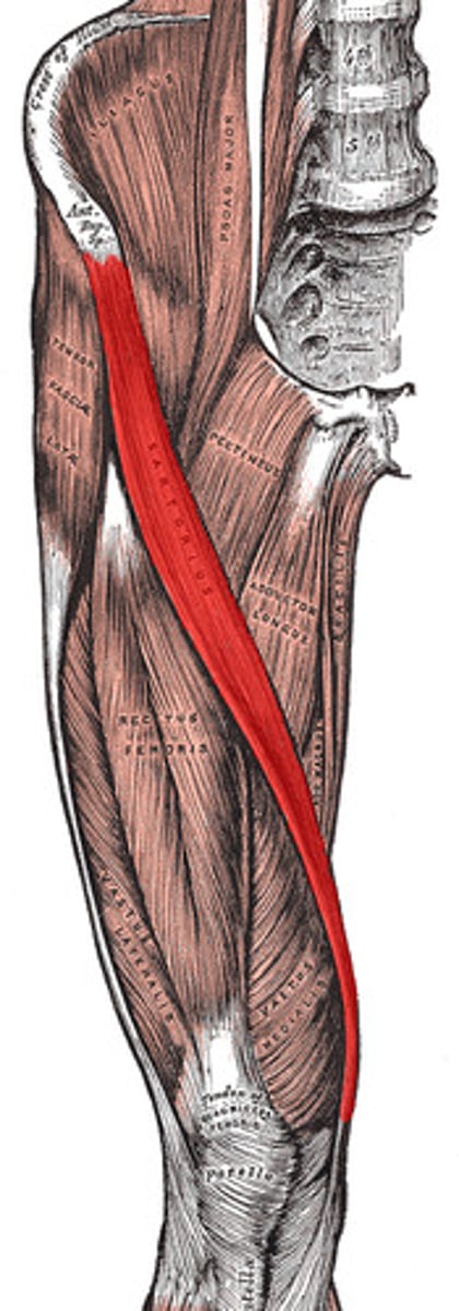
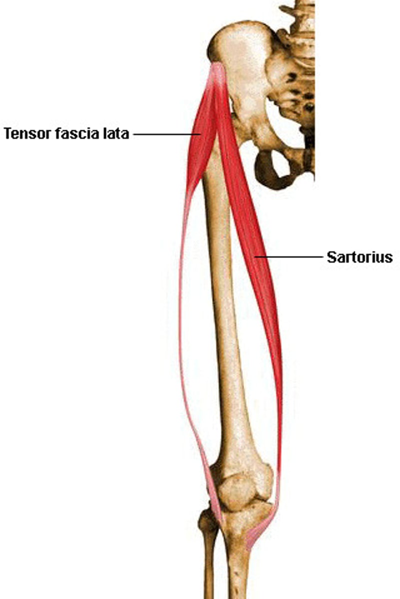
Sartorius
O: anterior superior iliac spine
I: Medial surface of tibia
A: Hip flexion, abduction, lateral rotation, knee flexion
N: femoral
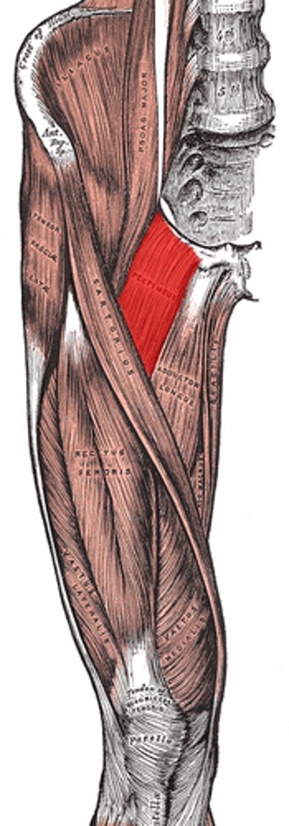
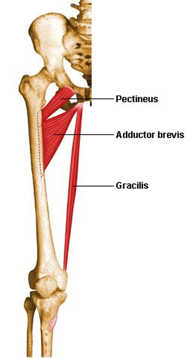
Pectineus
O: Pectineal line on superior pubic ramus
I: Pectineal line on the posteiror surface of the femur
A: Hip flexion, adduction, medial rotation
N: Femoral
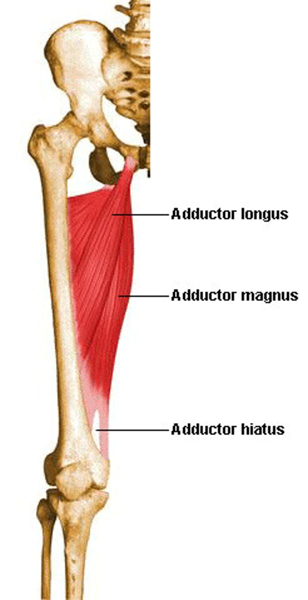
Adductor longus
O: body of pubis
I: Middle one third of linea aspera of the femur
A: Hip adduction, flexion, medial rotation
N: obturator
Adductor brevis
O: inferior pubic ramus
I: Proximal one third of the linea aspera of femur
A: Hip adduction, flexion, medial rotation
N: obturator
Adductor magnus
O: Ischial ramus and ischial tuberosity
I: linea aspera and adductor tubercle of femur
A: Hip adduction, extension and medial rotation
N: Obturator and tibial

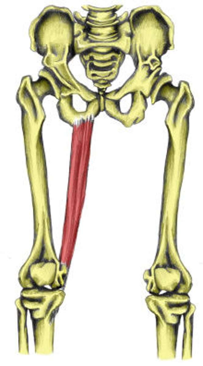
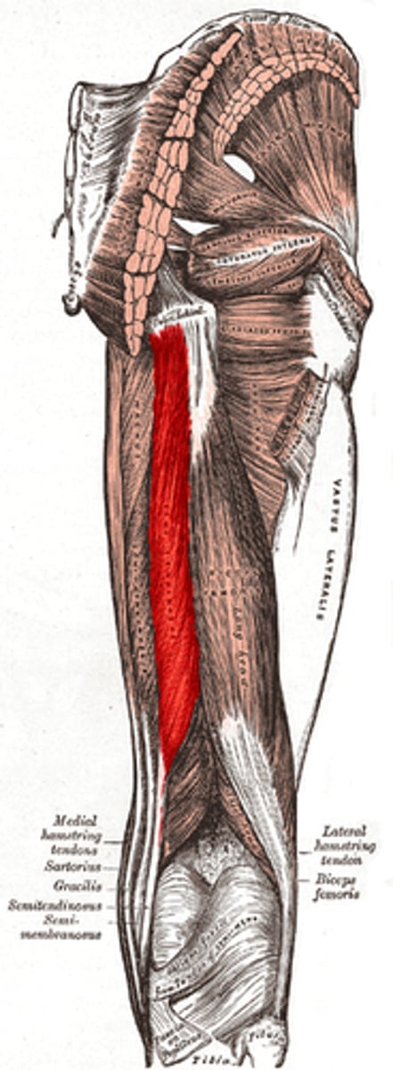
Gracilis
O: lower body of pubis and inferior ramus of pubis
I: Medial surface of tibia
A: Hip flexion, medial rotation, adduction, and flexion of knee
N: obturator
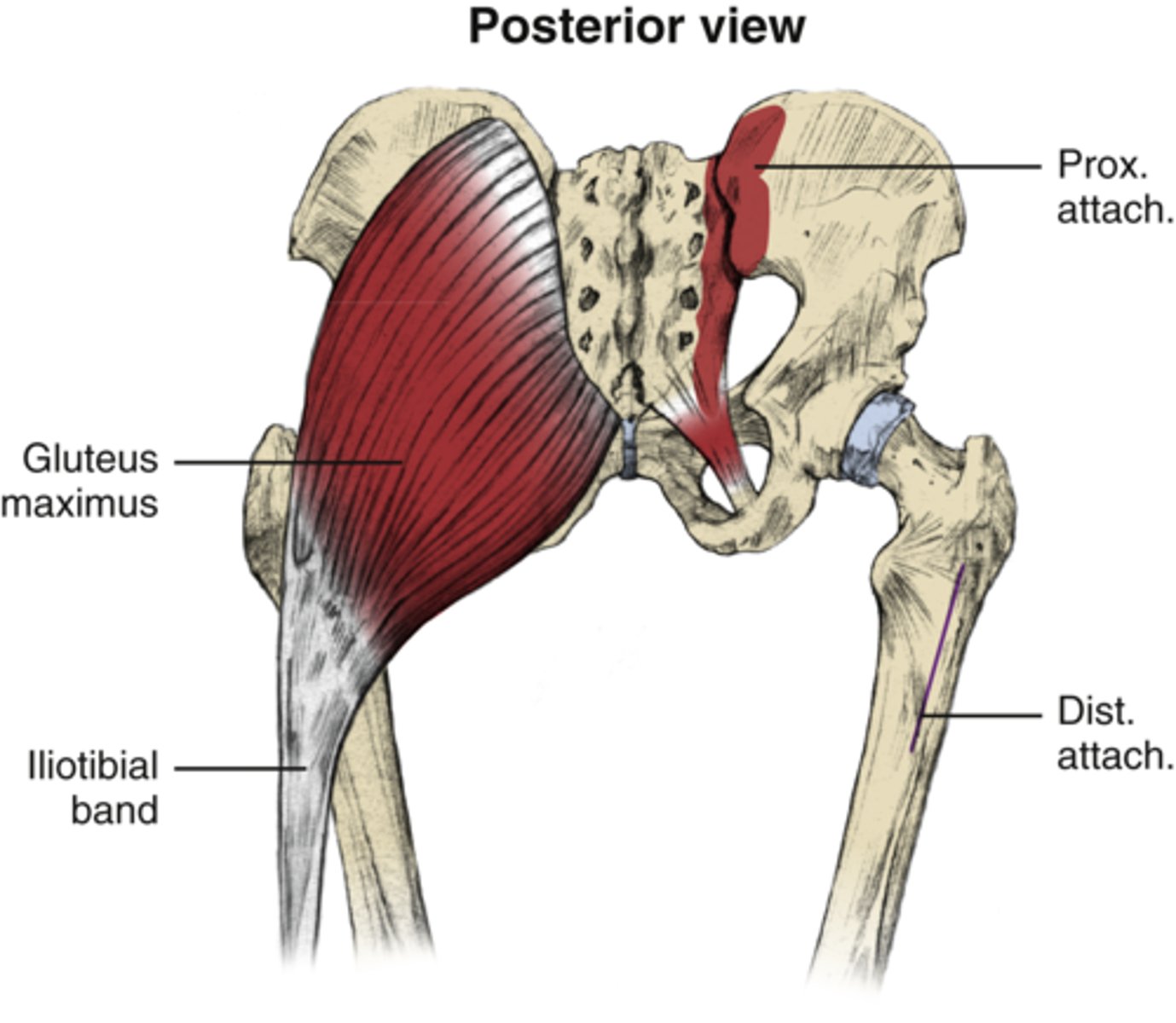
Glute max
O: Outer ilium, posterior gluteal line, thoracolumbar fascia, posterior side of sacrum and coccyx
I: Gluteal tuberosity and IT band
A: hip extension and external rotation
Secondary action: Hip adduction
N: inferior gluteal
Glute med/min
O: ilium
I: greater trochanter
A: abduction of hip, assist in medial and lateral rotation of hip
N: Superior gluteal
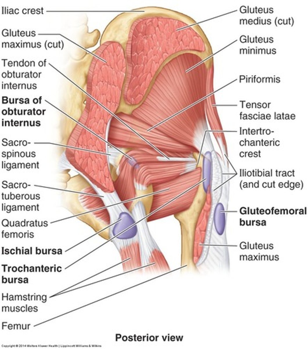
Tensor fasciae latae
O: Iliac crest and anterior superior iliac spine
I: Proximal one third of the iliotibial band of the fascia lata
A: Flex, abduct, medially rotate hip
N: Superior gluteal
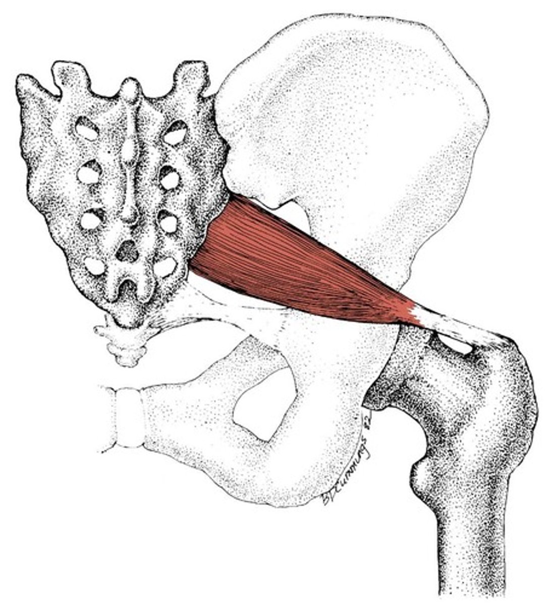
Piriformis
O: Anteiror side of sacrum
I: Apex of the greater trochanter
A: lateral rotator of hip: extension
medial rotator: flexion
N: Nerve to the piriformis
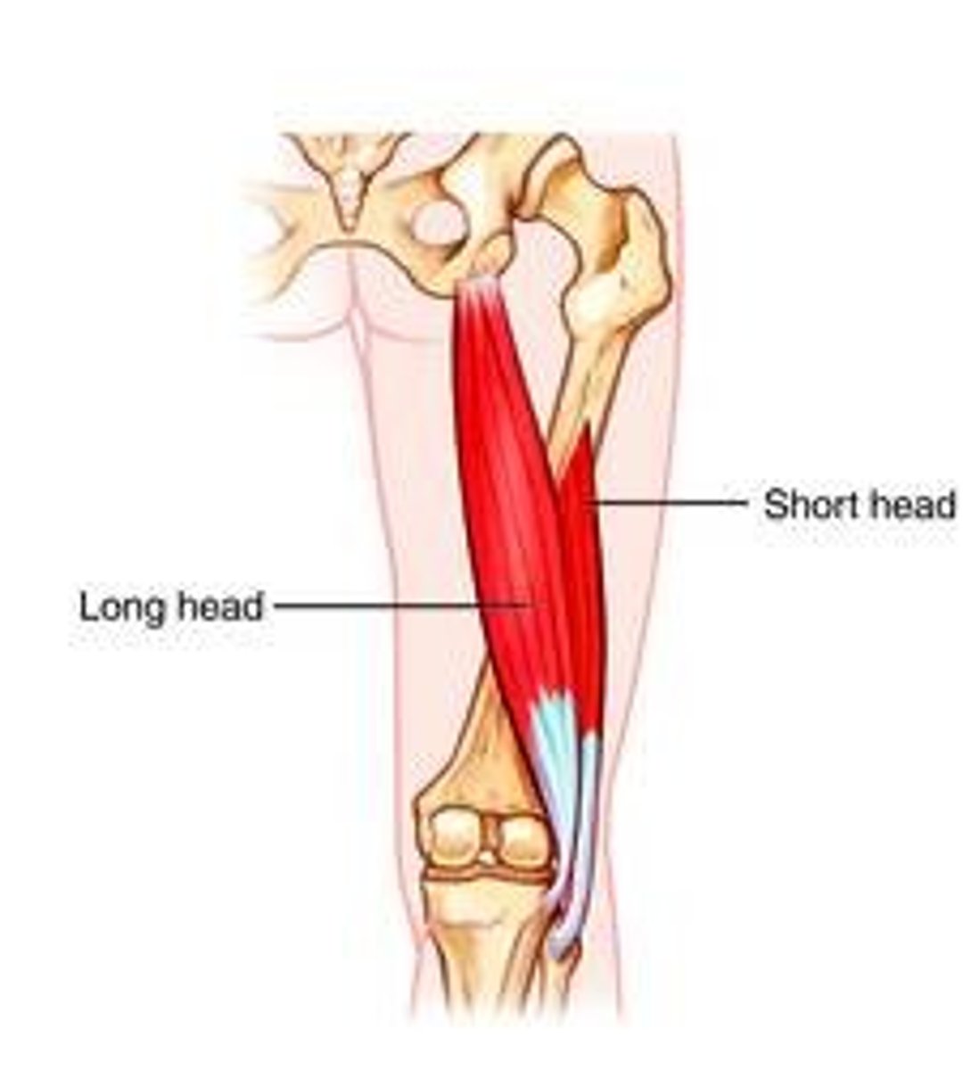
Biceps femoris
O: long head: ischial tuberosity
Short head: lateral lip of the linea aspera
I: Fibular head
A: flex knee, extend hip
N: sciatic
Semimembranosus
O: Ischial tuberosity
I: medial condyle of tibia
A: flex knee, extend hip
N: sciatic
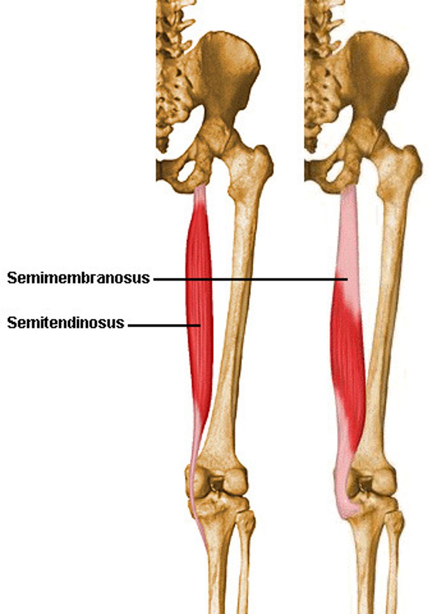
Semitendinosus
O: ischial tuberosity
I: medial surface of tibia
A: flex knee, extend hip
N: Sciatic
tib anterior
O: Lateral condyle and proximal 2/3 of lateral surface of tibia and interosseous membrane
I: Medial cuneiform and the base of the first metatarsal
A: Dorsiflexion, inversion
N: deep peroneal
Extensor digitorum longus
O: lateral condyle of tibia and medial surface of fibula
I: By four tendons that attach to the proximal base middle and distal phalanges via the dorsal digital expansion
A: Toe extension, ankle dorsiflexion, eversion
N: deep peroneal
Extensor hallicus longus
O: Medial surface of the fibula and adjacent interosseous membrane
I: Distal phalanx of great toe
A: first toe extension, ankle dorsiflexion, inversion
N: deep peroneal
Peroneus Tertius
O: Distal one third of the medial surface of the fibula and adjacent interosseous membrane
I: Base of 5th metatarsal
A: Ankle dorsiflexion, eversion
N: Deep peroneal
Peroneus longus
O: Lateral condyle of tibia, head, proximal two thirds of lateral surface of fibula
I: Lateral surface of the medial cuneiform and the lateral side of the base of first metatarsal
A: plantarflexion, eversion
N: Superficial peroneal
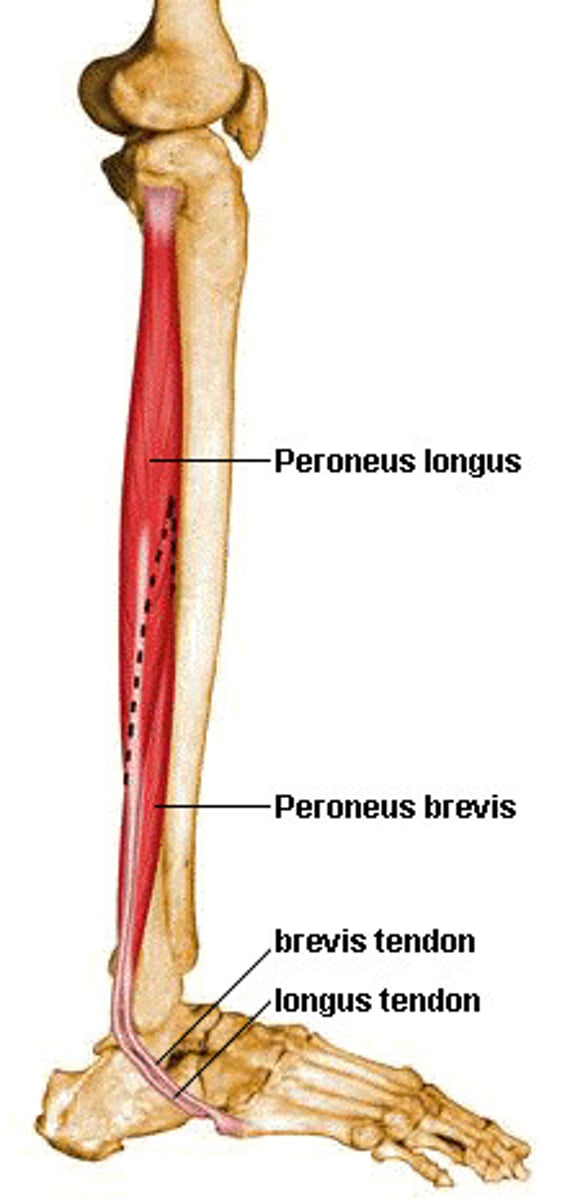
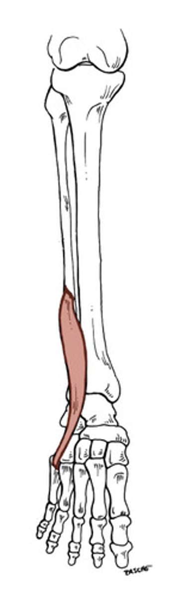
Peroneus brevis
O: lateral surface of fibula
I: styloid provess of the fifth metatarsal
A: plantarflexion, eversion
N: superficial peroneal
Gastroc
O: posterior aspect of the lateral and medial femoral condyle
I: calcaneal tuberosity via the achilles tendon
A: Plantarflexion, knee flexion
N: tibial
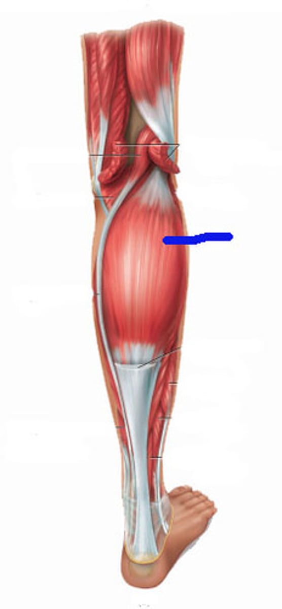
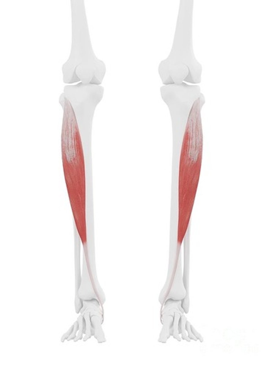
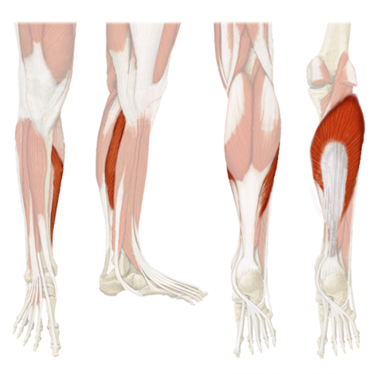
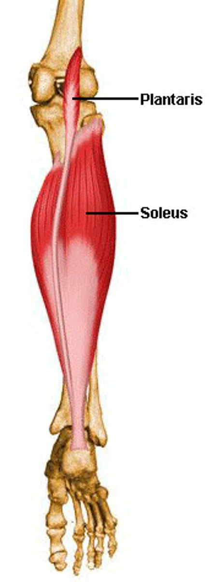
Soleus
O: Posterior surface of the fibula head and proximal 1/3 of its shaft and from the posterior side of the tibia
I: Calcaneal tuberosity via the achilles tendon
A: plantar flexion
N: tibial
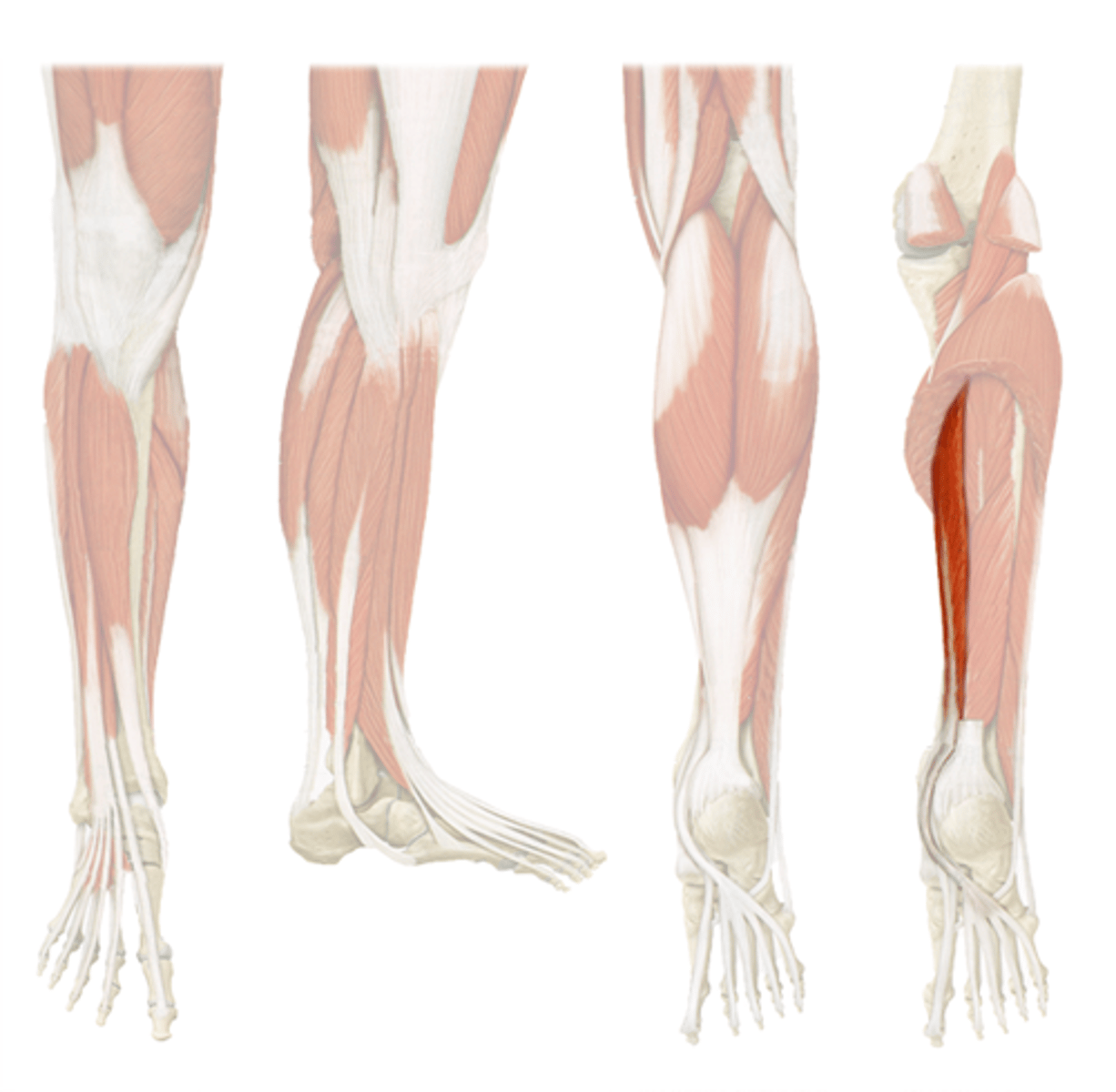
Plantaris
O: Inferior part of lateral supracondylar line of the femur
I: Joins the medial aspect of the achilles tendon to insert on the calncaneal tuberosity
A: slight knee flexion, plantarflexion
N: tibial
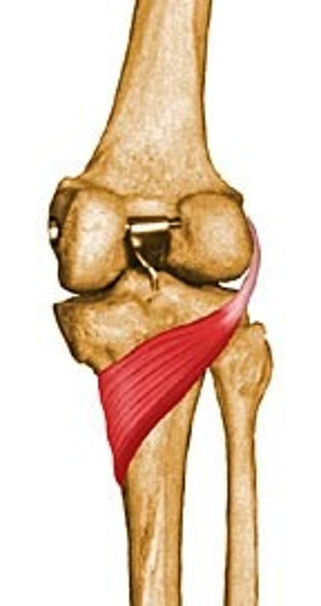
Popliteus
O: lateral condyle of femur
I: posteiror side of tibia
A: knee flexion and internal rotation
N: tibial
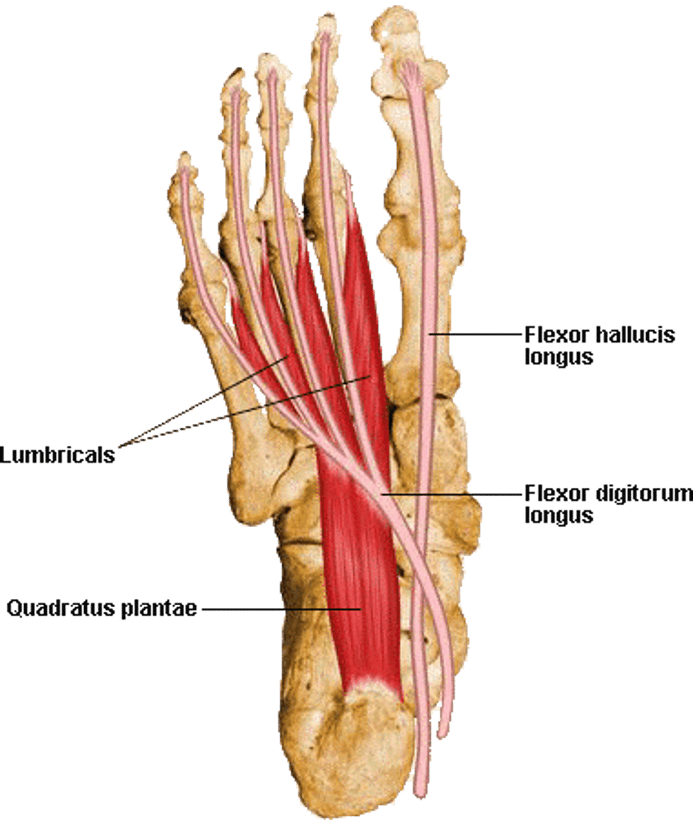
flexor digitorum longus
O: posterior surface of tibia
I: by four separate tendons to the base of the distal phalanx of the four lesser toes
A: 2-5 toes flexion, plantar flexion, inversion
N: tibial
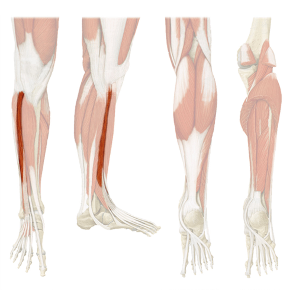
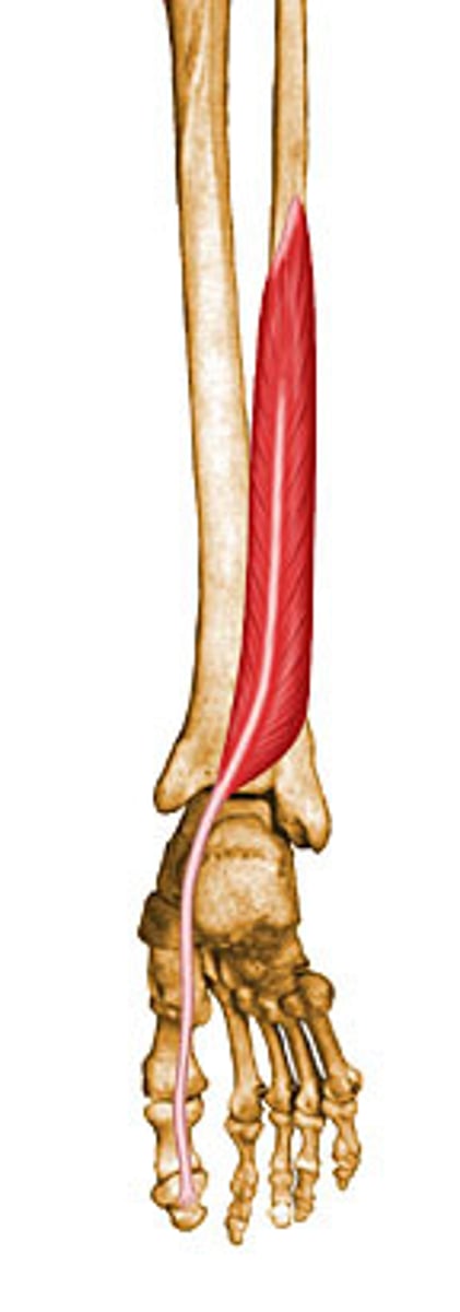
Flexor hallicus longus
O: posterior surface of fibula
I: plantar surface of the base of the distal phalanx of the great toe
A: Flexion of great toe, plantarflexion, inversion
N: tibial
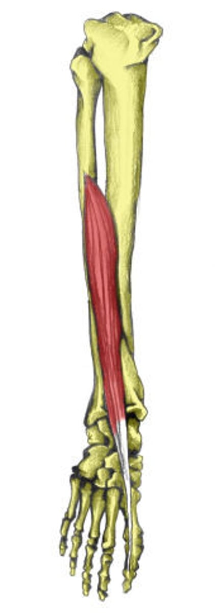
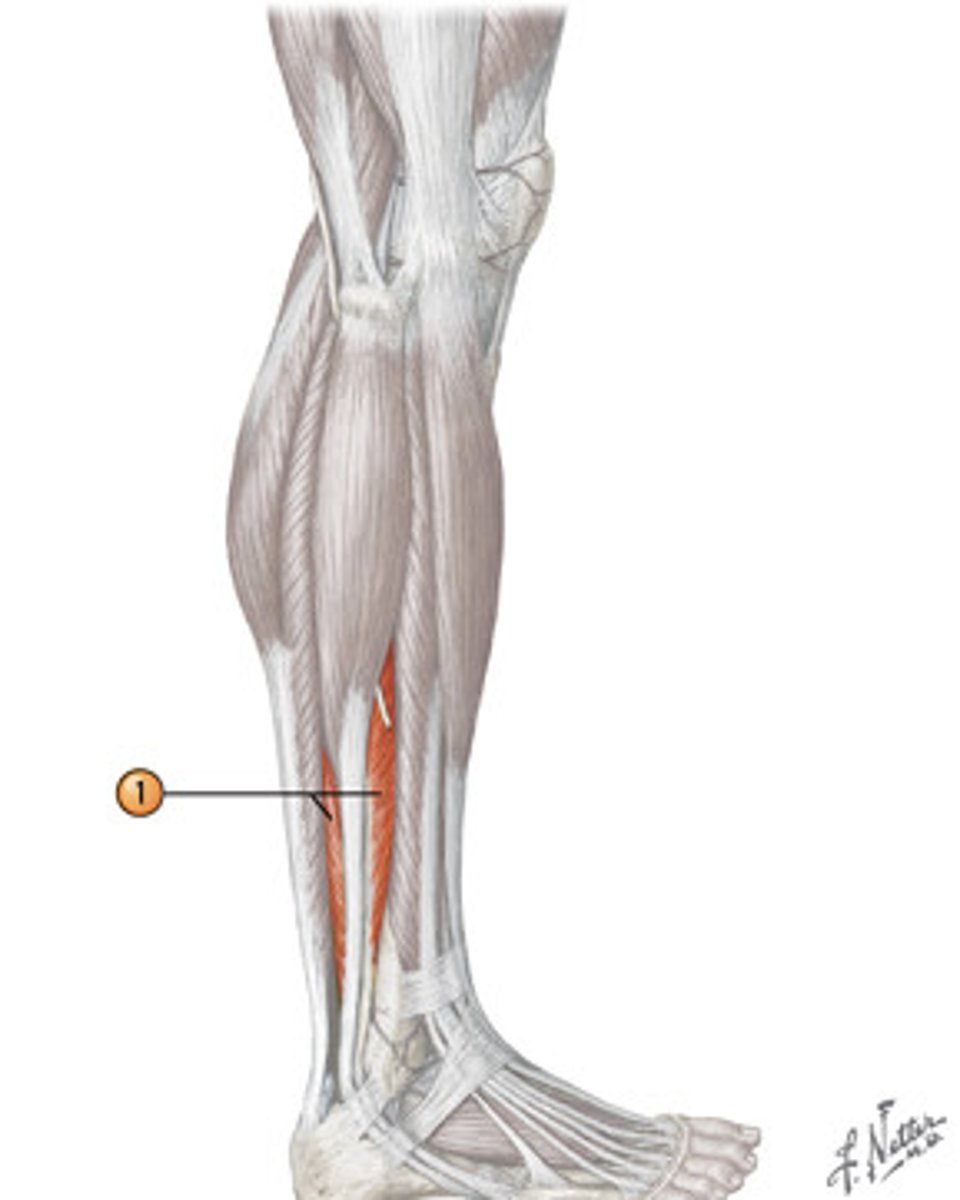
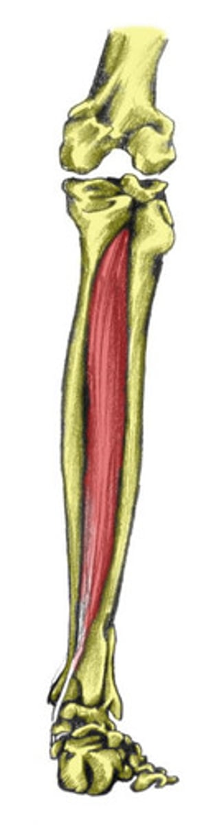
Tibialis posterior
O: posterior surface of the tibia and fibula and adjacent interosseous membrane
I: tuberosity of navicular and all tarsals except talus
A: plantarflexion, inversion
N: tibial
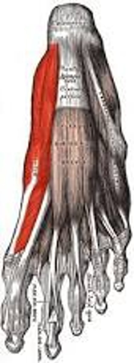
Abductor hallicus
1st layer
A: abduct the big toe
N: medial plantar
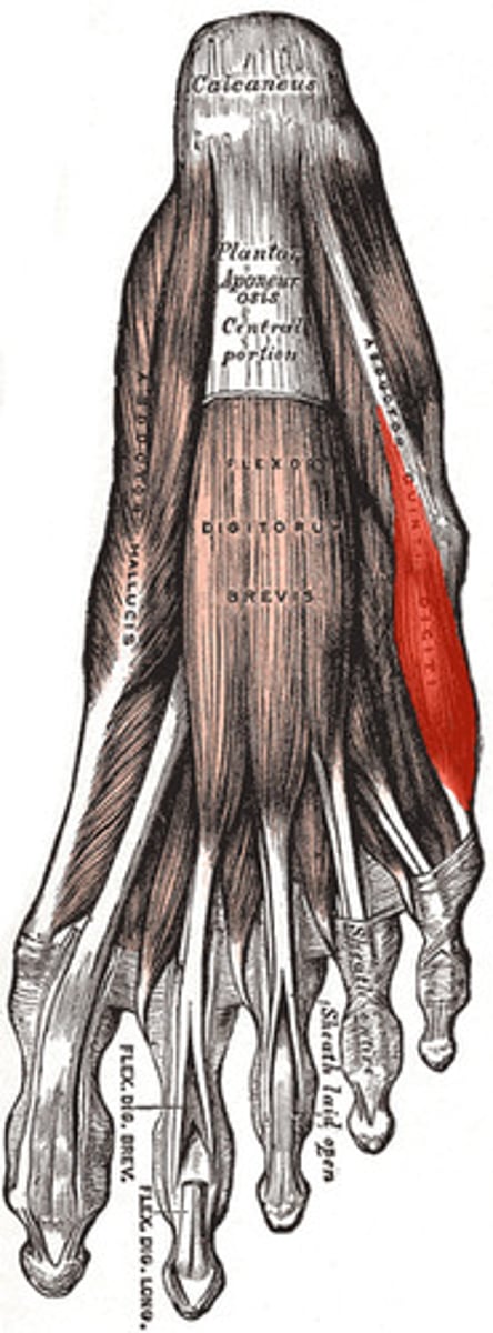
Flexor digitorum breves
1st layer
A: 2-4th toe flexion
N: medial plantar
Abductor digiti minimi
1st layer
A: 5th toe abduction and flexion
N: lateral plantar
Quadratus plantae
2nd layer
A: flexion of toes
N: lateral plantar

Lumbricals
2nd layer
A: MTP flexion, IP extension
N: toe 2: medial plantar
toe 3-5 lateral plantar
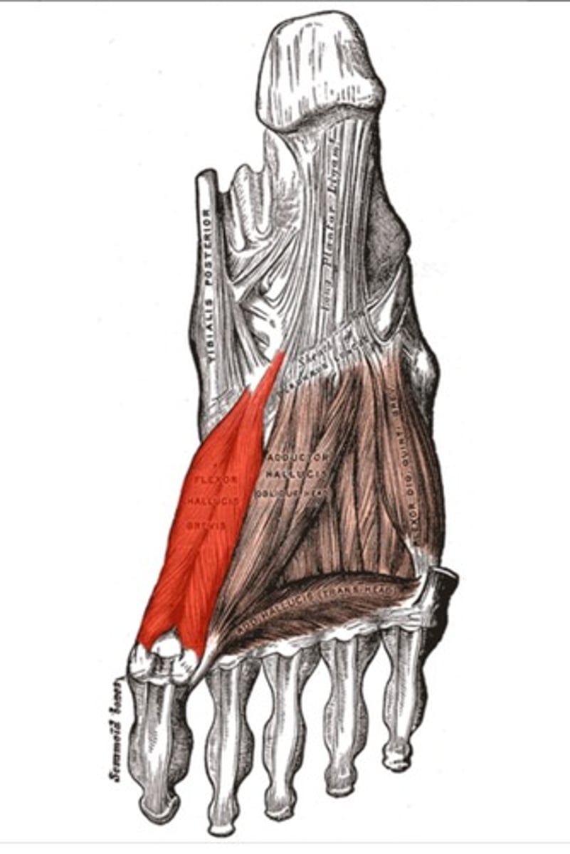
Flexor hallicus brevis
3rd layer
A: flexes big toe
N: medial plantar
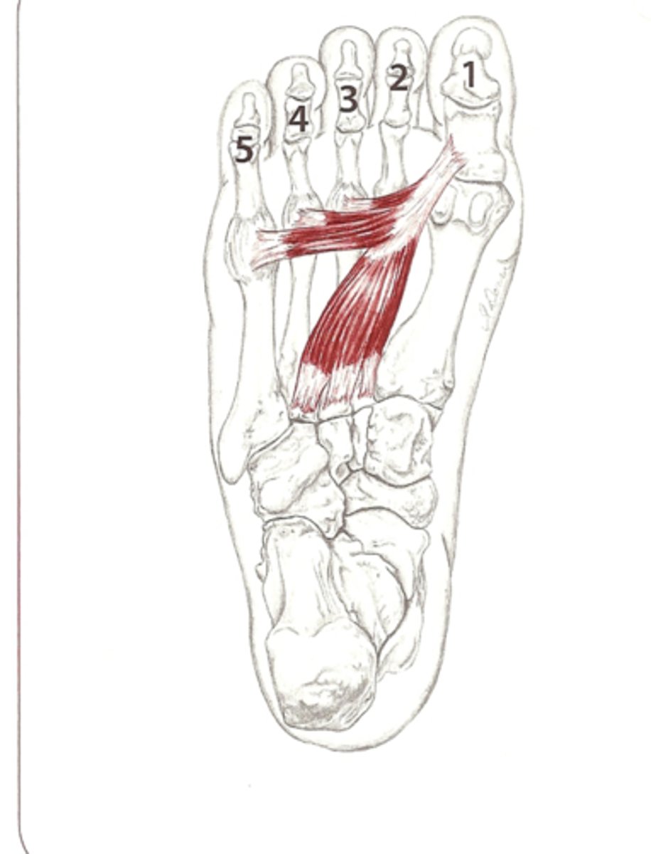
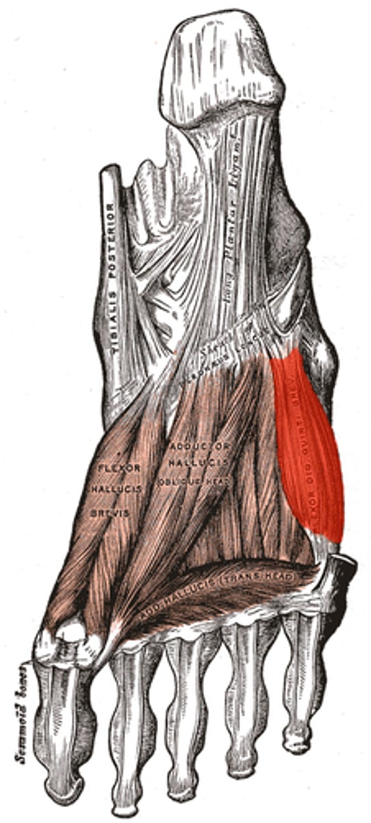
Adductor hallicus
3rd layer
A: adducts big toe
N: lateral plantar
Flexor digiti minimi
3rd layer
A: flexion of little toe
N: lateral plantar
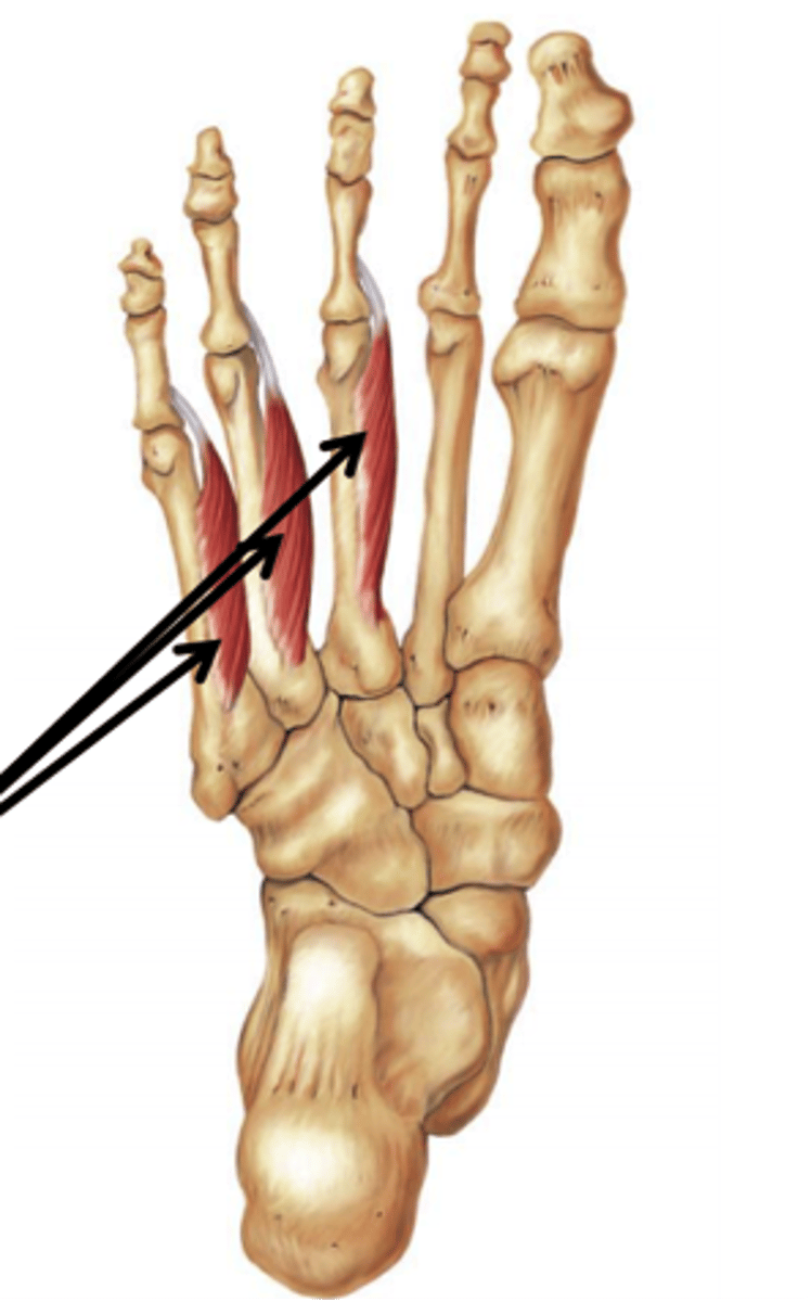
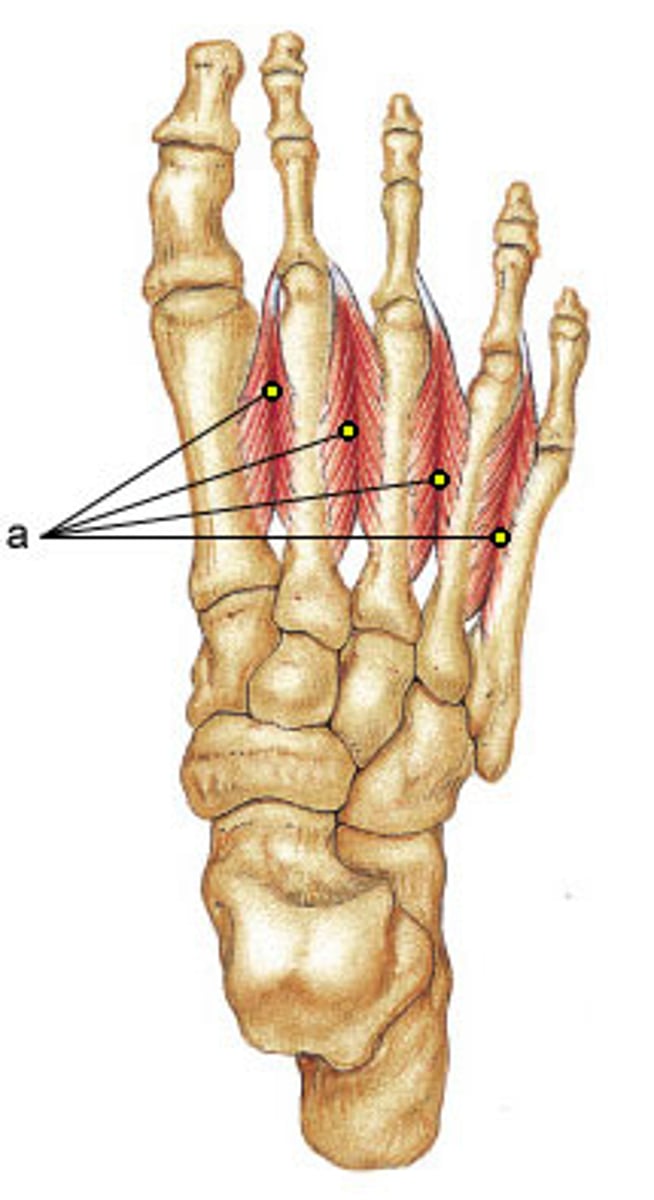
Dorsal interossei
4th layer
A: Abduction of 2-4 toes, flexion at MTP joint
N: lateral plantar
Plantar interossei
4th layer
A: adduction of 3-5 toes, flexion at MTP joints
N: lateral plantar