chapter 13
1/33
There's no tags or description
Looks like no tags are added yet.
Name | Mastery | Learn | Test | Matching | Spaced | Call with Kai |
|---|
No study sessions yet.
34 Terms
Overview of the Nervous System
Location & Origin
The nervous system is dorsal in position (toward the back of the body).
It is hollow inside — containing ventricles (in the brain) and a central canal (in the spinal cord).
It is ectodermal in origin, meaning it develops from the ectoderm, the outermost embryonic layer.
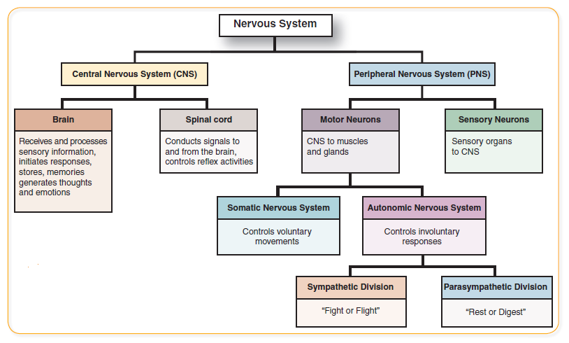
Main Divisions
System | Abbreviation | Major Components | Function |
Central Nervous System (CNS) | CNS | Brain and spinal cord | Processes and integrates information |
Peripheral Nervous System (PNS) | PNS | Cranial nerves and spinal nerves | Sends information to and from the CNS |
Afferent (Sensory) Pathway
Carries input toward the CNS.
Somatic Sensory – sensations from the body wall and limbs (e.g., touch, pain, temperature, pressure).
Visceral Sensory – sensations from internal organs (e.g., GI tract, blood vessels).
Efferent (Motor) Pathway
Carries output from the CNS to effectors.
Somatic Motor – voluntary control of skeletal muscles.
Visceral Motor (Autonomic) – involuntary control of smooth muscles, cardiac muscles, and glands.
Autonomic Nervous System (ANS)
Division | Function | State of Activity |
Sympathetic Nervous System | “Fight or Flight” – increases heart rate, blood flow, respiration | Active in stress |
Parasympathetic Nervous System | “Rest and Digest” – slows heart rate, increases digestion | Active in rest |
2. Embryonic Development of the Nervous System
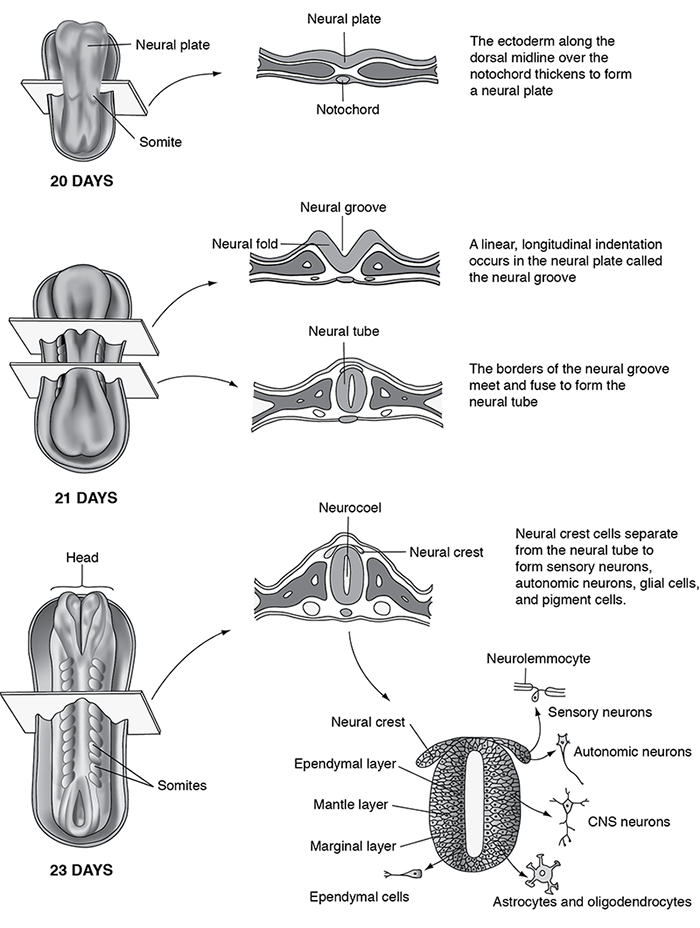
Formation Process
Neural Plate appears from ectoderm on the dorsal surface of the embryo (about week 3).
The plate folds to form Neural Folds and a Neural Groove.
The folds fuse in the midline forming the Neural Tube, which later becomes the CNS.
The hollow inside becomes the central canal (spinal cord) and ventricles (brain).
Notochord (below the neural tube) will guide the development of vertebral bones and spinal alignment.
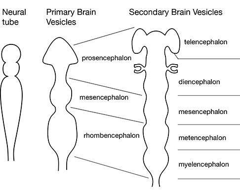
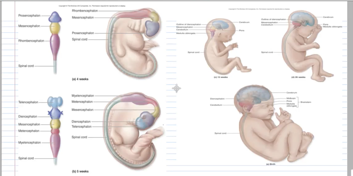
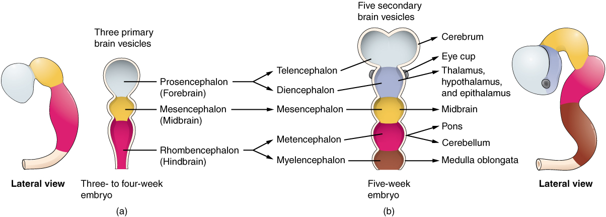
Brain Development: Vesicles and Flexures
Primary Brain Vesicles (3 weeks)
Prosencephalon → forebrain
Mesencephalon → midbrain
Rhombencephalon → hindbrain
Secondary Brain Vesicles (5 weeks)
Primary Vesicle | Secondary Vesicles | Adult Brain Structure |
Prosencephalon | Telencephalon and Diencephalon | Cerebral hemispheres, thalamus, hypothalamus |
Mesencephalon | (unchanged) | Midbrain |
Rhombencephalon | Metencephalon and Myelencephalon | Pons, cerebellum, medulla oblongata |
Major Brain Flexures
Cephalic flexure – dorsal bend between forebrain & midbrain.
Cervical flexure – dorsal bend between hindbrain & spinal cord.
Pontine flexure – ventral bend between midbrain & hindbrain.
[Embryo at 3 weeks showing brain flexures]
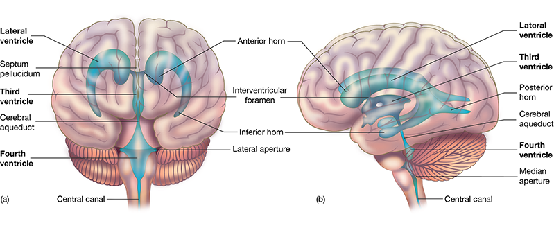
4. Ventricles and Cerebrospinal Fluid (CSF)
[Insert Diagram: Lateral, third, and fourth ventricles with CSF flow arrows]
Ventricles are cavities within the brain derived from the neural canal.
Lined by ependymal cells (glial cells that help circulate CSF).
CSF Production
Produced by choroid plexus (network of blood capillaries within ventricles).
Volume: ~150 mL in adults.
Functions:
Cushions and protects the brain and spinal cord.
Maintains stable chemical environment.
Provides buoyancy (reduces brain weight).
Removes waste.
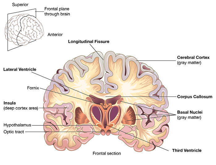
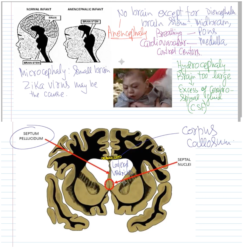
Septum pellucidum & corpus callosum
Septum pellucidum – thin membrane that separates the right and left lateral ventricles.
Corpus callosum – large commissural fiber tract connecting right and left cerebral hemispheres.
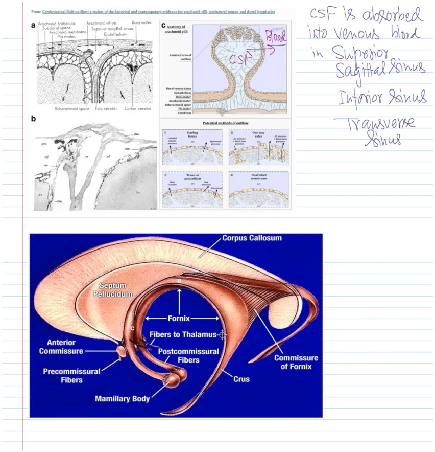
Pathway of CSF Flow
Lateral ventricles →
Interventricular foramina (of Monro) →
Third ventricle →
Cerebral aqueduct (of Sylvius) →
Fourth ventricle →
Median aperture (foramen of Magendie) + 2 lateral apertures (foramina of Luschka) →
Subarachnoid space →
Reabsorbed into venous blood via arachnoid villi in the superior sagittal sinus.
[Insert Diagram: CSF circulation and absorption through arachnoid villi]
![<ol type="1"><li><p><span><span>Lateral ventricles →</span></span></p></li></ol><ol type="1"><li><p><span><strong><span>Interventricular foramina (of Monro) →</span></strong></span></p></li><li><p><span><strong><span>Third ventricle →</span></strong></span></p></li><li><p><span><strong><span>Cerebral aqueduct (of Sylvius) →</span></strong></span></p></li><li><p><span><strong><span>Fourth ventricle →</span></strong></span></p></li><li><p><span><strong><span>Median aperture (foramen of Magendie) + 2 lateral apertures (foramina of Luschka) →</span></strong></span></p></li><li><p><span><strong><span>Subarachnoid space →</span></strong></span></p></li></ol><ol type="1"><li><p><span><span>Reabsorbed into </span><strong><span>venous blood</span></strong><span> via </span><strong><span>arachnoid villi</span></strong><span> in the </span><strong><span>superior sagittal sinus</span></strong><span>.</span></span></p></li></ol><p><span><strong><span>[Insert Diagram: CSF circulation and absorption through arachnoid villi]</span></strong></span></p>](https://knowt-user-attachments.s3.amazonaws.com/6b4ba189-26b4-4eab-83e3-f826ea541de7.png)
5. Meninges - layers
Layers
Dura mater – tough, fibrous outer layer.
Periosteal layer (attached to skull).
Meningeal layer (around brain & spinal cord).
Arachnoid mater – middle layer; web-like network connecting to blood vessels.
Pia mater – delicate inner layer adhering tightly to brain surface; rich in capillaries.
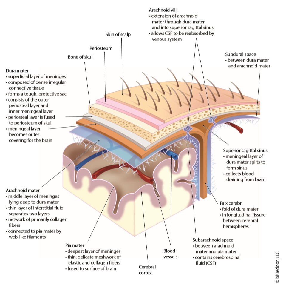
5. Meninges - spaces
Epidural space – outside dura; used for anesthesia.
Subdural space – between dura and arachnoid (potential space).
Subarachnoid space – between arachnoid and pia; contains CSF.

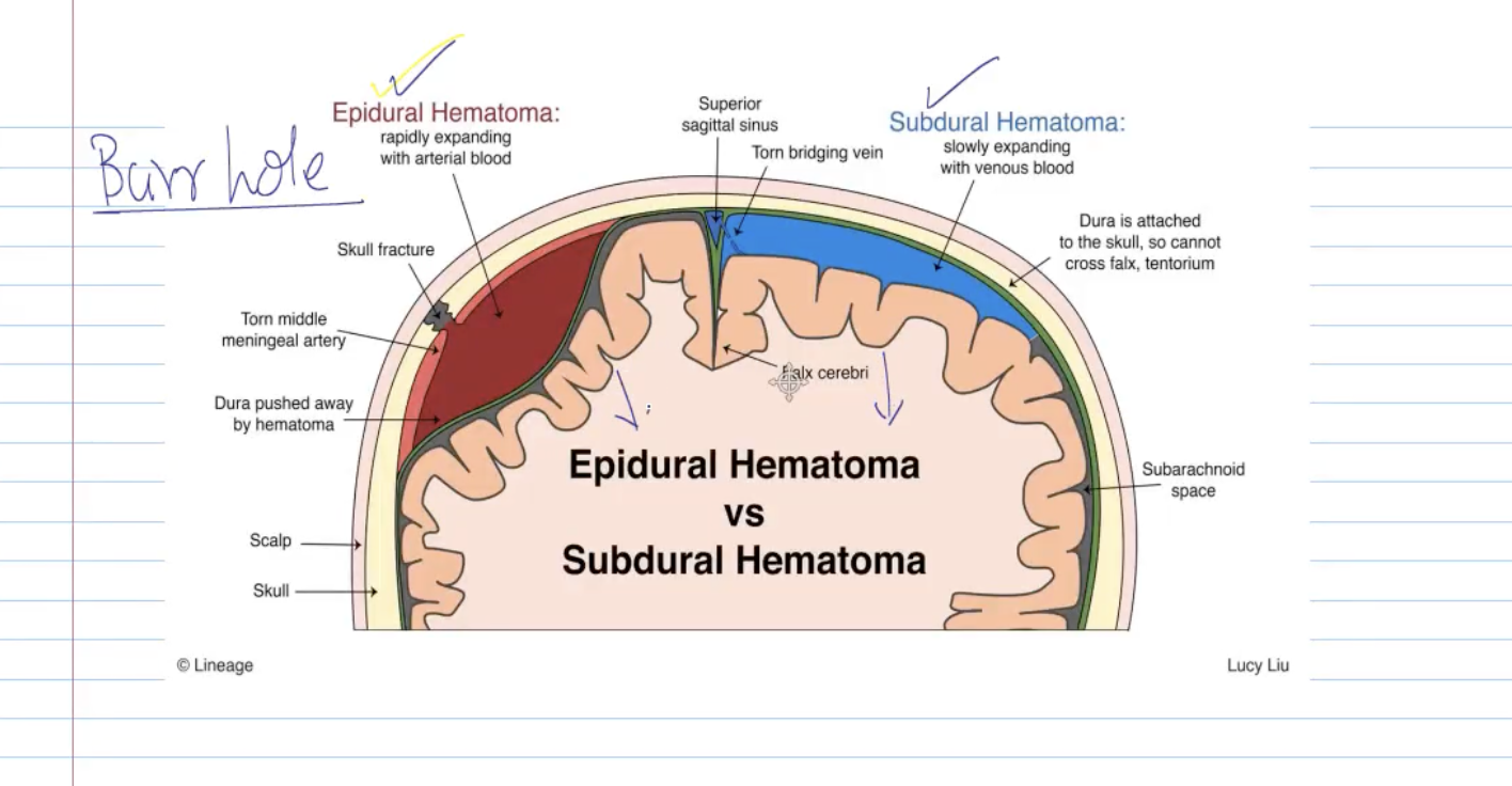
5. Meninges - clinical notes
Epidural hematoma – arterial bleeding between skull and dura.
Subdural hematoma – venous bleeding between dura and arachnoid.
Lumbar tap (spinal tap): 10–15 mL of CSF withdrawn from lumbar region to test for meningitis, infection, or pressure.
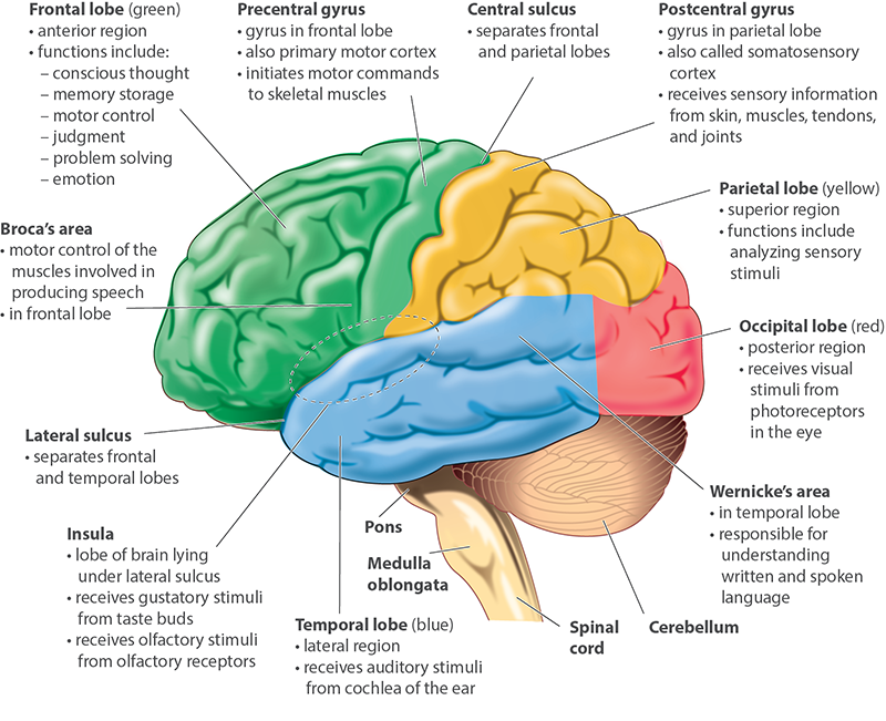
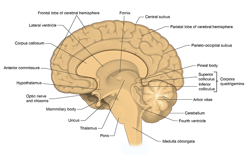
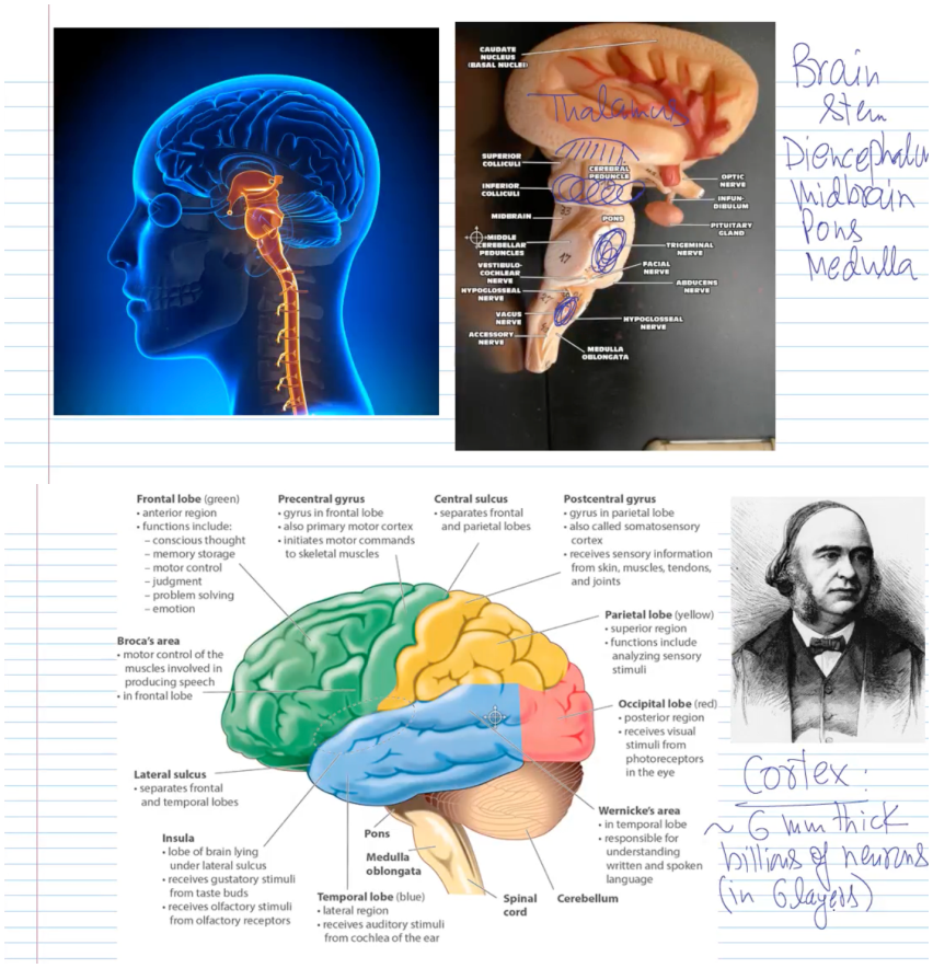
6. Brain Lobes and Fissures
Frontal lobe – planning, reasoning, movement, speech.
Parietal lobe – sensory perception (touch, pressure, pain, temp).
Temporal lobe – hearing, smell, language.
Occipital lobe – vision.
Insula – deep within lateral sulcus; taste, emotional context.
Cerebellum – coordination and balance.
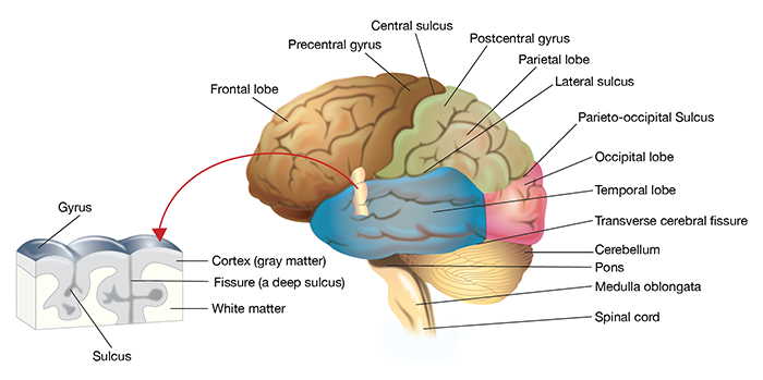
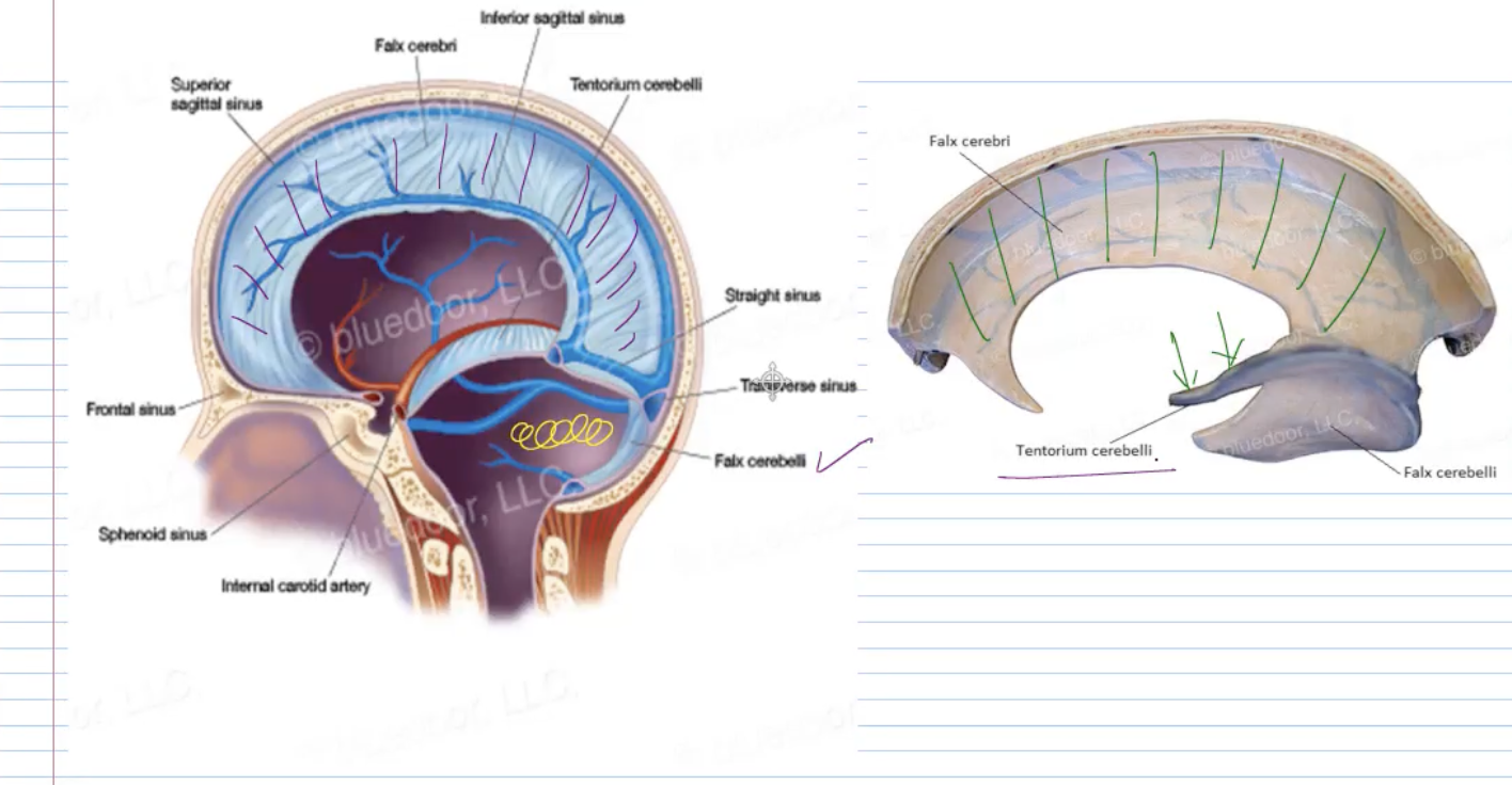
Major Fissures
Longitudinal fissure: separates right & left cerebral hemispheres.
Transverse fissure: separates cerebrum and cerebellum.
Central sulcus: separates frontal and parietal lobes.
Lateral sulcus (Sylvian): separates temporal lobe.
Parieto-occipital sulcus: separates parietal and occipital lobes.
[Insert Diagram: Falx cerebri and tentorium cerebelli positions]
![<ul><li><p><span><strong><span>Longitudinal fissure:</span></strong><span> separates right & left cerebral hemispheres.</span></span></p></li><li><p><span><strong><span>Transverse fissure:</span></strong><span> separates cerebrum and cerebellum.</span></span></p></li><li><p><span><strong><span>Central sulcus:</span></strong><span> separates frontal and parietal lobes.</span></span></p></li><li><p><span><strong><span>Lateral sulcus (Sylvian):</span></strong><span> separates temporal lobe.</span></span></p></li><li><p><span><strong><span>Parieto-occipital sulcus:</span></strong><span> separates parietal and occipital lobes.</span></span></p></li></ul><p><span><strong><span>[Insert Diagram: Falx cerebri and tentorium cerebelli positions]</span></strong></span></p>](https://knowt-user-attachments.s3.amazonaws.com/e879a917-bc3b-498b-983f-383d6dd1c1aa.png)
7. Functional Areas of the Brain
[Insert Diagram: Functional cortical areas, color-coded motor/sensory]
Brain Mapping (Korbinian Brodmann, 1909)
Identified 52 functional areas of the cerebral cortex (Brodmann areas).
Primary areas: handle direct sensory/motor input.
Association areas: integrate information from multiple regions.
![<p><span><strong><span>[Insert Diagram: Functional cortical areas, color-coded motor/sensory]</span></strong></span></p><p><span><strong><span>Brain Mapping (Korbinian Brodmann, 1909)</span></strong></span></p><ul><li><p><span><span>Identified </span><strong><span>52 functional areas</span></strong><span> of the cerebral cortex (Brodmann areas).</span></span></p></li><li><p><span><strong><span>Primary areas:</span></strong><span> handle direct sensory/motor input.</span></span></p></li><li><p><span><strong><span>Association areas:</span></strong><span> integrate information from multiple regions.</span></span></p></li></ul><p></p>](https://knowt-user-attachments.s3.amazonaws.com/b7b5077d-30b8-48a3-b04a-78adc84db9f2.png)
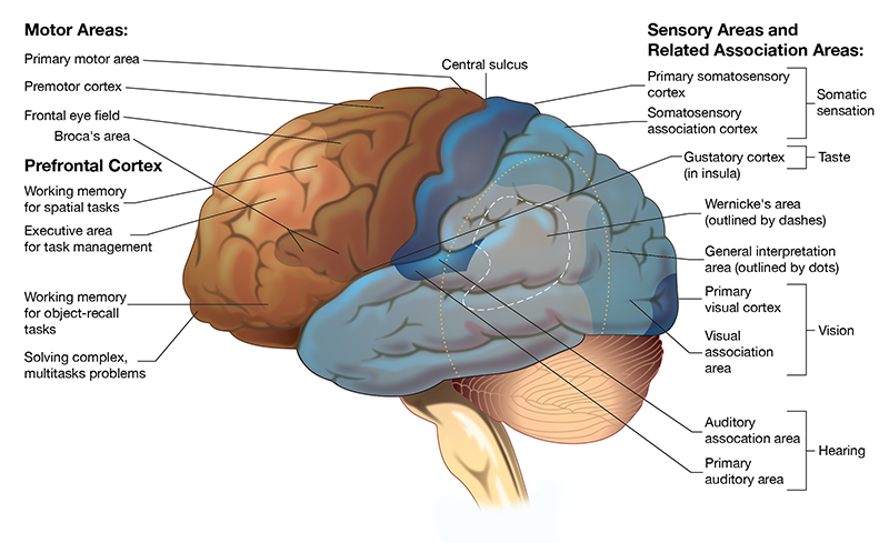
Major Functional Areas
# | Area | Location | Function |
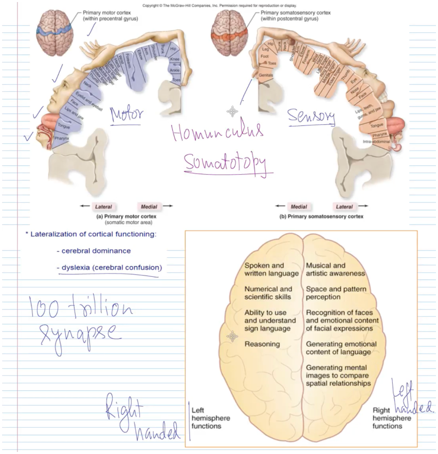
1 | Primary Motor Cortex | Precentral gyrus | Voluntary control of skeletal muscles |
2 | Premotor Cortex | Anterior to precentral gyrus | Controls learned, skilled movements |
3 | Frontal Eye Field | Frontal lobe | Controls voluntary eye movement |
4 | Prefrontal Cortex | Anterior frontal lobe | Cognition, personality, decision making |
5 | Primary Visual Cortex | Occipital lobe | Image processing (“see the object”) |
6 | Primary Auditory Cortex | Temporal lobe | Sound perception and recognition |
7 | Primary Gustatory Area | Insula | Taste perception |
8 | Primary Somatosensory Cortex | Postcentral gyrus | Touch, pressure, temperature, pain |
9 | Broca’s Area | Left frontal lobe | Motor speech – articulation of words |
10 | Wernicke’s Area | Left temporal/parietal junction | Comprehension of spoken/written language |
White matter fiber types & Reticular Activating System
Cerebral white matter tracts
Commissural fibers – connect right & left halves (e.g., corpus callosum, anterior commissure).
Association fibers – connect different areas within the same hemisphere.
Projection fibers – vertical fibers connecting cortex with lower centers (thalamus, brainstem, spinal cord).
Reticular Activating System (RAS)
Network in brainstem (midbrain, pons, medulla).
Active → awake, alert, conscious.
Inactivated → sleep, depression of CNS.
Severely damaged → coma, loss of consciousness.
Association Areas
General Interpretative (Gnostic) Area: integrates input from visual, auditory, and tactile senses into a common thought.
Visual Association Area: interprets what is seen.
Auditory Association Area: interprets sounds.
[Insert Diagram: Homunculus – motor & sensory representation on cortex]
![<ul><li><p><span><strong><span>General Interpretative (Gnostic) Area:</span></strong><span> integrates input from visual, auditory, and tactile senses into a common thought.</span></span></p></li><li><p><span><strong><span>Visual Association Area:</span></strong><span> interprets what is seen.</span></span></p></li><li><p><span><strong><span>Auditory Association Area:</span></strong><span> interprets sounds.</span></span></p></li></ul><p><span><strong><span>[Insert Diagram: Homunculus – motor & sensory representation on cortex]</span></strong></span></p>](https://knowt-user-attachments.s3.amazonaws.com/c02f136d-471f-47ce-94b1-01a380390448.png)
Convolutions, sulci, gyri, and neuron numbers
Convolutions: folds on the brain surface that increase surface area.
Sulci – shallow grooves.
Fissures – deep grooves (include all 3 meninges).
Gyri – elevated ridges between sulci.
Approximate neuron numbers:
Total brain: ~86 billion neurons.
Cerebrum: ~16 billion.
Cerebellum: ~70 billion.
Lateralization of Cortical Function
Left hemisphere: language, math, logic, reasoning.
Right hemisphere: creativity, spatial awareness, facial recognition, music.
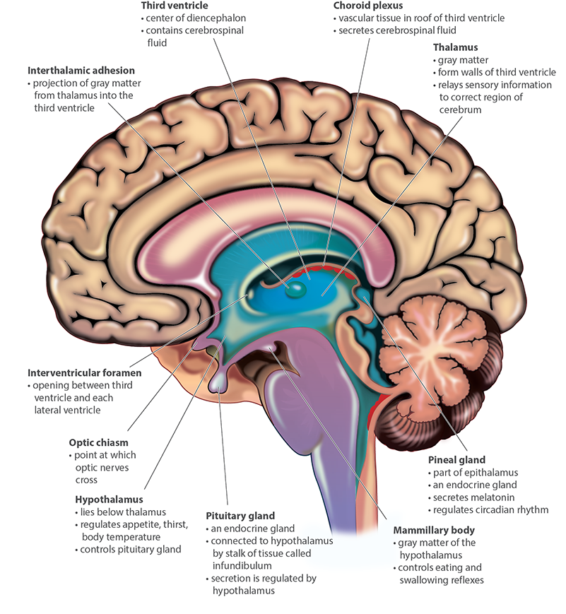
8. Diencephalon and Brainstem p1
Diencephalon
Thalamus: relay center for sensory information (80% of diencephalon).
Hypothalamus: controls autonomic functions (hunger, thirst, temperature, hormones).
Epithalamus (Pineal gland): secretes melatonin, regulates sleep and circadian rhythm.
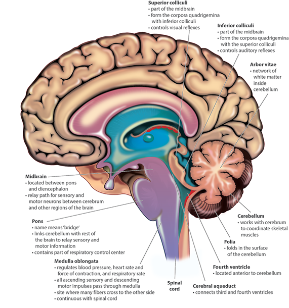
Midbrain (Mesencephalon)
Corpora quadrigemina:
Superior colliculi – visual reflexes, eye-head coordination.
Inferior colliculi – auditory reflexes, startle response.
Red nucleus: controls limb movement; high blood supply.
Substantia nigra: produces dopamine; regulates movement via basal ganglia.
Loss of dopamine → Parkinson’s disease.

8. Diencephalon and Brainstem p2
Basal Ganglia
Clusters of neurons for planning and execution of voluntary actions.
Includes caudate nucleus, putamen, and globus pallidus.
Part | Deficiency | Disorder |
Caudate | Overactive → OCD | Underactive → ADHD |
Putamen | Deficiency → Tourette’s syndrome |
|
Globus pallidus | Deficiency → Parkinson’s disease |
|
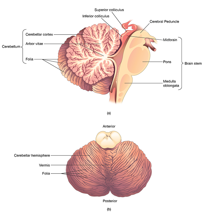
Pons and Cerebellum (Metencephalon)
Pons: “bridge” between cerebellum and medulla; regulates breathing, bladder, facial expressions.
Cerebellum: coordinates skilled muscle movement, balance, and posture.
Arbor vitae: white matter pattern (tree-like).
Gray matter: contains neuron cell bodies.
Medulla Oblongata (Myelencephalon)
Contains cardiovascular and respiratory control centers.
Regulates heart rate, blood pressure, and breathing rhythm.
Reflex centers for swallowing, coughing, vomiting.
9. Limbic System and Brain Disorders
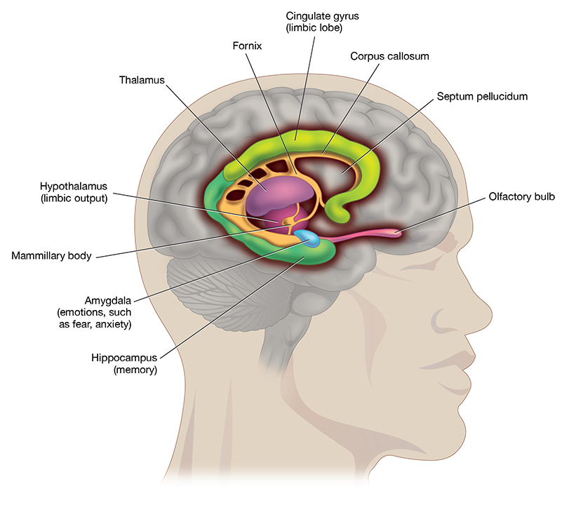
Limbic System
The “emotional brain.”
Includes: hippocampus, amygdala, hypothalamus, fornix, and cingulate gyrus.
Amygdala: emotions such as fear, anger, anxiety.
Hippocampus: memory formation.
9. Limbic System and Brain Disorders
Condition | Description |
Concussion | Sudden blow or jolt; brain moves violently but no tissue damage or bleeding. Causes dizziness or memory loss. |
Contusion | Tissue damage with hemorrhage (bleeding). More serious than concussion. |
Hemiplegia | Paralysis of one side of the body. |
Paraplegia | Paralysis from waist down. |
Quadriplegia | Paralysis from neck down. |
Cerebral Palsy | Permanent movement disorder due to oxygen deprivation at birth. |
Anencephaly | Absence of brain except brainstem. |
Microcephaly | Underdeveloped, small brain (can be caused by Zika virus). |
10. Brain Waves and Sleep
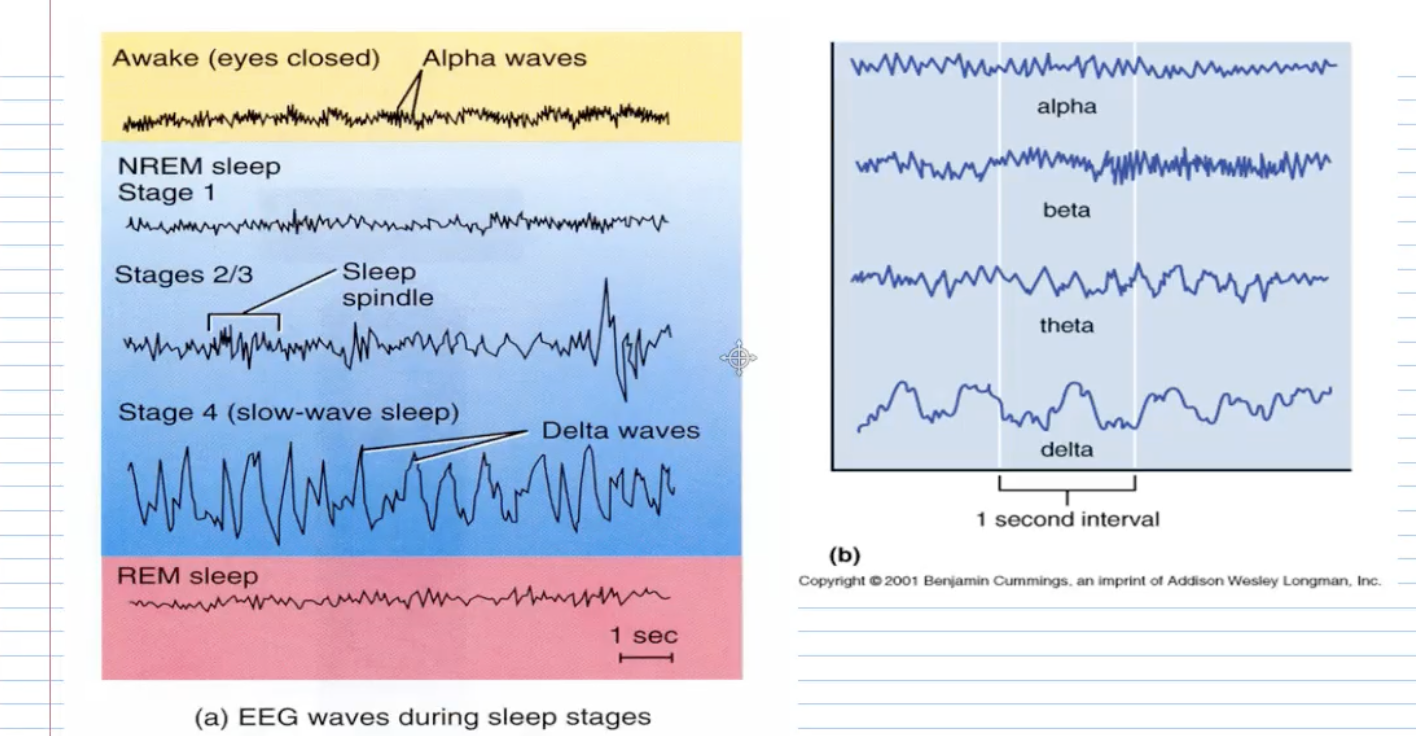
[Insert Diagram: EEG wave patterns – alpha, beta, theta, delta]
Electroencephalogram (EEG)
Measures electrical activity in the brain.
Wave Type | Frequency | State |
Alpha | 8–13 Hz | Relaxed, eyes closed |
Beta | 14–30 Hz | Alert, eyes open |
Theta | 4–7 Hz | Light sleep or daydreaming |
Delta | 3–5 Hz | Deep sleep (restorative) |
Gamma | ~100 Hz | Highly active brain |
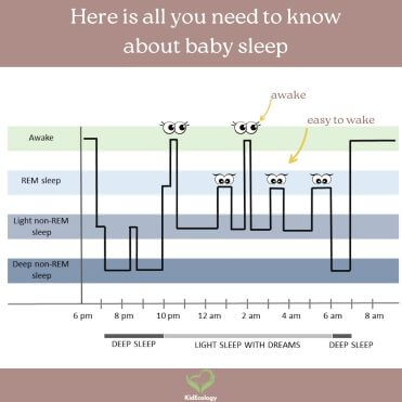
Sleep Stages
NREM Sleep (Non-Rapid Eye Movement) – 75% of total sleep
Stage 1: 5–10 min – light sleep, drifting.
Stage 2: 10–20 min – slower HR, body temp drops, sleep spindles.
Stages 3/4: Deep, restorative sleep; slow delta waves; muscles relax.
REM Sleep (Rapid Eye Movement) – 25% of total sleep
Brain highly active; vivid dreams; body largely paralyzed.
Occurs about 90 min after sleep onset.
Episodes lengthen toward morning.
[Insert Diagram: Sleep cycle timeline (10 pm – 6 am with REM stages labeled)]
Melatonin from pineal gland promotes sleep onset.
Insomnia: difficulty falling or staying asleep.
![<ol type="1"><li><p><span><strong><span>NREM Sleep (Non-Rapid Eye Movement) – 75% of total sleep</span></strong></span></p><ul><li><p><span><strong><span>Stage 1: 5–10 min – light sleep, drifting.</span></strong></span></p></li><li><p><span><strong><span>Stage 2: 10–20 min – slower HR, body temp drops, sleep spindles.</span></strong></span></p></li><li><p><span><strong><span>Stages 3/4: Deep, restorative sleep; slow delta waves; muscles relax.</span></strong></span></p></li></ul></li><li><p><span><strong><span>REM Sleep (Rapid Eye Movement) – 25% of total sleep</span></strong></span></p><ul><li><p><span><strong><span>Brain highly active; vivid dreams; body largely paralyzed.</span></strong></span></p></li><li><p><span><strong><span>Occurs about 90 min after sleep onset.</span></strong></span></p></li><li><p><span><strong><span>Episodes lengthen toward morning.</span></strong></span></p></li></ul></li></ol><p><span><strong><span>[Insert Diagram: Sleep cycle timeline (10 pm – 6 am with REM stages labeled)]</span></strong></span></p><ul><li><p><span><strong><span>Melatonin</span></strong><span> from pineal gland promotes sleep onset.</span></span></p></li><li><p><span><strong><span>Insomnia:</span></strong><span> difficulty falling or staying asleep.</span></span></p></li></ul><p></p>](https://knowt-user-attachments.s3.amazonaws.com/b5825278-651a-4df8-8610-76113314a687.png)
EEG
Theta waves in awake adults – can be seen in extreme emotional stress.
Delta waves in an awake adult – usually indicate brain damage.
Extra CSF details & hydrocephalus
CSF composition: similar to blood plasma but:
Very few WBCs.
No RBCs (unless hemorrhage).
Lower protein than plasma.
Rate: continuously produced, total ~500 mL/day, but only ~150 mL present at any moment (constant reabsorption).
Normal CSF pressure: about 7–10 mm (hydrostatic pressure).
Hydrocephalus:
Blocked CSF flow or absorption → ventricles enlarge → increased pressure.
In infants: head enlargement; in adults: brain compression.
Treated with ventriculoperitoneal (VP) shunt (tube from ventricle to abdominal cavity).
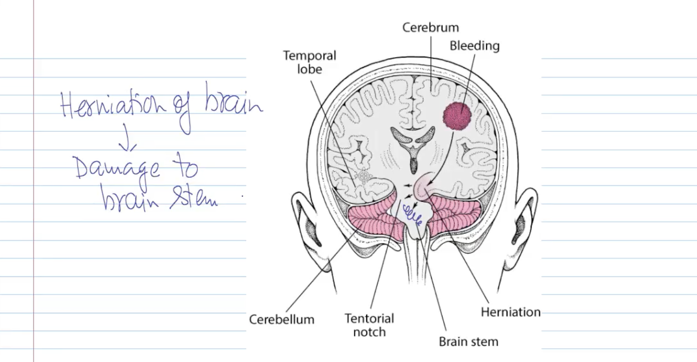
Cranial fossae & brain herniation
Cranial fossae:
Anterior cranial fossa – holds frontal lobes.
Middle cranial fossa – holds temporal lobes.
Posterior cranial fossa – holds cerebellum and brainstem.
Brain herniation:
Mass/hematoma (epidural or subdural) can force brain tissue downward through tentorial notch.
Can compress the brainstem, which is life-threatening.