BIO EXAM 2
1/113
There's no tags or description
Looks like no tags are added yet.
Name | Mastery | Learn | Test | Matching | Spaced | Call with Kai |
|---|
No analytics yet
Send a link to your students to track their progress
114 Terms
muscle tissue
able to stretch, able to shorten, able to respond to stimulus, able to return to original length.
Contractile
Able to become shorter or longer
Elastic
return to original shape
Muscle Cell
muscle fiber
Muscle fibers
contract in response to nervous signal (reflex), pace maker cells (heart), Hormones
Oxycontin
Contraction into labor
skeletal muscle tissue
responsible for voluntary movement
smooth muscle tissue
responsible for involuntary movement
What is a myofilament?
Filament that constitutes myofibrils.
What are the two types of myofilaments?
Actin (thin filament) and myosin (thick filament with heads and tails).
Sacromere
contractile unit of a muscle fiber
uncontracted smooth muscle
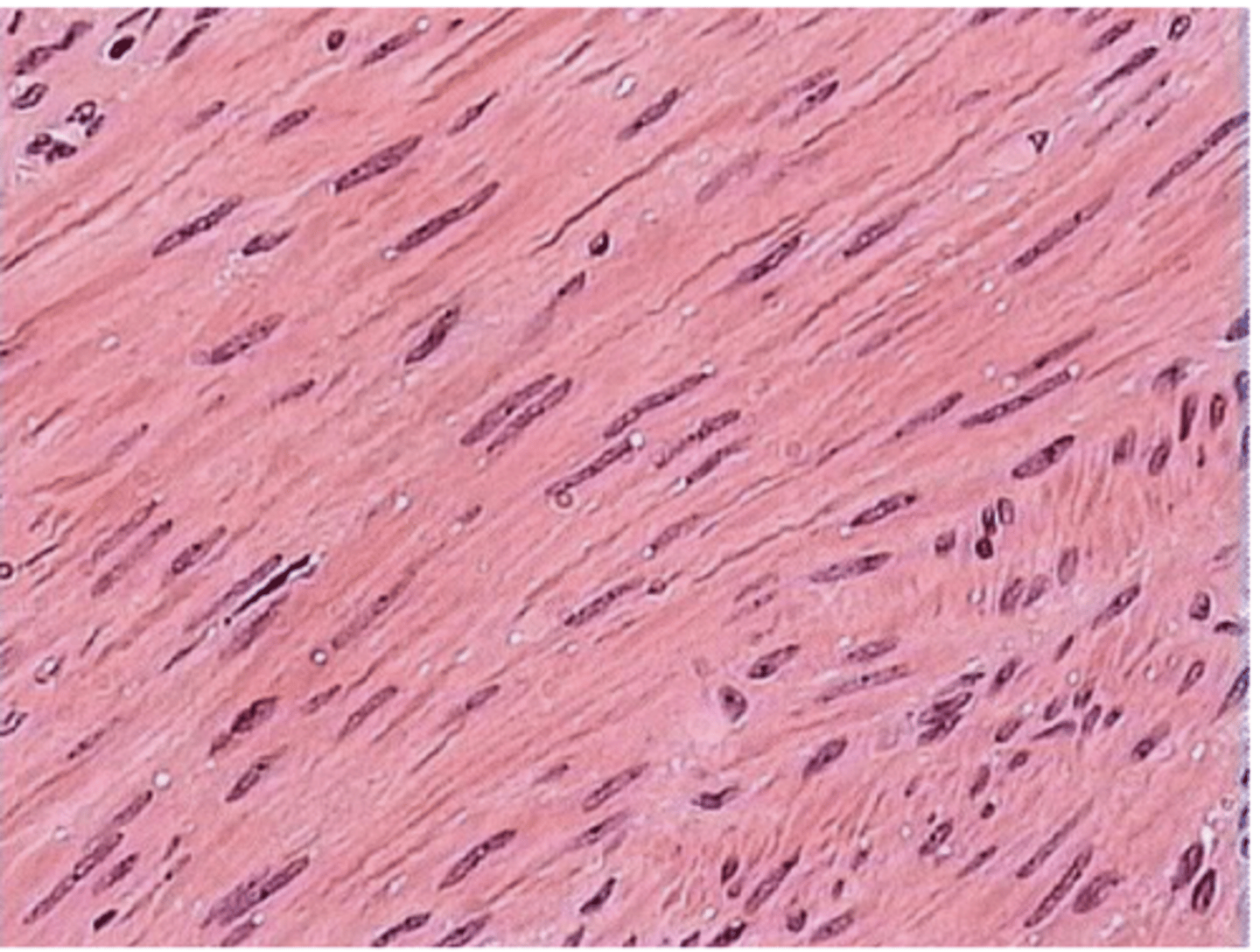
contracted smooth muscle
location of smooth muscle
walls of blood vessel, walls of lymphatic vessels, urinary bladder, uterus, GI Tract, iris of eye.
smooth muscle
Involuntary muscle found inside many internal organs of the body
Electrical signals travel through...
cell connected together so that cells function as a unit
What is required for skeletal muscle to contract?
Signal from a neuron
Exitable
Stimulated (Stimilus)
Extensible
capable of being extended/ stretch
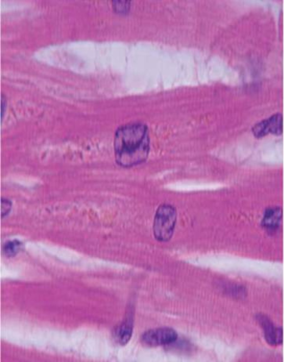
cardiac muscle tissue
responsible for contraction of heart
voluntary movement
movement that is under a person's control
involuntary movement
movement that is out of a person's control
cardiac muscle
Involuntary muscle tissue found only in the heart.
Cell Junctions
Adhesion junction keep cells together, gap junctions allo for united contraction.
How do skeletal muscles work in the body?
In pairs of antagonistic muscles
Name some examples of skeletal muscles in the body.
Triceps, biceps, quads, hamstrings
Skeletal "Muscle"
Skeletal muscle tissue, connective tissue, blood tissue, nerves
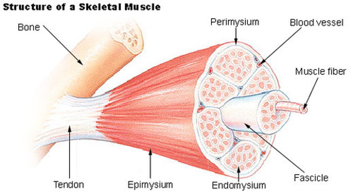
Muscle Hierarchy
- Muscles
- Fascicles
- Muscle fibers
- Muscle cells
- Myofibrils
- Sarcomeres
- Myofilaments
Tedons
connective tissue, extending from muscle, fascicle + fiber
Polymer (muscle)
Myofibril
Monomer (muscle)
Sarcomere
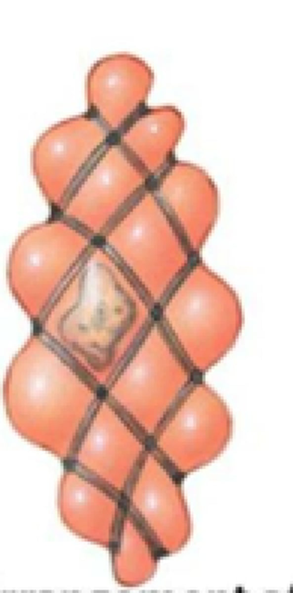
Muscle Fiber Structure
Cylindrical, long, multi-nucleated inward extensions of cell membrane, carry electrical signal inside, contains myofibrils, strores calcium.
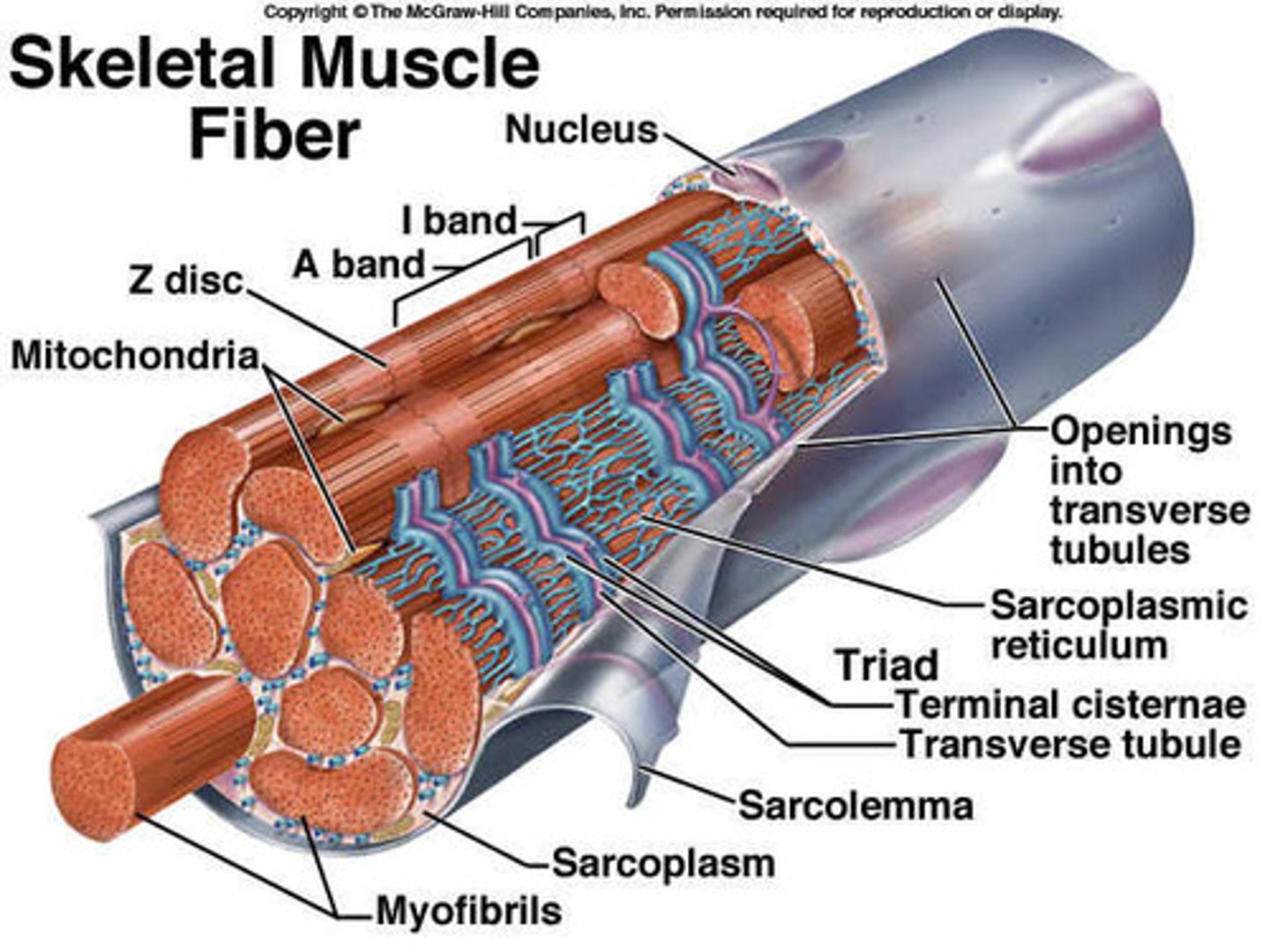
contractile proteins
Myosin filaments (thick,head tails), actin filament (thin)
Actin and myosin overlap from sarcomere
true
The length of the myosin is not decreased when contaction happens
True
z line
Adjacent sarcomeres are attached by joining thin filaments. Filaments DO NOT decrease in length
M line
stationary area where myosin bings to actin and pulls the actin toward the middle of sarcomere
nervous system functions
Senses external stimuli, senses internal stimuli, transmits signals from one organ to another organ, coordinates body activities.
Tissues
Groups of cells with a common structure that work together for a function.
Neurons
Send electrical signals, transmit nerve impulses
gilal cells (gila)
cells in the nervous system that support, nourish, and protect neurons. regulate nutrient/ion concentration.
Blood-brain barrier
Blood vessels (capillaries) that selectively let certain substances enter the brain tissue and keep other substances out. Protects from toxins
Myelin Sheath
Produce cerebrospinal fluid (crushion), scavenge dead cells, pathogen in brain, structural support for synapse, stimulate nerve growth, outnumber neurons 10:1
Astrocytes
form blood brain barrier, Provide structural and metabolic support for neurons.
Glioblastomas
the most aggressive type of primary brain tumor in adults, Uncontrolled growth of Astrocytes.
Glioblastomas life expency
3 months w/out treatment, 12-16 w/ treatment
Why is it difficult to treat Glioblastomas?
Almost impossible to remove entire tumor, many drugs cant cross the blood-brain barrier.
What are microglia?
Immune cells of the CNS
How do microglia differ from other glia?
During embryogenesis, they emerge from the same family of stem cells that become white blood cells
microglia functions
Look for dead cells, toxins, pathogens, etc. Check if neurons are functioning.
Microglia behavior
Can be altered by many factors; stress, depression, autism.
Where do we find the greatest concentration of neurons and gila?
The brain is considered a part of the peripheral nervous system
3 types of neurons
sensory (form senses), motor (muscle), interneurons (brain spinal)
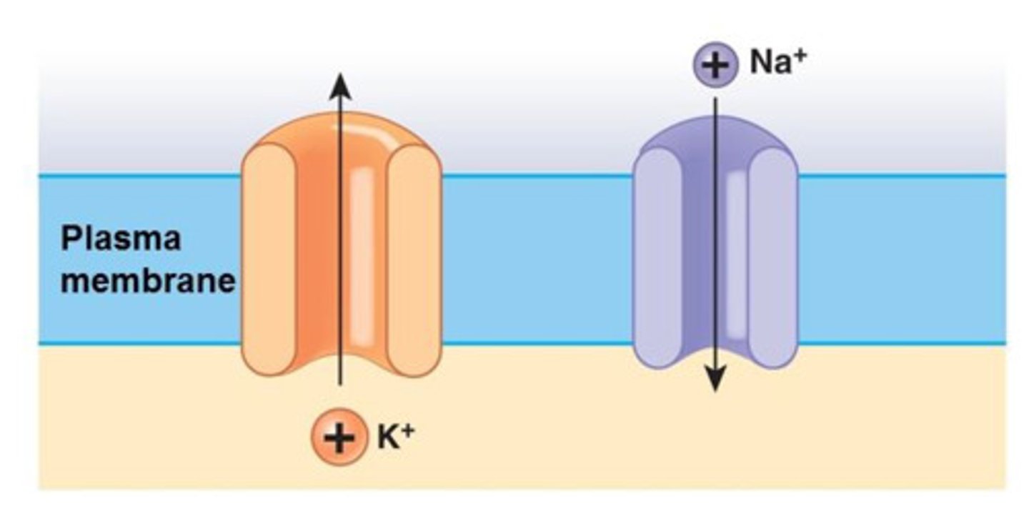
Change in electrical charge (voltage) across a membrane
makes a neuron work
Nerve signal
movement of ions (Na+, K+) across a membrane all the way down a axon.
Structure and Function of Neurons
Neurons are the fundamental units of the nervous system, responsible for transmitting information throughout the body.
Two types of branches extend from the cell body: axons and dendrites
The resting membrane potential
approximately -70 mV, maintained by the distribution of ions (Na+, K+, Cl-) across the membrane.
Depolarization
occurs when sodium ions enter the neuron, leading to a reversal of the intracellular charge, which is crucial for action potential generation
What structures protect the Central Nervous System (CNS)?
Bone (skull and vertebrae), meninges, and cerebrospinal fluid (CSF)
What do the protective structures of the CNS provide?
Physical and chemical barriers
CSF functions
CSF is produced by glial cells and serves multiple functions: nutrient delivery, waste removal, buoyancy, and shock
blood-brain barrier
selectively allows substances to enter the CSF, impacting how drugs like caffeine and alcohol affect brain function.
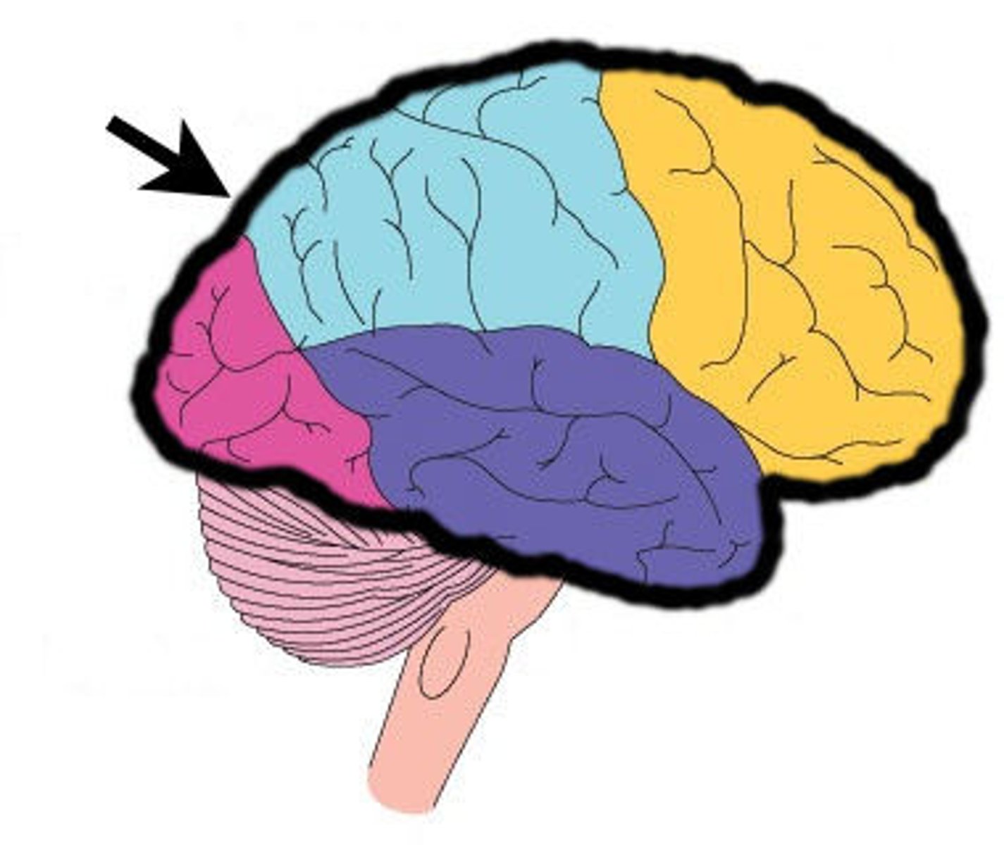
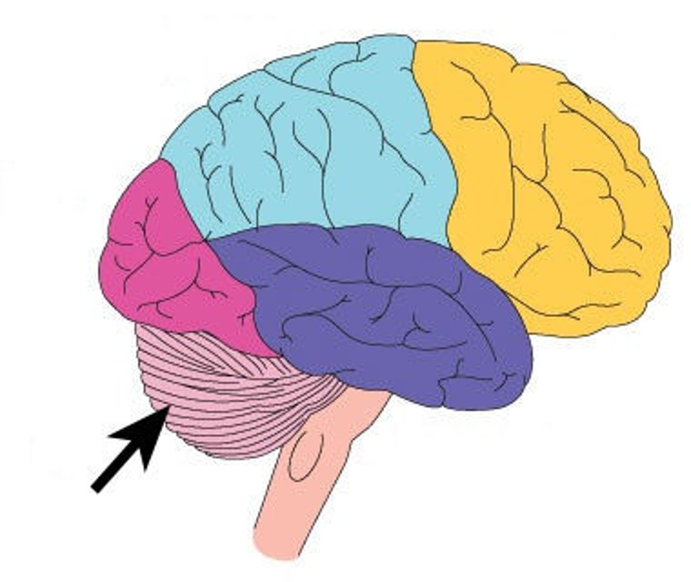
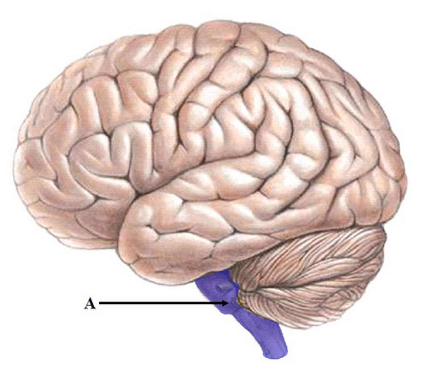
The brain is divided into
the cerebrum, cerebellum, and brainstem, each responsible for different functions.
cerebrum
Area of the brain responsible for all voluntary activities of the body
cerebellum
A large structure of the hindbrain that controls fine motor skills.
brainstem
The oldest part and central core of the brain, responsible for automatic survival functions. regulates vital functions such as heart rate, breathing, and reflexes
Neuron Communication Mechanisms
Neurons communicate through synapses, where neurotransmitters are released from axon terminals into the synaptic cleft.
What opens in response to an electrical signal?
Calcium channels
What is the role of calcium channels in neurons?
Facilitating neurotransmitter release
Why is neurotransmitter release essential for signal transmission between neurons?
allow the neuron to communicate with other neurons
depolarization
Loss of a state of polarity; loss or reduction of negative membrane potential
hyperpolarization
membrane potential becomes more negative
central nervous system (CNS)
the brain and spinal cord
peripheral nervous system (PNS)
motor and sensory nerves that connect the CNS to the rest of the body.
organization of the nervous system
Central Nervous System (CNS) and Peripheral Nervous System (PNS)
What are the components of a neuron?
Dendrites, cell body, and axon
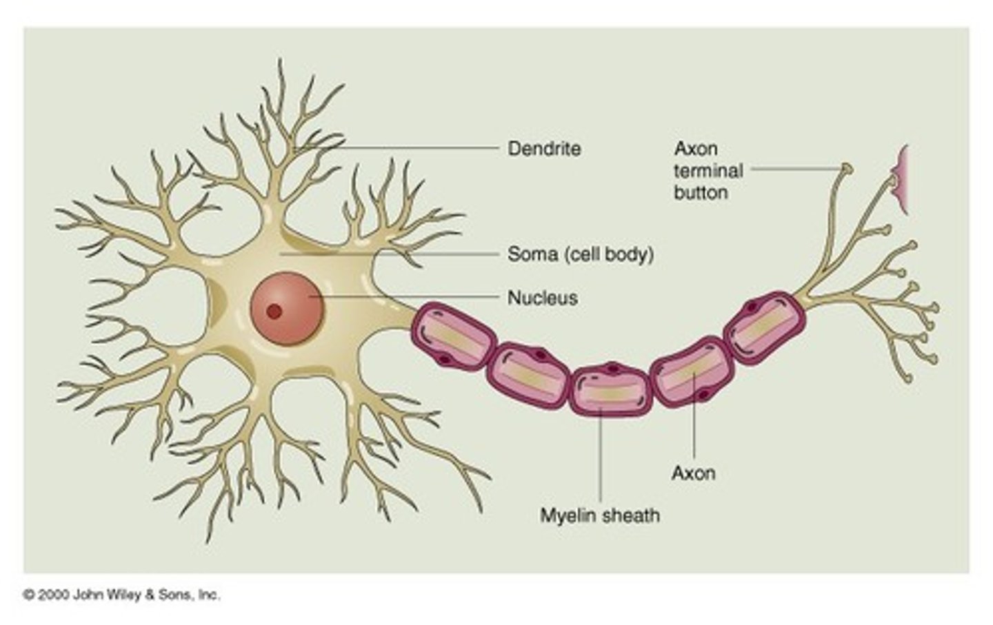
What is the role of dendrites in a neuron?
Receiving signals
What is the role of the cell body in a neuron?
Processing signals
What is the role of the axon in a neuron?
Transmitting signals
Function of Neurons
process and transmit information
Dendrites receive
incoming signals through gated ion channels
the axon transmits
signals away from the cell body.
Neurons communicate through
synapses
neurotransmitters are released to
transmit signals to other neurons.
resting membrane potential of neuron
-70mV
What ions does the Na+/K+ pump transport?
3 sodium ions out and 2 potassium ions into the cell
How does the Na+/K+ pump contribute to the negative charge inside the neuron?
By actively transporting ions in and out of the cell
A stimulus can cause
gated ion channels to open, leading to depolarization if the inside of the cell becomes more positive (e.g., -60mV).
Excitatory signals
increase the likelihood of an action potential
inhibitory signals
decrease likelihood that neuron will fire
Nerve Impulses
electrochemical changes that neurons use to transmit information
voltage-gated sodium channels open
allowing Na+ ions to rush into the neuron, further depolarizing the membrane.
depolarization travels along the axon
as a nerve impulse, with adjacent segments of the membrane being depolarized in a wave-like manner.
Myelin sheaths
insulate the axon, allowing for faster signal transmission through saltatory conduction.
Inactivated sodium channels
prevent the impulse from traveling backward, ensuring unidirectional flow.
Synaptic Transmission
The relaying of information across the synapse by means of chemical neurotransmitters.
When a nerve impulse reaches the axon terminal
it triggers the opening of voltage-gated Ca2+ channels, allowing calcium ions to enter the neuron.
What is the role of Ca2+ in synaptic transmission?
Ca2+ causes synaptic vesicles to fuse with the presynaptic membrane and release neurotransmitters into the synaptic cleft.
What happens when Ca2+ enters the presynaptic terminal?
It triggers the fusion of synaptic vesicles with the presynaptic membrane, leading to the release of neurotransmitters into the synaptic cleft.
Neurotransmitters bind to
receptors or chemically-gated ion channels on the postsynaptic neuron, leading to either excitatory or inhibitory effects.