A&P Chapter four notes
1/99
There's no tags or description
Looks like no tags are added yet.
Name | Mastery | Learn | Test | Matching | Spaced | Call with Kai |
|---|
No analytics yet
Send a link to your students to track their progress
100 Terms
What are the two main components of ECM
Ground Substance and Protein Fibers
What are some of the functions of the ECM
providing the tissue with strength to resist stretching and compressive forces
directing cells to their proper places within a tissue
regulating the development, mititic activity, and sirvival of cells
holding cells in their proper positions
Ground Substance - some notes
ECF (fluid) with water, ions, nutrients, other solutes, and three families of macromolecules: glycosaminoglycans, proteoglycans, and glycoproteins
Characteristics of Glycosaminoglycans (GAGs)
long, straight, polysaccharide chains
the negative charge of some of these sugars in a chains attract positively charged ions in the ECF, creating a concentration gradient that draws water out of cells and “traps” it in the ECM, which helps the ECM to resist compression
Characteristics of Proteoglycans
GAGs bound to a protein core. Thousands of proteoglycans can join together and cann bind into a very long GAG, forming huge proteoglycan aggregates. This makes the ECM firmer.
Tissues that contain more of these are more solid and resistant to compression
Act as a barrier to diffusion (immunity from invading microorganisms)
What are the three protein fibers found in the ECM?
collagen
elastic
reticular
Collagen Fibers characteristics
20-25% of all protein in body
very resistant to tension and pressure and stretching
Elastic Fibers Characteristics
may be stretched to 1.5 times their resting length without breaking (Distensibility)
the ability to return to their normal shape after being stretched (elasticity)
glycoproteins support and organize the elastin
Reticular Fibers characteristics
type of collagen fiber
thinner and shorter than collagen fibers
interweave to form a meshwork (netlike) that supports the cells and ground substance of many tissues. Also forms webs in certain organs, such as the spleen, to help trap foreign invaders
What is Marfan Syndrom?
results from defects in the gene that codes for a glycoprotein called fibrillin-I
This glycoprotein is a component of the ECM that is required for the normal deposition of elastic fibers. With defective fibrillin-I, elastic fibers cannot function properly because they are not correctly distributed and anchored in the ECM.
characteristic signs and symptoms including tall stature with long limbs and fingers; multiple skeletal abnormalities such as malformation of the sternum (breastbone); recurrent joint dislocations due to weak ligaments; abnormalities of the heart valves and the lens of the eye; and dilation of the aorta
What does it mean when tight junctions are ‘Leaky”
Note that some tight junctions are “leaky” and do not form a complete seal, unlike regular tight junctions, which allows certain substances to pass between the cells.
What do desmosomes do?
integral proteins that link two cells
allow components of the ECF to pass through
increase the strength of the cell by making sure stress is evenly distributed
attatched to intermediate filaments in the cytoskeleton that reinforce them
cells subject to mechanical stress have many of these
Gap Junctions
small pores in adjacent plasma membranes formed by protein channels
allows substances to pass freely from the cytosol of two cells
prominent in cells that can communicate with electrical signals like those of the cardiac muscle.
Identify:
Tissue Type
Locations found
location specific functions
SIMPLE SQUAMOUS EPITHELIUM
Lining the alveoli in lungs (Diffusion)
Forming outer boundary of serous membranes such as the pluera membrane (secretion)
Forming certain parts of Kidney tubules (diffusion/filtration)
Lining blood vessels (diffusion)
Identify:
Tissue Type
Locations found
location specific functions
SIMPLE CUBOIDLE EPITHELIUM
(relatively thin so substances can diffuse quickly)
Kidney tubules (absorption and secretion)
Glandular ducts such as thyroid (secreation into blood or ecf)
Identify:
Tissue Type
Locations found
location specific functions
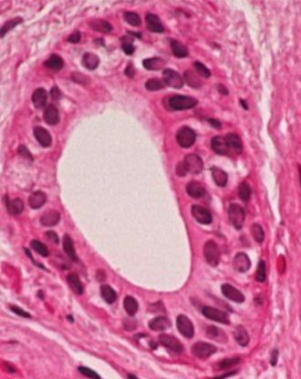
NONCILIATED SIMPLE CLUMNER EPITHELIUM (with microvilli)
Small intestine (absorption) (and secreation - digestive enzymes)
Gallbladder (absorption)
kidney tubules (absorption)
Tissue Type
Locations found
location specific functions
CILIATED SIMPLE COLUMNER EPITHELIUM
Uterine Tube (propel an oocyte toward uterus)
Identify:
Tissue Type
Locations found
location specific functions
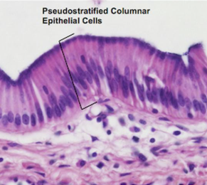
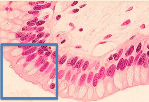
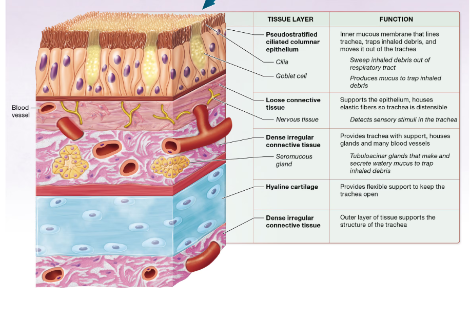
PSUEDOSTRATIFIED COLUMNER EPITHELIUM (ciliated)
Larger respiratory passages like the trachea and the nasal cavity (protective function)
Have goblet cells that seccrete mucous between ciliated cells
Tissue Type
Locations found
location specific functions
PSUEDOSTRATIFIED COLUMNER EPITHELIUM (nonciliated)
Vas Defernes (protection)
Epididymis (protection)
Parts of the urethra (protection)
Tissue Type
Locations found
location specific functions
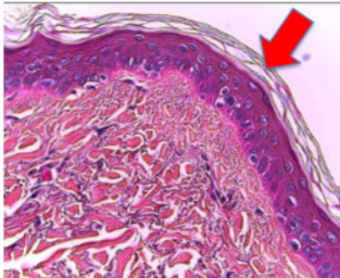
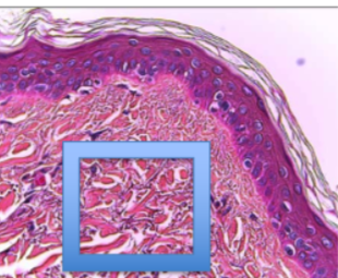
KERITINIZED STRATIFIED SQUAMOUS EPITHELIUM
Epidermis - outter layer of skin (Protection from mechanical stress)
Tissue Type
Locations found
location specific functions
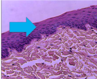
NONKERATONIZED STRATIFIED SQUAMOUS EPITHELIUM
lines mouth, pharynx, esophagus, anus, and vagina (protection but kept moistness nnot found in keratonized stratified squamous epithelium)
Tissue Type
Locations found
location specific functions
STRATIFIED CUBOIDAL EPITHELIUM
(relatively rare)
Sweat Glands (secretion)
Tissue Type
Locations found
location specific functions
STRATIFIED COLUMNER EPITHELIUM
(relatively rare)
ducts of certain glands - including salivary (secretion)
parts of the male urethra (protection)
conjuctiva (clear membrane on eye) (protection)
Tissue Type
Locations found
location specific functions
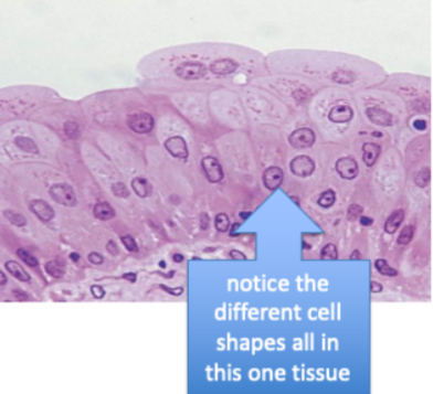
TRANSITIONAL EPITHELIUM
(found ONLY in urinary system)
lining of interior of kidneys, ureters, urinery bladder, and urethra (barrier and distensibility)
True or Flase:
Epithelial cells are vascular
False,
Epithelial cells are avascular, meaning that oxygen and other nutrients from the blood must diffuse up to the cells from tissues that are deep to it
(this is why there is a limit to the amount of layers epithelials can be in)
Where is the ECM of epithelial cells located?
beneath the cells in the basement membrane
(which consists of the basil lamina from the epithelials consisting of mostly collagen fibers and ground substance, and the underlying reticular lamina which is produced by the connective tissue and consists mostly of fibers and ground substnace.)
what is the role of the basement membrane?
the two layers of the basement membrane “glue” the epithelial tissue to the underlying connective tissues.
anchors underlying blood vessels in place and provides a barrier between the epithelial tissue and the underlying tissue.
provides a barrier that can slow the spread of carcinomas to other tissues.
What are corcinogens?
What Are Carcinomas?
agents inducing changes in DNA that can lead to cancer
Cancers of epithelial tissues are called carcinomas
Often cancer cells produce enzymes that degrade components of the basement membrane, which facilitates their spread into neighboring or distant tissues.
what is a gland?
A structure that makes and secretes a product, arising from epithelial tissue that grew inward.
Have secretory cells that manufactor a product and release it.
What is the difference between exocrine and endocrine glads?
exocrine releases secretions to apical surface of epithelium, while endocrine lacks ducts and secretes their products, which are usually hormones, directly into the bloodstream
exocrine has local actions only, while endocrine can reach distant targets through the bloodstream
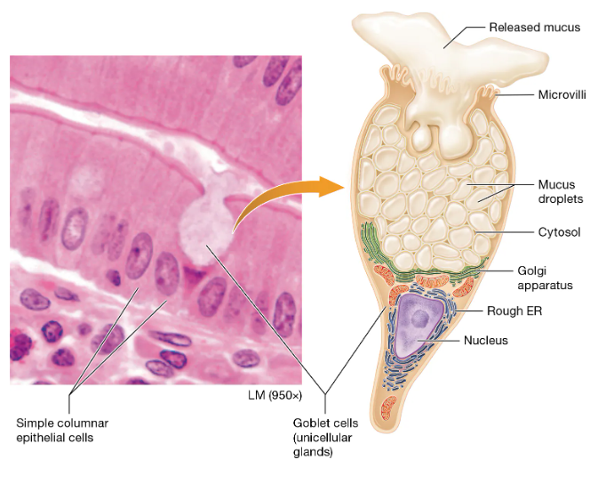
What is the most comon unicellular exocrine gland?
the goblet cell -
secretes mucus (protects the underlying epithelium
found abundantly in the epithelium lining the digestive tract and respiratory tract
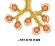
what is the name of the multicellular exocrine gland that is spherical
Acinar (compound acinar pictured, but simple acinar is not branched)
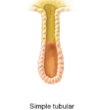
What is the name of the multicellular exocrine gland that long and straight?
Tubular (simple tubular pictured)
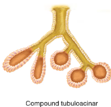
What is the name of the compound branched exocrine gland that has both shapes?
compound tubuloacinar
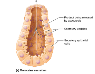
How does the merocrine exocrine secretion method work
they package their products into secretory vesicles for release by exocytosis
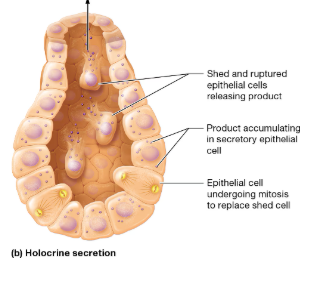
How does the holocrine exocrine secretion method work
In this type, the secretory cells accumulate their product in their cytosol, and the product isn’t released until the cell ruptures and dies
How does the apocrine exocrine secretion method work (rare)
portions of the cytoplasm are pinched off with the product being secreted.
What are the three main carcinomas?
lung adenocarcinoma (ah-den-oh′-kar-sih-NOH-muh), a cancer of the lung;
ductal and papillary carcinoma (PAP-ih-lehr-ee), cancers of the breast; and
basal cell carcinoma, a cancer of the skin, shown here:
What do fibroblasts do and where are they found?
they are a cell within connective tissue proper
produces protein fibers and ground substance and other elements of ECM
Usually lay close to collagen fibers, as they continuously produce collagen.
what stem cell is the begining of all connective tissue?
Mesenchymal Stem Cells
What are adiposcytes and where are they found?
(fat cells)
cells dominated mosstly by a single large inclusion containing lipids, with nuclei squished towards perimeter.
mainly found in adipose tissue
What are mast cells and where are they found?
Largest resident cells in connective tissue
cells of the immune system that have granules containing inflamitory mediators (histamine).
When released, it incites a protective response that activiates the immune system.
What are phagocytes
Cells of the immune system that ingest foreign substances, microorganisms, and dead and damaged cells by phagocytosis
(resident or migrant cells)
What is hypertrophic Obesity
most common type of obesity
lipid inclusions in the adipocytes accumulate excess fatty acids and increase in size. Adipocytes may grow to as much as four times their normal size in cases of severe obesity
the number of adipocytes remains the same.
What is hypercellular obesity?
actual number of adipocytes increases
generally severe
correlated with the development of obesity in infancy or early childhood
adults lack the ability to divide and form new adipoc
What is Osteoarthritis?
caused by a variety of factors, including age, joint trauma, genetic disorders, and infection.
developes in hyaline cartilage lining joint degenerates
leads to destruction of proteoglycans and collagen fibers
makes cartilage less resistant to stress
What is a popular dietary supplement to treat osteoarthritis?
glucosamine
Glucosamine is a sugar derived from shellfish and certain fungi that is used by chondroblasts in their synthesis of proteoglycans
Hypothetically should work
Studies on the effectiveness of glucosamine supplementation, however, have yielded mixed results.
Tissue Type
Locations found
location specific functions
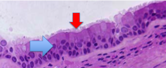
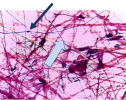
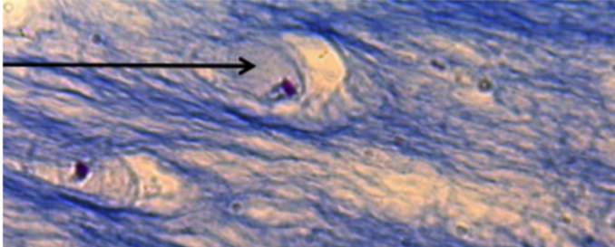
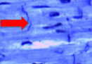
What is the black arrow pointed to and what is its function?
What is the EC component at the blue arrow
LOOSE CONNECTIVE TISSUE
(also known as areolar connectie tissue)
composed primarily of ground substance and all three types of protein fibers, fibroblasts, and other cells such as adiposcytes suspended in ground substance
functions: support, contains many blood vessels whose nutrients diffuse up to superficial epithelial, houses numerous immune cells (which refelects the fact that it is the first tissue to come into cantact with epithelium)
Locations: deep to the epithelium of the skin (papillary layer of the dermis), in membranes lining body cavities, and as layers in the walls of hollow organs
the black arrow is pointing to a fibroblast
the blue arrow is pointing to a elastic fiber
Tissue Type
Locations found
location specific functions
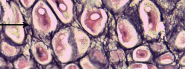
DENSE IRREGULAR CONNECTIVE TISSUE
ECM is course and tough
predominant fiber - collagen
Function: the organization of the collagen (the pink swirls) makes this tissue strong and allows it to resist tension on all three planes
Location: Found in organs that are subject to tension, such as the dermis, as well as around organs and joints.
Tissue Type
Locations found
location specific functions
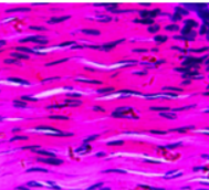
DENSE REGULAR COLLAGENOUS CONNECTIVE TISSUE
contaians thick collagen fibers, arranged in parallel to one another to from bundles.
Function: VERY STRONG because oriented in a single direction, but only resist tension on 1 plane
Location: Tendons (muscle to bone), ligaments (bone to bone)
Tissue Type
Locations found
location specific functions
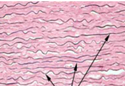
what are the black arrows pointing to?
DENSE REGULAR ELASTIC CONNECTIVE
consists mainly of parallel elastic fibers with randomly oriented collagen fibers
Function: allows certain organs to stretch
Location: lining of large blood vessels (like aorta), certain ligaments (like in the spine)
Tissue Type
Locations found
location specific functions
RETICULAR TISSUE
populated by nurmerous fibers produced by surrounding fiberblasts (often called reticular cells) that interweave to form a mesh-like network
function and location:
supports small structures such as blood vessels and lymph vessels,
also found in lymph nodes and in spleen where the “nets” help trap old and foreign cells
house white blood cells
forms part of the basement membrane that supports epithelia and internal structure of liver and bone marrow
Tissue Type
Locations found
location specific functions
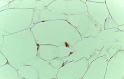
ADIPOSE TISSUE
(consisting of adipocytes and their surrounding ECM containing coarse collagen)
Locations:
White adipose tissue is found deep to the skin in a layer known as the hypodermis,
abdomen, breasts, hips, buttocks, and thighs
Functions:
insulation, warmth, shock absorption, protection, major energy reserve
What are characteristics of brown adipose tissue
small percentage in body, mainly in infants and young children.
have many mitochondria (reason they apper brown) also an extensive blood supply
oxidize fatty acids 20 times faster than white adipose tissue, can easily generate heat in cold temperatures
used for fuel and insulation
What does the ECM of cartilage look like?
gel like
contains glycosaminoglycans, proteoglycans, collagen fibers, and elastic fibers
contains chondroblasts - makes most of ECM cartilage
contains chondrocytes - matured chondroblasts that rest in lacunae (a cavity). they mature as they become surrounded by their ECM
True or false: cartilage is avascular
true,
as few, if any, blood vessels course through the cartilage itself. The blood supply to this tissue is mostly limited to an outer sheath of dense irregular connective tissue called the perichondrium (around)
the perichondrium supplied the chondroblasts and the chondrocytes
The three cartilages an be classified based on the composition of their ecm
Tissue Type
Locations found
location specific functions
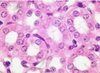
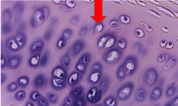
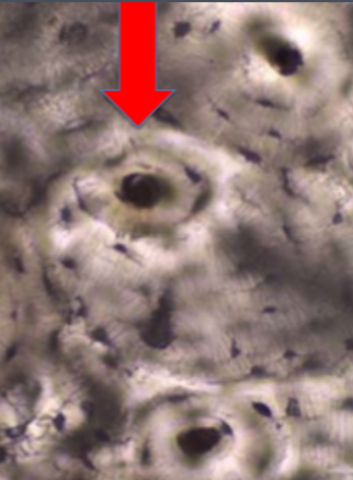
what is the opening at the arrow?
HYALINE CARTILADE CONNECTIVE TISSUE
(most abundant in body)
large amounts of ground substance
fine type of collagen fiber that forms small bundles (forms glassy appearance)
covers ends of bones where they form joints (where it is called articualr cartilage where it forms a smooth surface for bones to slide with little friction
protects bone by equally distributing stresses
found where strong, yet flexible support is needed, most of the fetal skeleton begins as hyaine cartilage
Red Arrow is pointing to a lacunae
Tissue Type
Locations found
location specific functions
ELASTIC CARTILAGE CONNECTIVE TISSUE
Locations : external ear and epiglottis (part of the larynx).
ECM: filled with distensible elastic fibers that allow the tissue to vibrate to detect or make sound
Tissue Type
Locations found
location specific functions
what cell is the arrow pointing to?
FIBROCARTILAGE CONENCTIVE TISSUE
ECM: dominated by collagen bundles (as compared to hyaline cartilage that is dominated by ground substance).
Fibroblasts fill ECM with dense cartilage and some elastic finers
FUNCTION: shock absorption, great strenghth with some elasticity.
LOCATION: found in joints and intervertebral disks. Forms articular discs that imrove the fit of two bones.
Fibrocartilage differs from the other two types in that it lacks a perichondrium.
What is bone tissue also called?
Osseous Tissue
Tissue Type
Locations found
location specific functions
what is the arrow pointing to?
COMPACT BONE
Location: Skeletal System
Functions: supporting the body, protection of organs, place of attatchment for muscles, storing calcium, housing bone marrow (which produces blood cells)
ECM: has two components organic and inorganic
organic: consists of collagen fibers and a ground substance called osteoid
inorganic: (65% of bone mass) composed of calcium and phosphate crystals making bone hard.
RED ARROW: pointing to osteon
What are the three cells of a mature bone and their functions?
osteoblasts - bone builders. synthesize the organic portion of the ECM by exocytosis. Also produce chemicals required for calcium to be deposited. found on outer surface of bone, closely associated with the dense irregular tissuecovering called the periosteum.
Osteocytes - Mature osteoblasts that are surrounded by their ECM and reside in the lacunae. Maintain Bone by exocytosis.
Osteoclasts - multinucleated bone destroyers. carry out bone resorption by secreting hydrogen ions and enzymes.
True or False: Tension on the bone will increase osteoclast activity?
False,
It will result in increased osteoblast activity and bone deposition,
PRESSURE placed on bone can result in increased osteoclast activity, causing bone resorption.
Tissue Type
Locations found
location specific functions
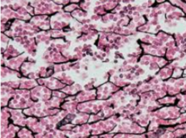
what is the ECM called?
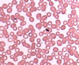
Identify WBC,RBC, and Platelets
BLOOD CONNECTIVE TISSUE
FLUID
consists of mainly water, dissolved solutes, and proteins, doesnt contain protein fibers
RBC (erythrocytes) - Light pink circles
WBC (Leukocytes) - Bigger light pink circles with blue in them
Platelets- tiny dots
plasma - Space around blood cells
What is the main component of muscle tissues?
Myocytes (muscle cells)
What does it mean when it is said: “muscle cells are excitable cells”
they respond to chemical and electical stimulation
What is the cytoplasm of muscle cells filled with?
Myofilaments
What are the characteristics of striated muscle cells?
there are regions where the myofilaments overlap (dark bands) and where they dont (light bands),
as compared to smooth muscle which doesn’t overlap, but does have irregularly scattered bundles of myofilaments throughout the cytoplasm
What is the endomysium?
Small amount of ECM around each muscle cell
sometimes called the external lamina —> similar to basil lamina in function, as it helps to hold muscle tissue together.
Tissue Type
Locations found
location specific functions
SKELETAL MUSCLE TISSUE
found attached to skeleton and produces voluntary body movement through contraction
striated
1 muscle cell usually spans the entire length of the muscle. so muscle cells are also called muscle fibers
multinucleated, with the nuclei located near the outer edge of the muscle.
Why is it adventageous for skeletal muscle to be multinucleated?
This helps with constant production of proteins like enzymes, structural proteins, and contractile proteins
How are skeletal muscles formed?
by the fusion of embryonic cells called myoblasts. The nucleus of each myoblast is retained in the mature skeletal muscle fiber, causing it to be multinucleate.
Tissue Type
Locations found
location specific functions
CARDIAC MUSCLE TISSUE
found only in the heart
striated muscle cells called cardiac muscle cells (which are branched and joined by intercalated discs, which contain tight juctions, gap juctions, and desmosomes)
relatively short and thick
1 nucleus at the center
Tissue Type
Locations found
location specific functions
SMOOTH MUSCLE TISSUE
involuntary
not striated
LOCATIONS: in the wall of every hollow organ, blood vessels, eyes, skin, ducts of certain glands.
single ovoid nucleus in center of cell
most of these cell contain gap junctions in their plasma memranes that link them with other smooth muscle cells
What are the two main types of cells in nervous tissue?
Neurons and neuroglial cells (perform various supportive functions)
What does the ECM of nervous tissue look like?
contains few protein fibers, mostly made up of ground substance with unique proteoglycans
Tissue Type
location specific functions
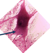
NERVOUS TISSUE
parts of the neuron -
cell body (soma): large portion of neuron where nucleus and other organells are housed
Axon: responsible for moving a nerve impulse from soma to target cell
Dendrites: recieve messages from other axons and bring impulses to the cell body
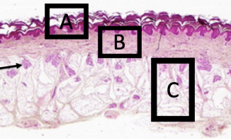
Identify each structure labeled
a. epidermis
b. dermis
c. hypodermis
arrow - secretory portion of sweat gland
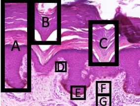
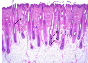
Identify each labeled structure
a- epidermis
b- stratum corneum
c. stratum lucidum
d. dermal papilla
e - stratum Basale
f- papilliary layer of dermis
g - reticular layer of dermis

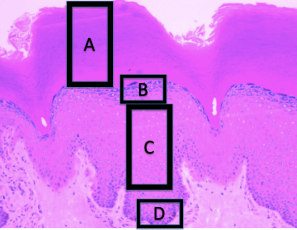
identify each structure
a- adipose tissue
b- hypodermis
c- dermis
D- epidermis
identify each structure
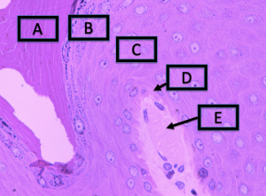
identify each structure
a - stratum corneum
b - stratum granulosum
c- stratum spinosum
d - stratum basale
e - Messiener’s corpuscle
identify each structure
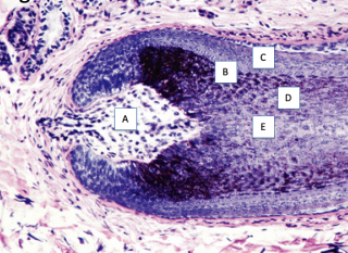
identify each structure labeled
a - dermal papilla
b - cutical
c- root sheath
d - cortex
e - medulla
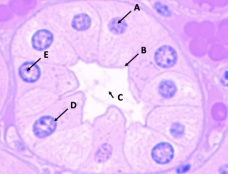
Identify each structure
a- chromatin
b- plasma membrane
c - lumen
d - nuclear membrane
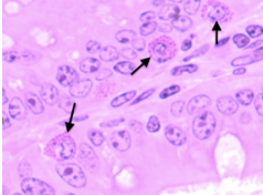
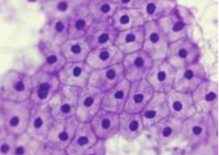
Identify The structures
secretory granuels in cytoplasm
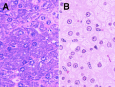
Identify the dark blue structures in panel A
the dark blue structures in panel are are rough er. This means that they most likely have a secretory function (of proteins)
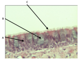
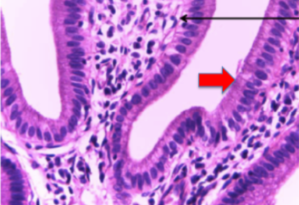
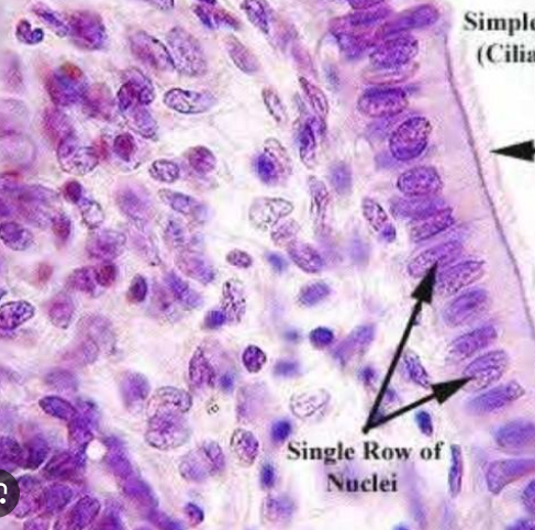
Identify each structure
a - cilliated pseudostratified columner epithelium
b - goblet cell
c - cillia
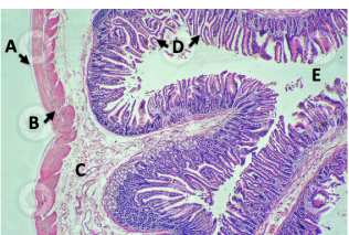
Identify each structure
a - serous membrane
b- muscle
c- submucose
d - intestinal villi
helpful picture of the tissue of the lung (apical surface is facing lumen of trachea)
Membranes
Most membranes consist of a superficial layer of secretory cells and a layer of connective tissue on which they rest;
FUNCTIONS: Membranes anchor organs in place, serve as barriers, function in immunity, and secrete various substances.
What are the four differnet types of membranes?
Serous, synovial (TRUE MEMBRANES)
mucous, and cutaneous. (MEMBRANE LIKE STRUCTURES)
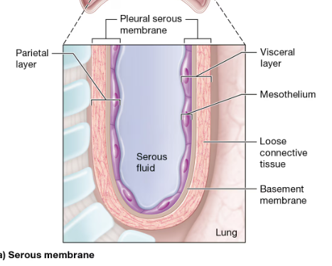
Serous membrane notes
line pleural, pericardial, and peritoneal cavities
consist of a layer of simple squamous epithelium called mesothelium, its basement membrane, and a layer of loose connective tissue
continuous sheet that folds on itself
visceral and parietal layers
mesothelium cells secrete serous fluid that fills the space between the 2 layers.
serous fluid provides lubrication so organs can move without friction
What is a “friction rub”
Certain viral and bacterial infections can cause inflammation of the serous membranes of the pleural and pericardial cavities.
When these serosae are inflamed, the thin layer of serous fluid produced by the mesothelium is inadequate to reduce friction as the organs move within their cavities.
This causes the parietal and visceral layers to rub together as the organs expand and contract
produces a grating sound called a friction rub that can be heard with a stethoscope. Friction rubs are typically quite painful and cause chest pain that worsens with inhalation, body movement, and swallowing. They usually resolve with treatment of the underlying condition.
Synovial Membranes Notes
line the cavities surrounding freely moveable joints, including the hip, knee, elbow, and shoulder
don’t have a layer of epithelial cells.
Instead, they’re made up of two connective tissue layers: The inner layer consists of modified fibroblasts called synoviocytes ,
and the external layer is generally a mixture of loose and dense irregular connective tissue.
Synoviocytes secrete a fluid called synovial fluid, which is a watery, slippery fluid that lubricates the joint.
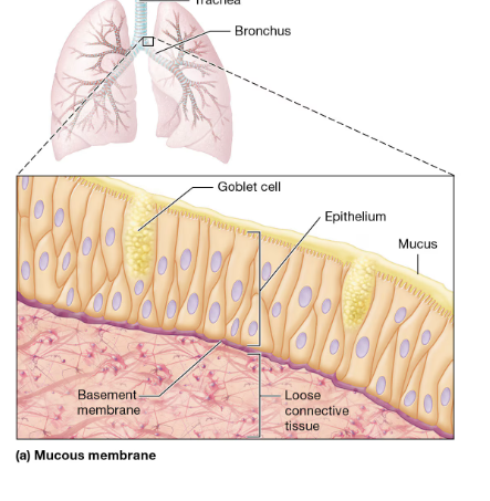
Mucous Membranes Notes (membrane - like)
line all body passages as part of the walls of hollow organs that open to the outside of the body,
LOCATIONS: including the respiratory passages, the mouth, the nasal cavity, the digestive tract, and the male and female reproductive tracts
Mucosae consist of a layer of epithelium, its basement membrane, a layer of loose connective tissue called the lamina propria
FUNCTIONS :
Not surprisingly, mucosae contain glands with goblet cells that secrete mucus. As we discussed earlier, mucus serves many functions, most of which are protective in nature.
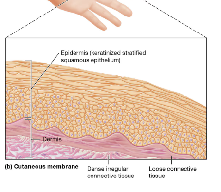
Cutaneous Membrane notes
refers to the skin
(epidermis and dermis)
Tissue Repair
tissue repair occurs differnetly in different tissues
some are capable of regeneration - damaged/dead cells are replanced with the same type, tissues function is gerally completely restores
Some are not capable of regeneration and must go through fibrosis - fibroblasts divide by mmitosis and produce collagen to fill in the defect left and tissue does not regain functon - end result is scar tissue
Capacity of Specific Tissues for Tissue Repair
epithelials typically go through regeneration, certain epithelials like those of the skin and digestive tract contian stem cells that divide continuously. Epithelials in other places use mature cells to go through mitosis.
Most connective tissue goes thorugh regeneration.
- CARTILAGE IS AN EXCEPTION - has little capacity to divide and usually result in fibrosis
smooth muscle usually regerates
Mature skeletal muscle and cardiac muscle cannot undergo mitosis due to their large size and complicated structure,
HOWEVER - skeletal muscle has a limite degree of regeneration, as there are satellite cells that can divide and become skeletal muscle fibers, alloeing a limited degree of regeneration
Neurons of nervous tissue generally do not regenerate. -
neuroglial cells retain the ability to divide, neurons are generally unable to undergo mitosis.
For this reason, damaged neurons in the brain and spinal cord are typically replaced by neuroglial cells that divide to produce a scar. However, if the cell body of a neuron is intact and only the axon is damaged, there is some chance that the axon will regenerate
What other two factors effect a tissue’s ability to repair?
NUTRITION
Tissue repair involves production of proteins such as collagen, so protein (amino acid) intake must be adequate for repair to occur. Another dietary requirement is sufficient vitamin C intake, which is needed by the fibroblasts to produce collagen.
BLOOD SUPPLY
the blood supply to the damaged area must be taken into consideration, as blood brings in oxygen, nutrients, and cells of the immune system that are needed for tissue repair. This is why people suffering from diseases of the arteries often have nonhealing wounds, even in tissues capable of regeneration