HMI30`1: Pathology + Anatomy for each Organ
1/56
There's no tags or description
Looks like no tags are added yet.
Name | Mastery | Learn | Test | Matching | Spaced | Call with Kai |
|---|
No analytics yet
Send a link to your students to track their progress
57 Terms
Aorta Main Pathology
AAA
Dissections
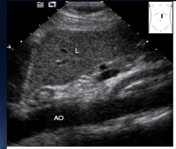
What is this an image of + Label
Longitudinal Proximal Aorta
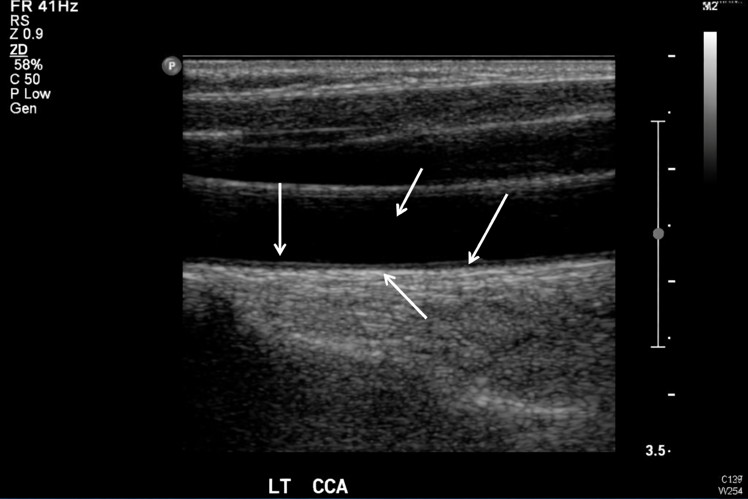
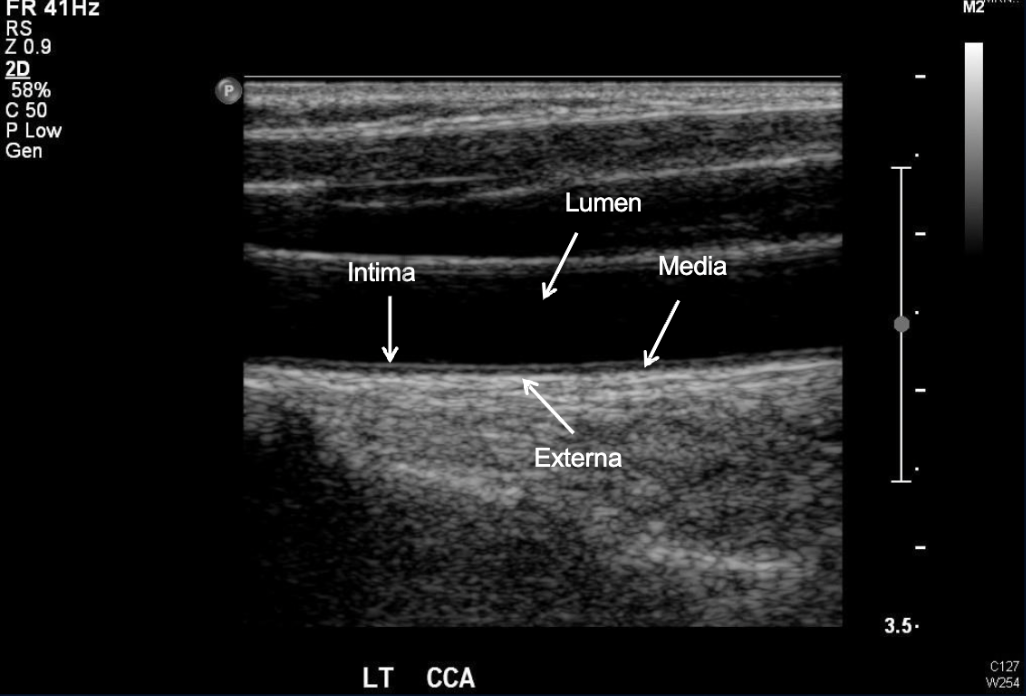
Label the anatomy of this Carotid Artery
What is this + Label
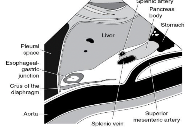
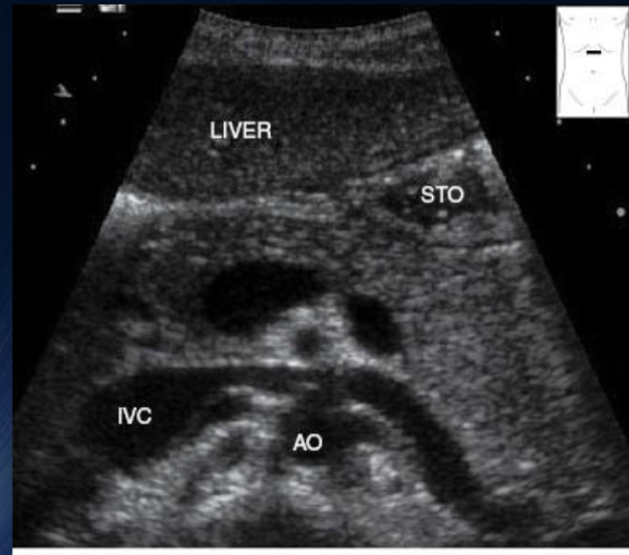
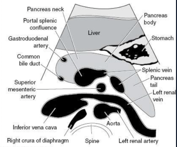
Trv Proximal Aorta (SMA lvl)
Size of AAA?
Diameter = 3mm or larger

Pathology?
AAA
Pathology?
Dissection
IVC Pathology
DVT (Deep Vein Thrombosis)
Thrombosis
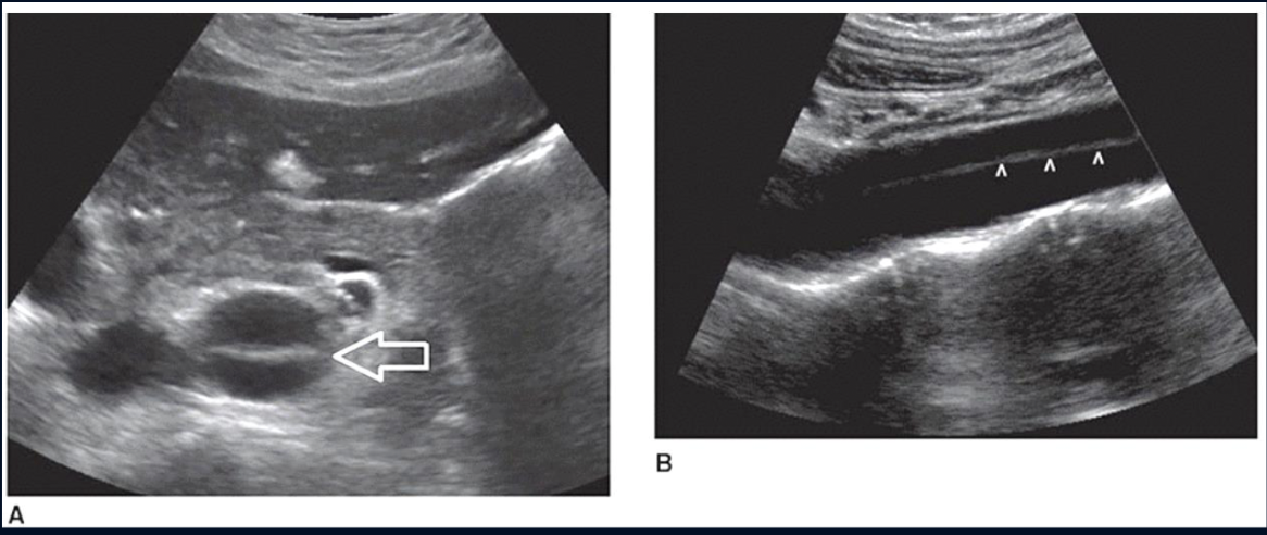
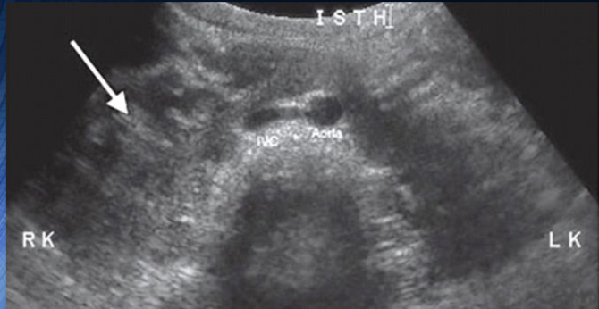
what is the arrow pointing to
Normal IVC (as it can be compressed)
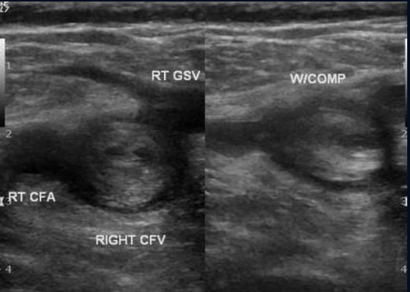
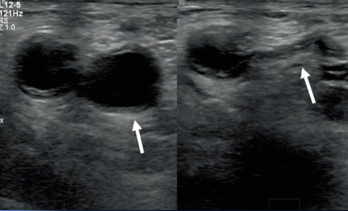
Pathology?
DVT in the Common femoral vein
Pathology?
IVC Thrombosis
Liver Pathology
Hepatitis
Haemangioma
HCC
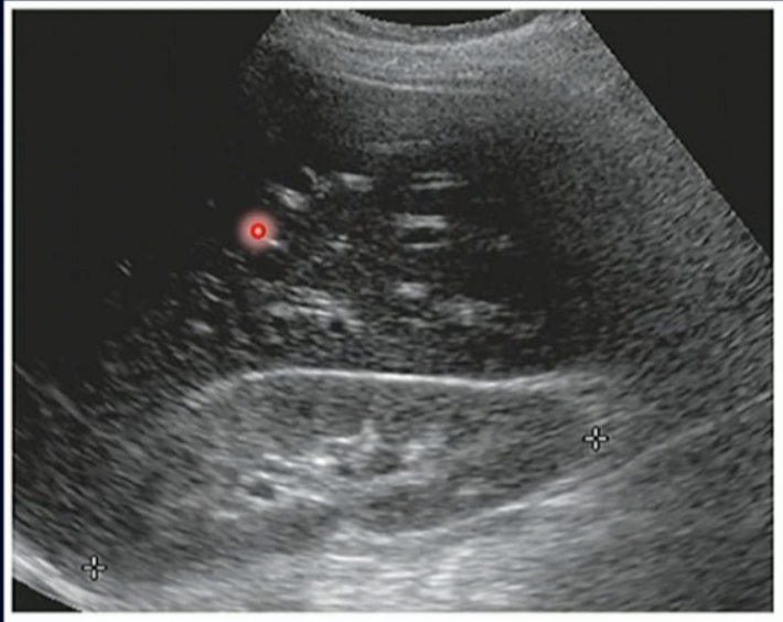
Fatty Infiltration
Different Lines/Ligaments of Liver
Cantlile’s Line: Runs from GB to IVC
Falciform Ligament: Splits Right + Left Liver lobe
Caudate Lobe above | Quadrate Lobe Below
Pathology?
Fat Infiltration
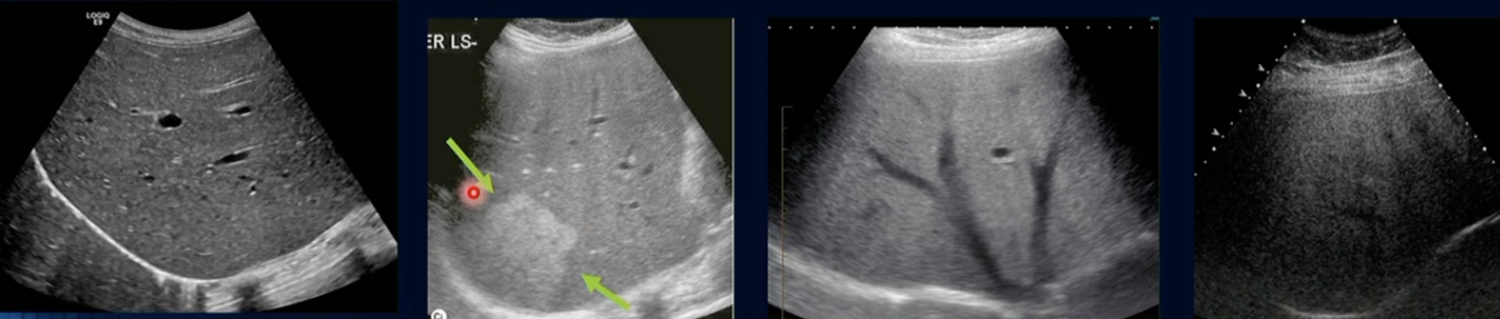
Pathology? Which is Normal and whats the worst
Fatty Infiltration
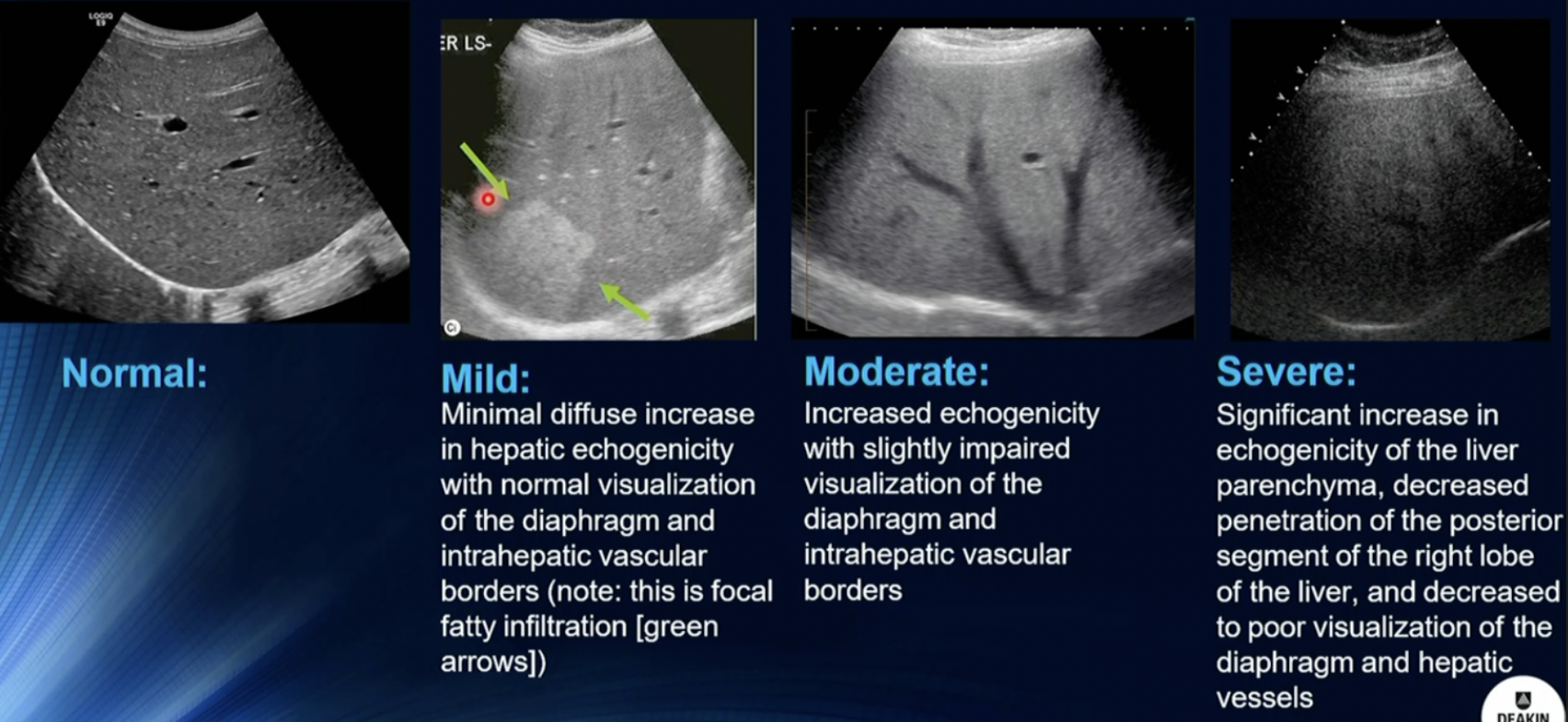
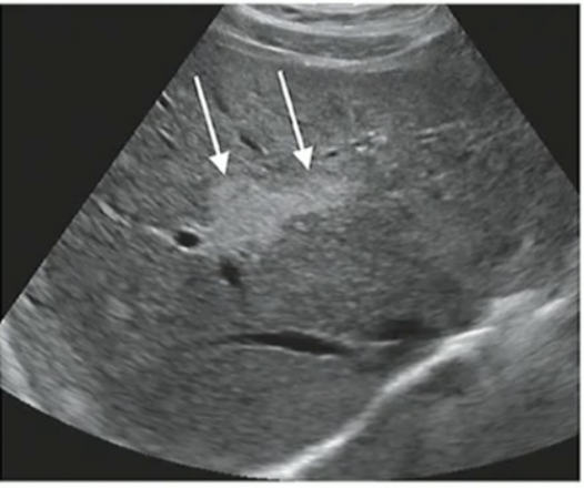
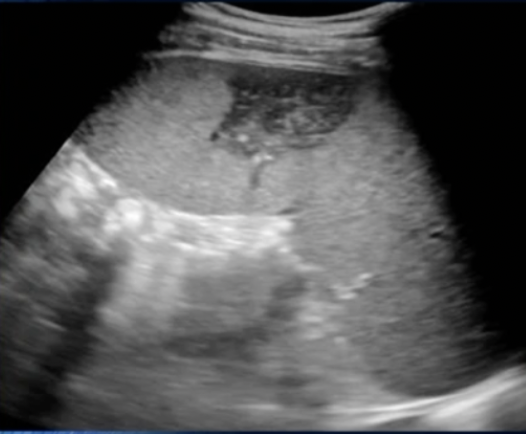
Pathology?
Hepatitis (Inflammation of Liver due to Echogenic areas)
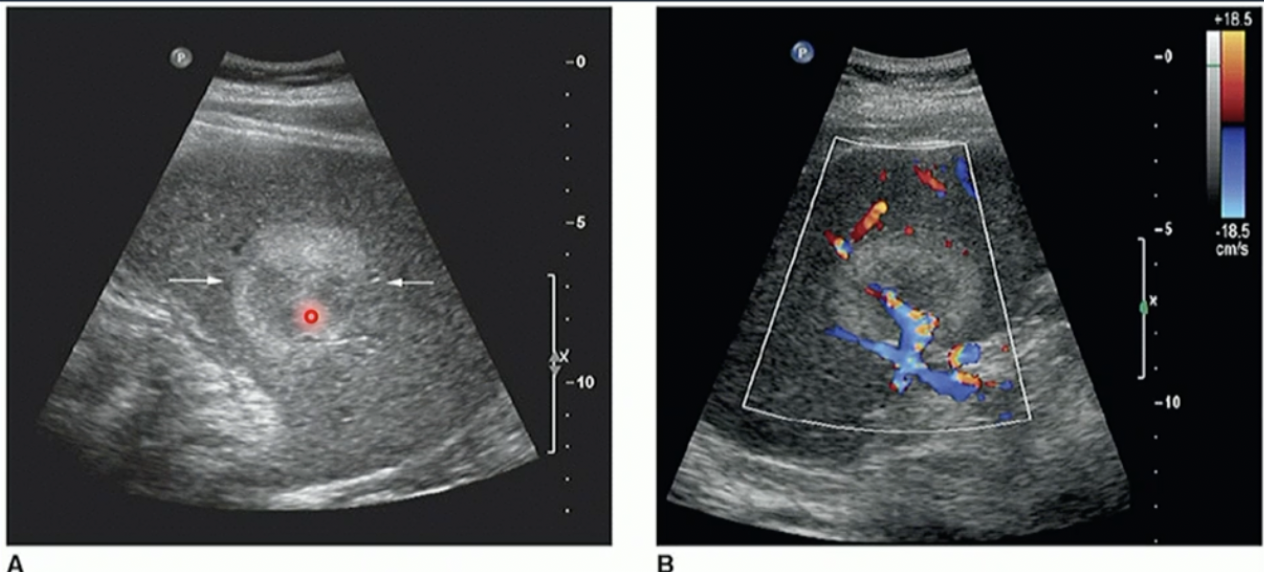
Difference between Haemangioma + HCC
Haemangioma has no blood vessels/lack of
HCC has blood vessels/blood flow
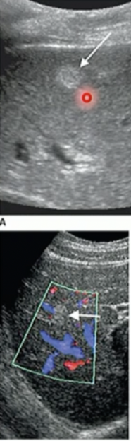
Pathology?
Haemangioma (due to no colour on it)
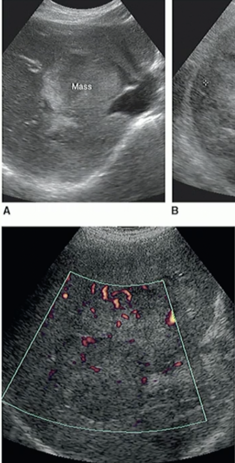
Pathology?
HCC due to colour/bloodflow
Spleen Pathology
Spenunculi (Parts of the Spleen that are separated, its normal)
Calcification
Haemangioma
Infarction
Splenomegaly (20cm on US is large)
What is this?
Splenuculi

Pathology?
Splenic Calcification (Post. Acous. shadowing + Echogenic appearance)
Path?
Haemangioma of Spleen (no B/F)
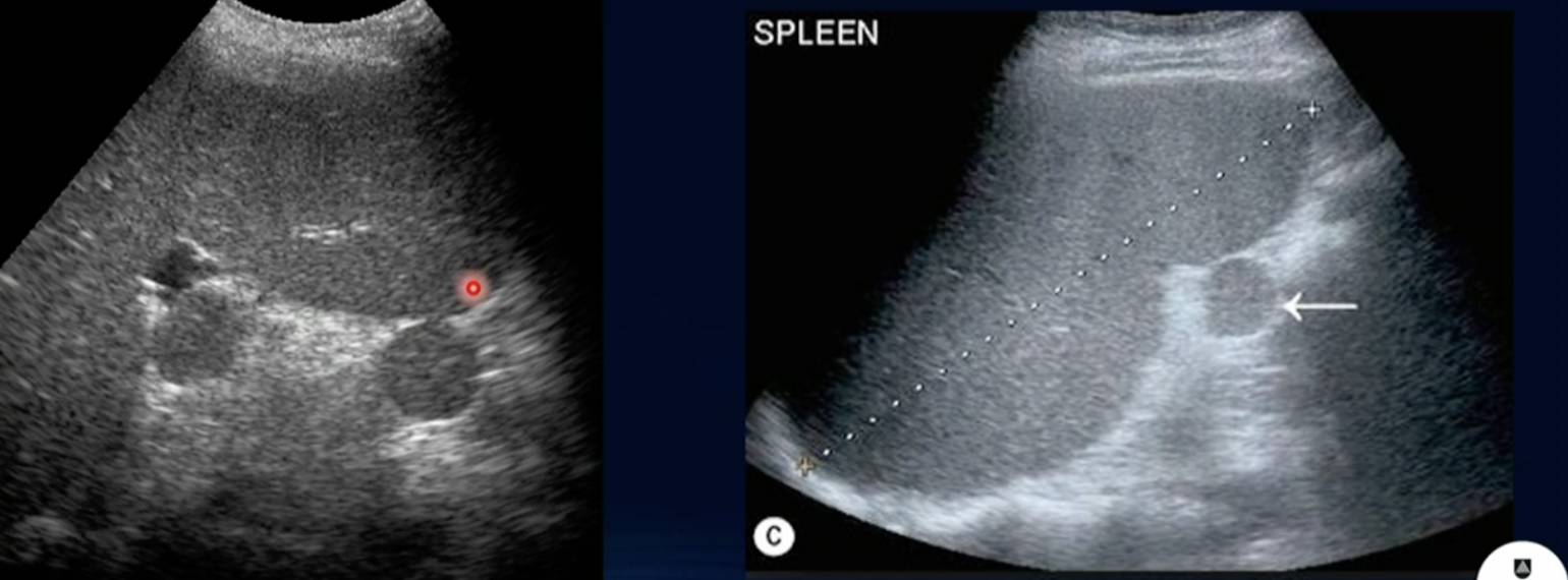
Path?
Splenic Infarction (Anechoic Region)

Path? (hint… SPLEEN)
Splenomegaly
Path?
Malignant Lymphoma of Spleen
Kidney Pathology
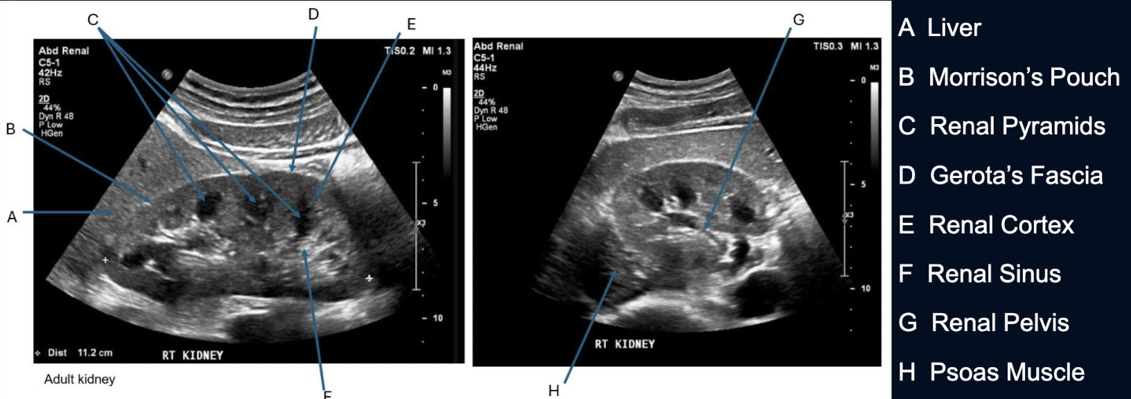
Normal
Dromedary Hump (Normal Enlargement of Cortex) (use colour to see theres no blood flow)
Extrarenal Pelvis (Renal Pelvis outside of Kidney)
Horseshoe Kidney (fusion of right+left kidney at the poles)
Duplex Kidney (2 Renal Pelvis’s)
Path:
Urolithiasis (Renal Calcification)
Hydronephrosis
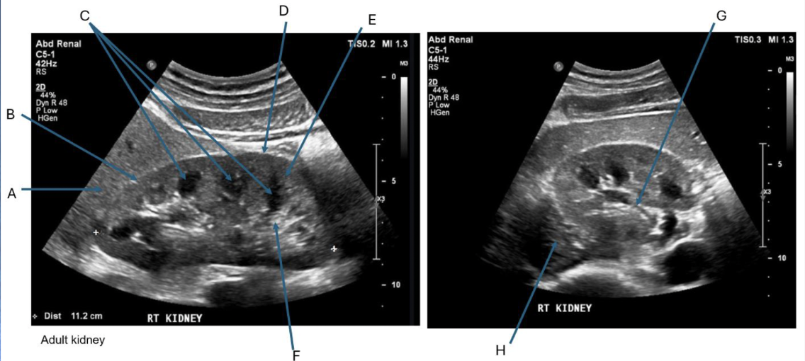
Label + Which is Long/TRV
Long then Trv
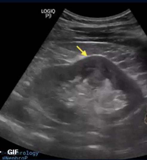
What is this?
Dromedary Hump
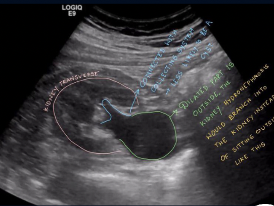
What is this?
Extrarenal Pelvis
What is this?
Horseshoe Kidney (hard to see on US)

What is this?
Duplex Kidney
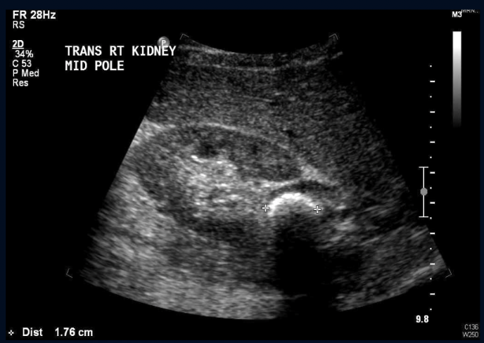
Path?
Urolithiasis in the Rt Middle Pole of Kidney
Path?
Staghorn Calculus
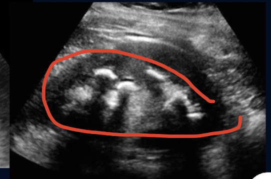
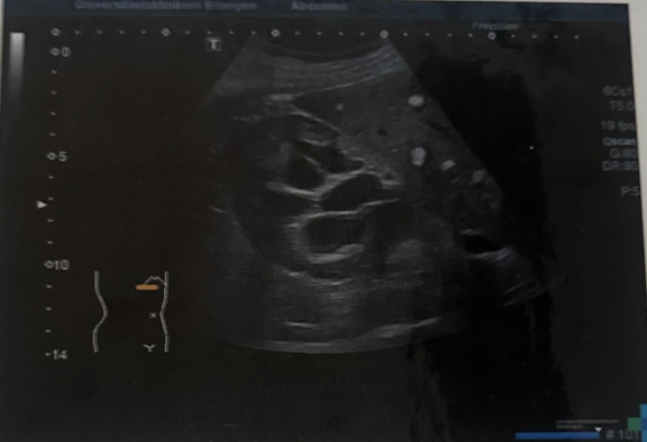
What is this?
Moderate Hydronephrosis (As it is one coherent structure there is not segmented Echogenic Structures)
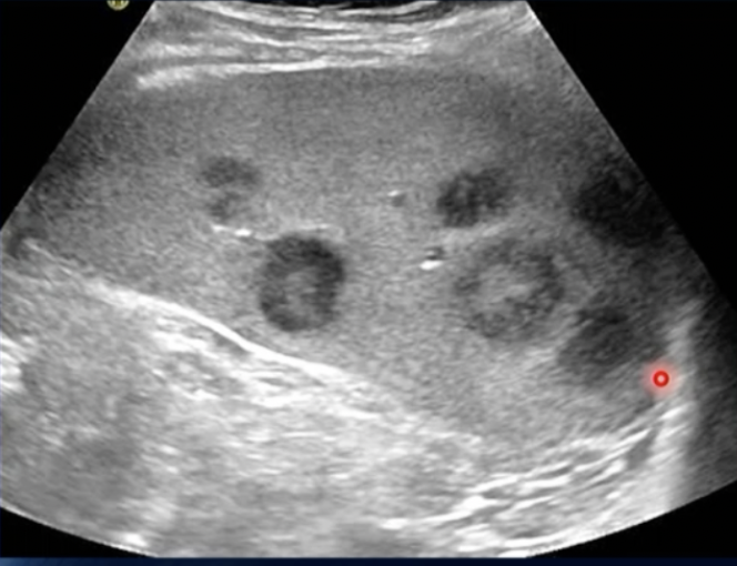
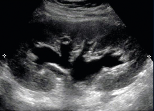
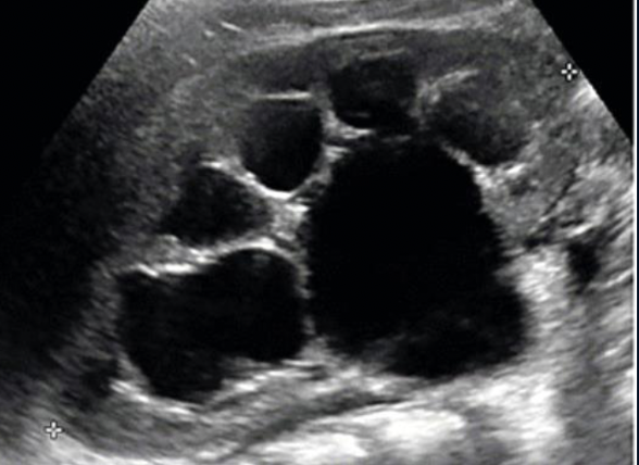
Path?
Parapelvic Cysts (as it is segmented)
Path?
Severe Hydronephrosis
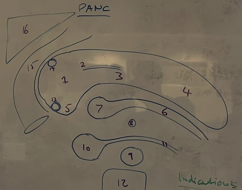
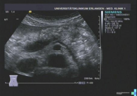
Label Pancreas
Head
Major Pancreatic Duct
Body
Tail
Uncinate Process
Splenic Artery
Celiac Trunk
Superior Mesenteric Artery (SMA)
Superior Mesenteric Vein (SMV)
Portal Vein
(Likely a vessel or duct branching)
Vertebral Body
…
…
Duodenum (Specifically the C-loop or descending part)
Liver
Pancreas Pathology
Pancreatitis (Acute = >3.5mm | Chronic = Scarring on Panc Body)
Tumour
Path?
Acute Pancreatitis
Path?
Chronic Pancreatitis (Scarring on body)
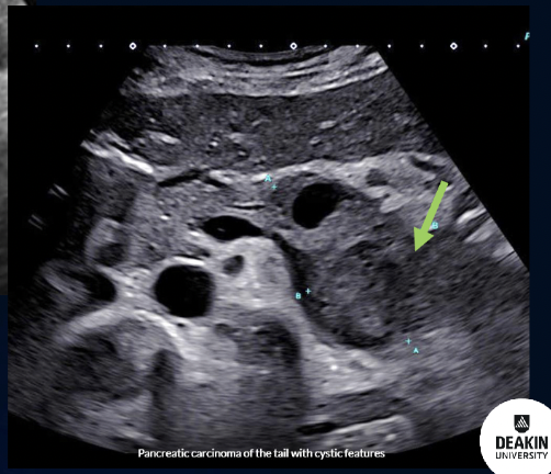
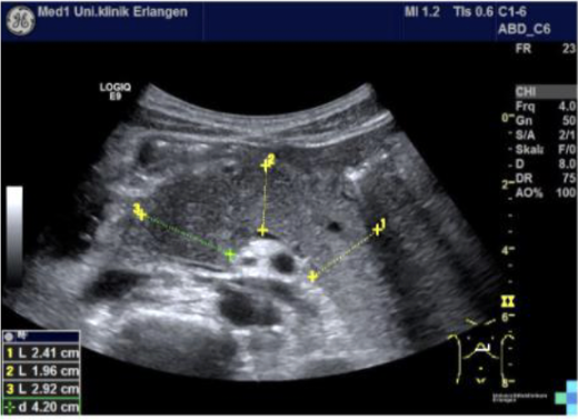
Path?
Tumour on the TAIL of Pancreas
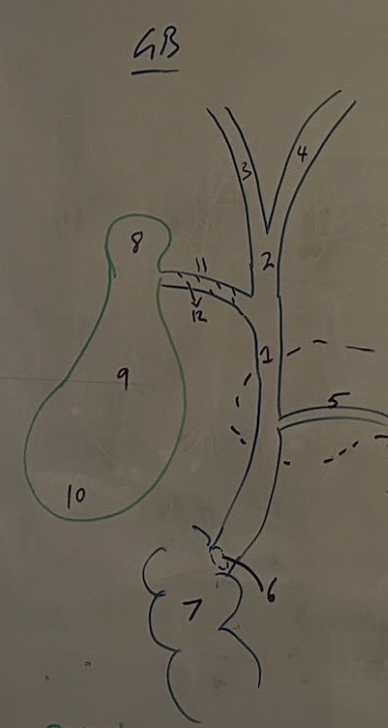
GB Anatomy
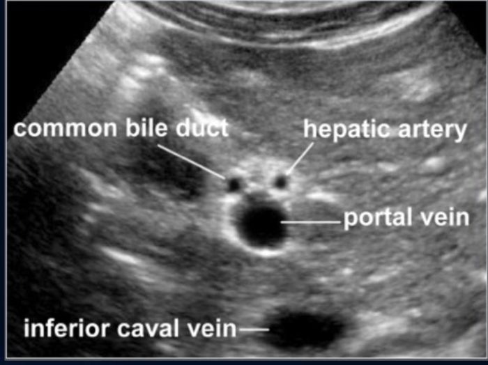
Common Bile Duct (CBD)
Common Hepatic Duct (CHD)
Right Hepatic Duct
Left Hepatic Duct
Major Pancreatic Duct
Ampulla of Vater
Duodenum
Neck
Body
Fundus
Cystic Duct
Heister
GB Path
Cholelithiasis
Cholecystitis (Larger than 3.5mm)
Polyps
Tumefactice Sludge
Adenomyosis (Comet Tail Artefact)
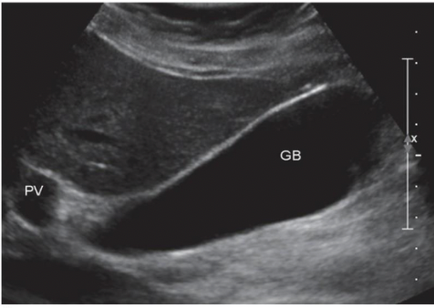
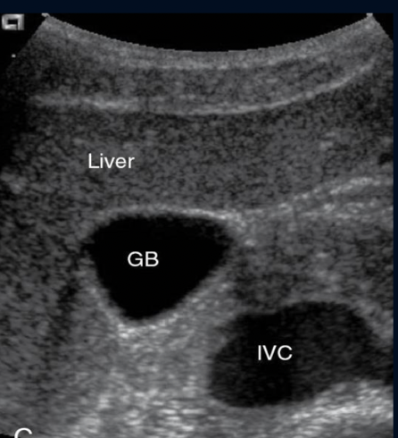
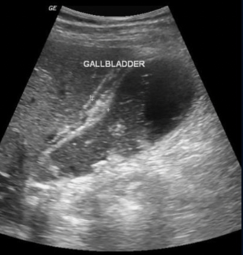
What view of GB?
Long, Portal Vein on the Left and Liver
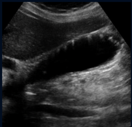
View of GB?
Trv
Neck Post, Fundus Superior
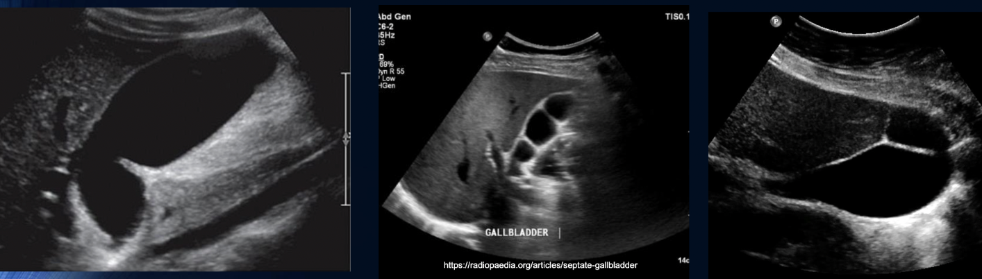
are these pathology or congenital?
Congenital
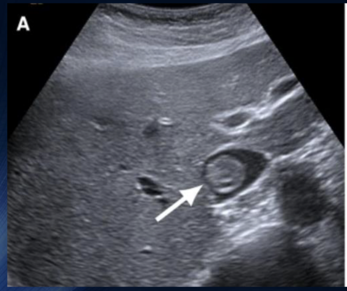
Path?
Cholelithiasis
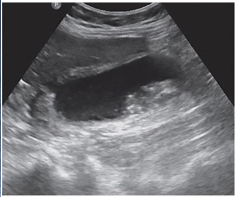
Path?
Tumefactive Sludge
Path?
Cholecystitis (thickened wall)
Path?
Adenomyosis
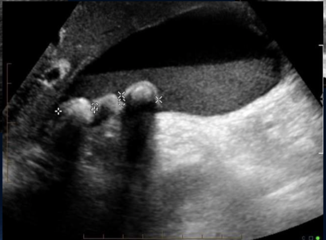
Path?
Polyps (NO ACOUSTIC SHADOW)
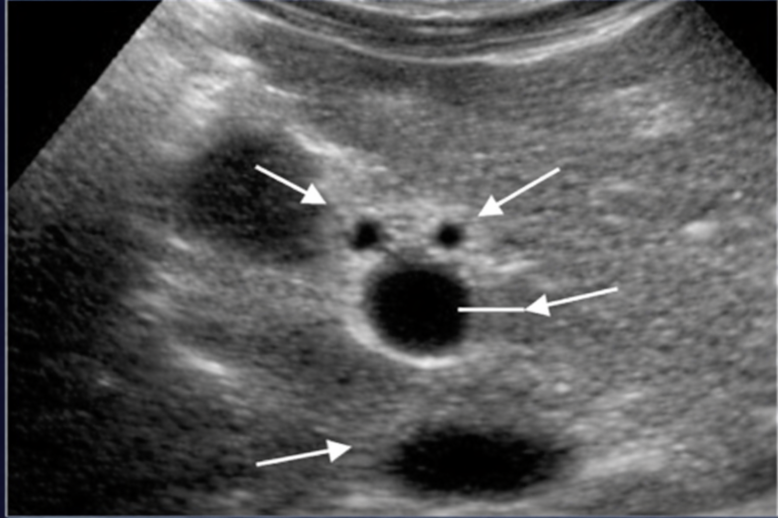
Label Anatomy of PortaHepatis TRV
Portahepatis Pathology
Choledolithiasis
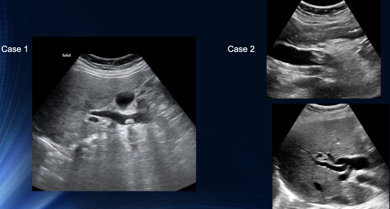
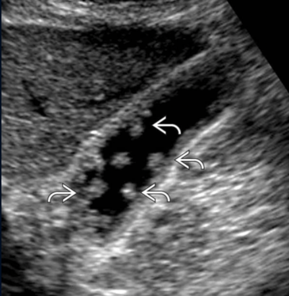
Path?
Case 1: Calculus presen with acoustic shadowing
Case 2: Common Bile Duct is Dilated + Echogenic Material Present within
Both Choledocholithiasis