(1) GENERAL ORGANS
1/17
There's no tags or description
Looks like no tags are added yet.
Name | Mastery | Learn | Test | Matching | Spaced |
|---|
No study sessions yet.
18 Terms
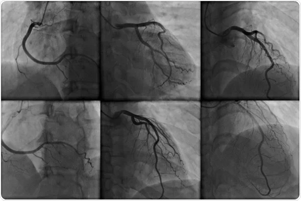
Angiography
- Is a radiographic test used to evaluate blood vessels and the circulation.
- Radiopaque material is injected through a catheter inserted in the blood vessel, and images are recorded using standard radiographic techniques.
Biopsy
Involves the removal and evaluation of tissue.
Computed Tomography
- Uses a computerized x-ray system to produce detailed sectional x-ray images.
- The system is very sensitive to differences in tissue density and produces detailed two-dimensional planar images; the use of contrast agents increases attenuation.
- In spiral or helical CT scanning pictures are taken continuously, which decreases the time needed to obtain images.
TRUE
TRUE OR FALSE
CT scans use X-rays to create detailed images, while MRIs utilize magnetic fields and radio waves
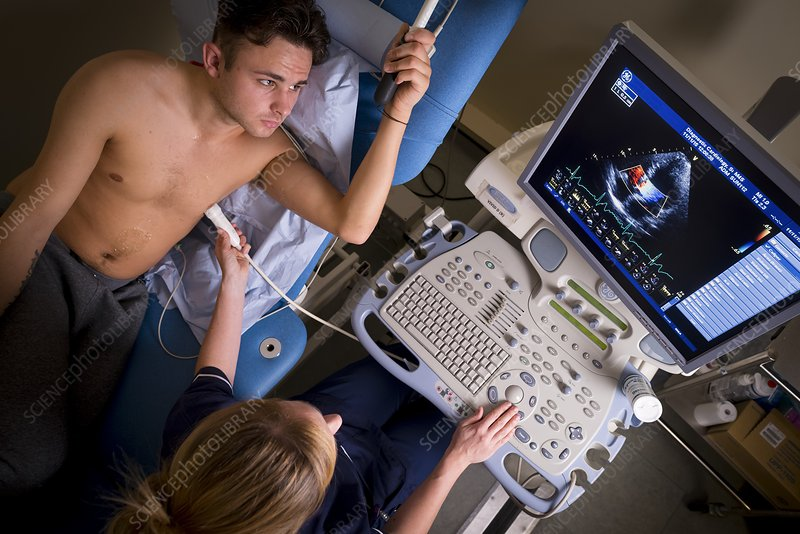
Doppler Echography
- Uses ultrasound technology to measure shifts in frequency caused by object movement.
- For example, it s used to evaluate blood flow velocity and turbulence in the heart (Doppler echocardiography) and peripheral circulation.
Endoscopy
Is used to examine the interior of a hollow viscus (e.g., digestive, respiratory, and urogenital organs and the endocrine system) or canal (e.g., bile ducts, pancreas).
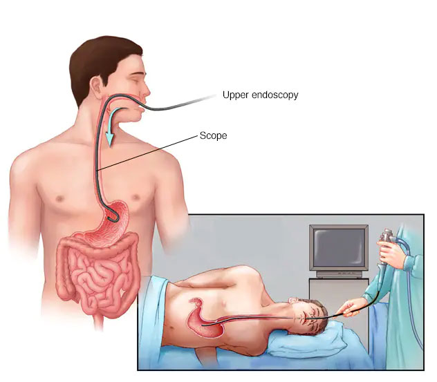
Endoscope
- A flexible or inflexible tube with a camera and light source, is inserted into a body orifice.
- Still and/or video images are recorded and biopsy specimens are obtained for tissue examination or other laboratory diagnostic tests
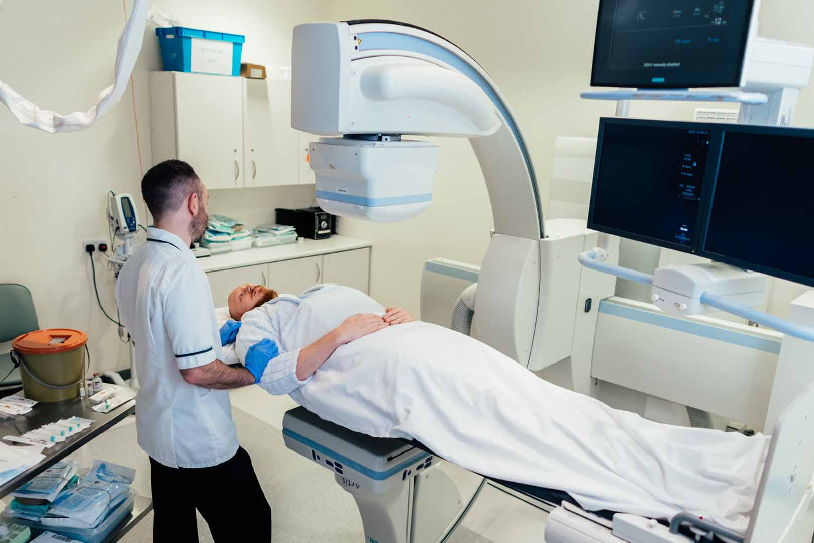
Fluoroscopy
- Uses a fluoroscope, a device that makes the shadows of x-rays visible, to provide real-time visualization of procedures.
- Exposes a patient to more radiation than routine radiography but often is used to guide needle biopsy procedures and nasogastric tube advancement.
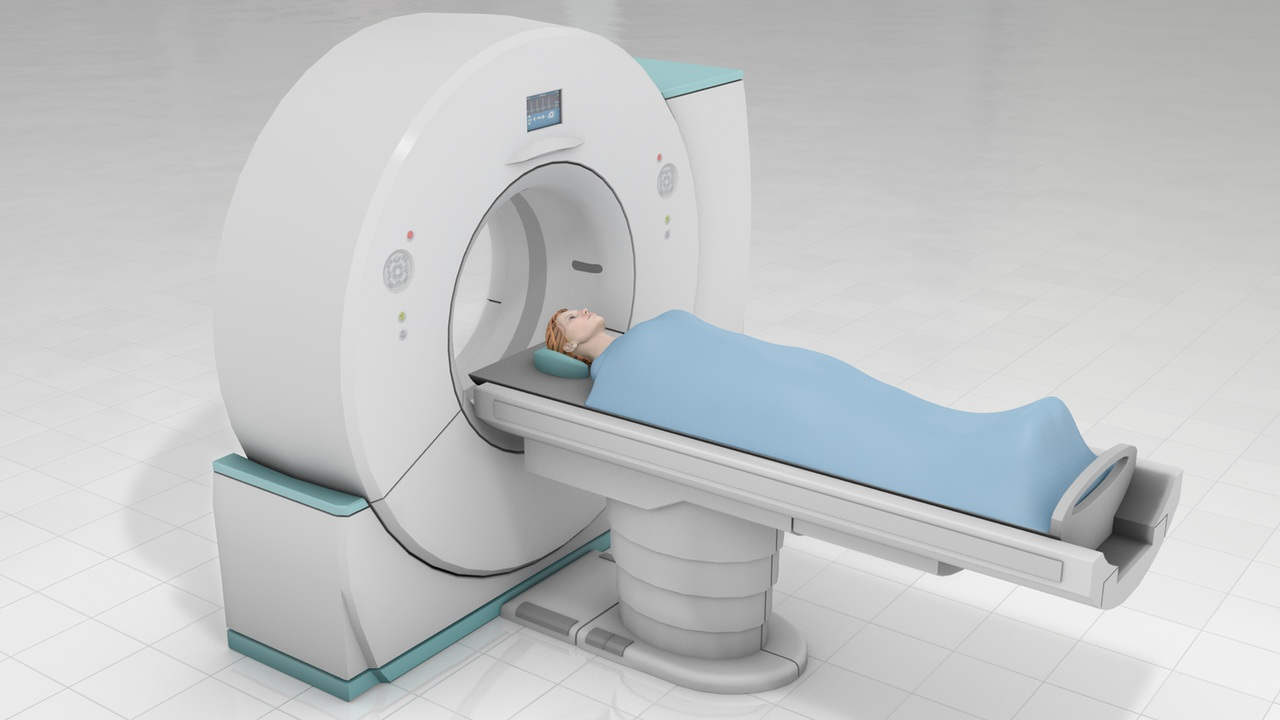
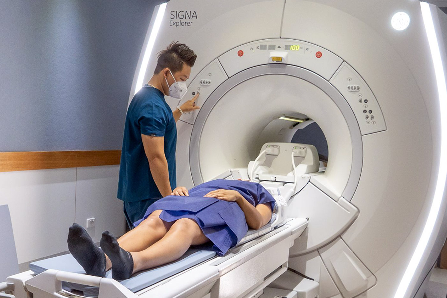
Magnetic Resonance Imaging (MRI)
- Uses an externally applied magnetic field to align the axis of nuclear spin of cellular nuclei.
- The patient is surrounded by the magnetic field.
- Brief radiofrequency pulses are applied to displace the alignment.
- The energy emitted when the displacement ends is detected, which allows finely detailed planar and three-dimensional images to be produced; the use of contrast agents increases the attenuation
Molecular Imaging
- Is an investigational technology that assesses biologic processes at the cellular and subcellular level in living tissue.
- Areas of interest include the biology of cancer and cardiovascular disease.
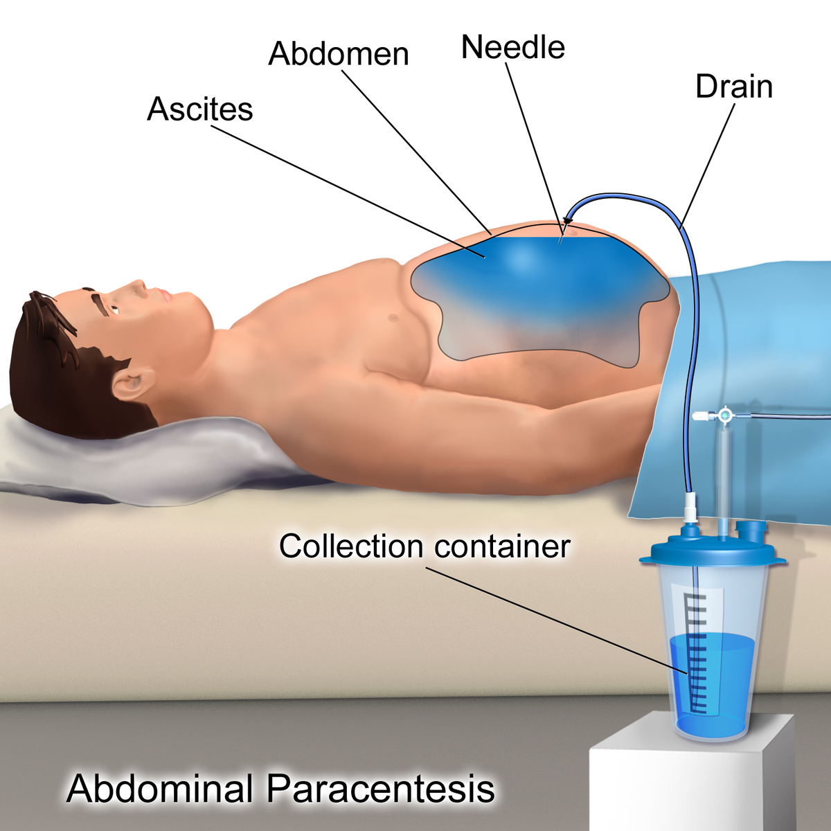
Paracentesis
- Is the removal and analysis of fluid from a body cavity.
- In abdominal paracentesis fluid is removed from the abdominal cavity.
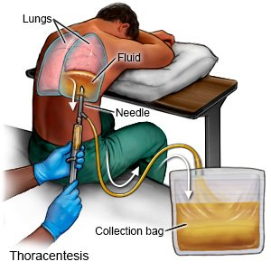
Thoracocentesis
Fluid is removed from the pleural space.
Plethysmography
- Assesses changes in the size of vessels and hollow organs by measuring displacement of air or fluid from a containment system (Figure 5-13).
- Body type is used to assess pulmonary function.
Positron Emission Tomography (PET)
- Uses positron-emitting radionuclides (e.g., fluorodeoxyglucose F 18) to visualize organs and tissues of the body.
- The radionuclides decay, producing positrons that collide with electrons.
- A special camera detects photons released when the positrons and electrons collide.
- Provides quantitative information regarding the structure and function of organs and tissues.
- Is commonly used to detect and monitor malignancies.
Radionuclide Studies
- Involve the administration of oral, parenteral, or inhaled radioactive chemicals or pharmaceuticals.
- X-ray images, usually serial, record the collection and dispersion of the radioactive material.
- The ventilation-perfusion scan of the lungs and the bone scan are examples of radionuclide studies.
Single-Photon Emission Computed Tomography
- Is similar to PET but involves the administration of radionuclides that emit gamma rays.
- Is less expensive than PET but provides more limited image resolution.
- Is commonly used to assess the coronary arteries, bone, brain, prostate, and thyroid.
Standard Radiography (Plain Films, X-Ray Films)
- Images are produced on photographic plates by passing x-rays (roentgen rays) through the body.
- These films are sometimes difficult to interpret because the three dimensionality is lost on the planar images.
- Film-based radiography is being replaced by digital imaging, in which a flat-panel imager containing thousands of independent semiconductor detectors is used instead of film.
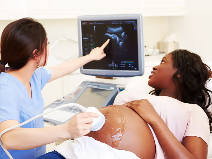
Ultrasonography (Echography)
- Uses ultrasound (high-frequency sound waves imperceptible to the human ear) to create images of organs and vessels.