Exam questions- Cardiology
1/43
Earn XP
Description and Tags
From all avalible exam papers
Name | Mastery | Learn | Test | Matching | Spaced |
|---|
No study sessions yet.
44 Terms
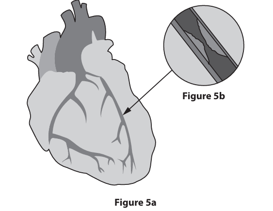
Figure 5a shows the external structure of a human heart. Figure 5b shows a blocked coronary artery.
State what is meant by the term myogenic. (2)
(muscle/heart/ventricle) beat/contracts/pumps (1)
on its own/spontaneously/rhythmically (1)
without input/signal from {nerves/brain} (1)
Describe the function of the coronary artery. (2)
{carries/delivers/takes/supplies} blood containing {oxygen/glucose/lipids} (1)
to {heart muscle/ventricle wall/myocardium} (1)
One risk factor for cardiovascular disease (CVD) is a high fat diet. This may lead to fatty deposits in the wall of the coronary artery. Fatty deposits could eventually lead to the coronary artery becoming blocked. Explain how a blocked coronary artery may lead to a heart attack. (4)
less oxygen/ oxygenated blood to heart {walls/tissue/muscle/cells} (1)
so {less/no} respiration (1)
so {less/no} ATP/energy(1)
so {part of heart/heart cells} die/scar tissue (1)
so heart cannot {beat/pump/contract} (1)
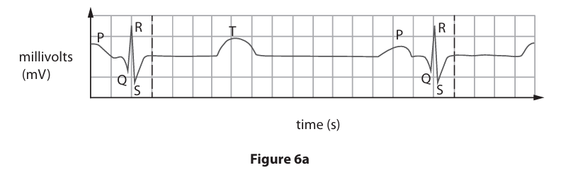
Figure 6a shows an electrocardiogram (ECG) trace during the cardiac cycle.
Identify the letter on Figure 6a that represents when the atria contract. (1)
A- P
B- R
C- S
D- T
A- P
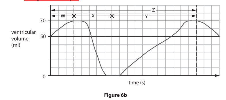
Figure 6b shows the changes in the volume of blood in the left ventricle during the cardiac cycle. Identify the letter on Figure 6b that represents ventricular systole. (1)
A- W
B- X
C- Y
C-Z
B- X
The duration of a heartbeat in Figure 6b is 0.8 seconds.
Calculate, using information from Figure 6b, the cardiac output for this heart.
Cardiac output = stroke volume (ml) X heart rate (beats per minute)
Show your working. (3)
stroke volume (1)
70 (ml) heart rate (1) 60/0.8
cardiac output (1)
70 x 75= 5250
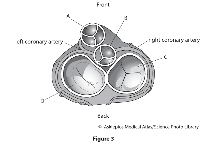
Figure 3 shows a transverse (cross) section of a human heart, seen from above. The section shows the four heart valves.
Identify the aortic valve in Figure 3. (1)
-A
-B
-C
-D
B
Describe the function of the bicuspid valve. (2)
prevent backflow (of blood) (1)
from (left) ventricle to (left) atrium (1)
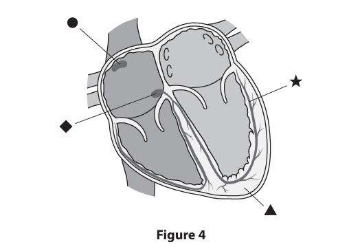
Figure 4 shows a longitudinal section of a human heart.
Identify the atrioventricular node (AVN) in Figure 4. (1)
A circle
B triangle
C diamond
D star
C- diamond
The fibrous layer separating the atria from the ventricles does not conduct electrical impulses.
Describe how the electrical impulses spread from circle to star in Figure 4. (3)
(from) SAN/sinoatrial node/pacemaker (1)
(spread) {across/from} the {atria/muscle/ tissue/wall} (1)
(through/to) {AVN/ diamond} (1)
reference to time delay (1)
(down) {bundle of His/bundle branch/septum} (1)
(up) {Purkinje/Purkyne} fibres (1)
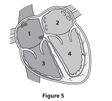
Cigarette smoke contains many harmful chemicals, such as nicotine. When a smoker inhales cigarette smoke into their lungs, these chemicals may be absorbed into the blood.
Identify the heart chamber in Figure 5 that would first receive nicotine from the lungs. (1)
A- 1
B- 2
C- 3
D- 4
B- 2

Figure 6 shows the changes in blood pressure throughout the circulatory system.
Explain two changes in arterial blood pressure, shown between points W and X, in Figure 6. (4)
Blood pressure drops/reduces/falls (1)
as it gets {near {arterioles/capillaries/smaller vessels}/further from heart} / (due to) less (peripheral) {resistance/friction} (1)
OR
when the pressure is increasing/high/120 (1)
(it is because the) ventricles are contracting/systole/heart beats/ heart contracts/contraction phase of cardiac cycle/aortic valve is opening/has to overcome (peripheral) resistance (1)
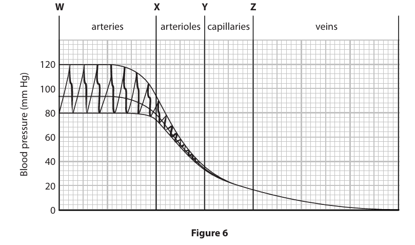
Calculate, using information in Figure 6, the percentage decrease in blood pressure in the capillaries from point Y to point Z. Show your working. (3)
Difference (1)
(their) reading for Y minus (their) reading for Z
Division (1)
(their) difference / (their) value for Y
Percentage (1)
X100
any number within range 40 to 64
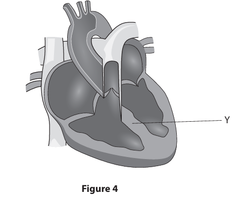
Figure 4 shows the structure of a human heart.
Name the structure labelled Y on Figure 4. (1)
septum
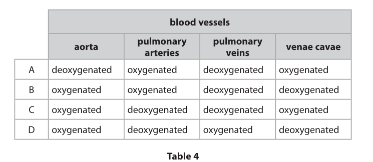
Identify which row of Table 4, A, B, C or D, correctly describes the type of blood present in each blood vessel. (1)
-a
-b
-c
-d
D
Sentence 1 gives an incomplete definition. Identify the missing words from Sentence 1 to complete the definition. (1)
The volume of blood pumped out of the heart in one minute is the ............................................... .
A cardiac output
B heart rate
C stroke volume
D ventricular systole
A cardiac output
A patient visits their doctor. The doctor determines they have a reduced stroke volume. The doctor tells the patient they have congestive heart failure. The doctor finds that the patient has a resting heart rate of 105 beats per minute.
(i) Which term describes this patient’s resting heart rate? (1)
A atrial fibrillation
B bradycardia
C tachycardia
D ventricular fibrillation
C tachycardia
After the patient received treatment for congestive heart failure, their resting stroke volume increased from 40 ml to 60 ml. Calculate the percentage increase in this patient’s stroke volume after treatment.
Show your working (3)
Subtraction (1)
60 – 40 or 20
Division (1)
20/40
Evaluation (1)
X 100 = 50 (%)

A student investigated the effect of caffeine on the heart rate of the water flea, Daphnia.
Figure 3 shows a Daphnia and the location of its heart.
The student placed a Daphnia in a Petri dish containing pond water.
The student observed the Daphnia under a light microscope. The student counted the number of Daphnia heartbeats per minute when the Daphnia was in pond water containing differing concentrations of caffeine.
The results are shown in Table 2.
Calculate the mean number of heartbeats for the Daphnia in 0.05% caffeine concentration. Show your working. (2)
addition (1)
384 + 393 + 405 + 390 + 405 + 390 or 2367
division (1)
(2367) / 6 or
Answer= 394.5 or 395 bpm
Calculate the percentage increase in the mean Daphnia heart rate between 0.00% caffeine concentration and 0.01% caffeine concentration.
Show your working. (3)
difference (1)
358.0 – 240.5 (= 117.5 or 118)
division (1)
117.5 ÷ 240.5 or 118 ÷ 240.5
percentage (1)
0.488 X 100= 48.86% - 49%
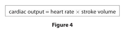
Figure 4 shows the equation for cardiac output. Explain how caffeine changes the cardiac output of the Daphnia. (4)
(caffeine) increases cardiac output (1)
• (because) heart rate is increased/more heartbeats (per minute) (1)
• (caffeine) is a stimulant/it stimulates (1)
• {binds/fits/complementary shape} to receptors (1)
• {increases electrical activity in/acts on} SAN (1)
• increases {strength/force/power) of contraction of {heart/ventricle(s)/heart muscle} (1)
• increases stroke volume (1)
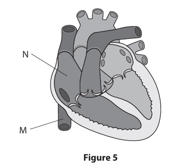
Figure 5 shows the human heart in longitudinal section, together with some of the main blood vessels.
Identify the structures labelled M and N in Figure 5. (2)
(M =) vena cava (1)
(N =) (right) atrium (1)
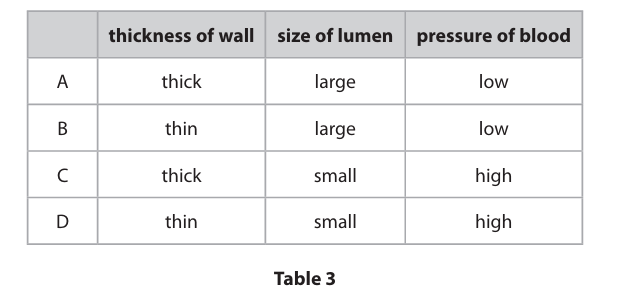
Blood leaves the heart, in the aorta, to travel around the body.
Which row in Table 3 correctly outlines the features of the aorta? (1)
-a
-b
-c
-d
C thick, small, high
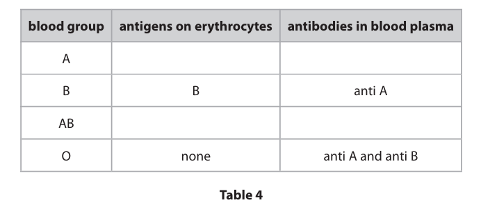
The two most important blood grouping systems for humans are the:
● ABO system
● rhesus system.
Table 4 lists the antigens and antibodies present in people of different ABO blood groups. Complete Table 4 for blood groups A and AB. (4)
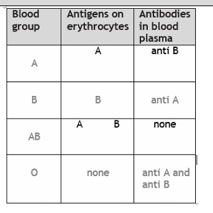
Explain what happens if group B blood is transfused into a patient who is blood group O. (2)
anti-B (antibodies)/antibodies in {recipient/patient/group O} (1)
• {bind to/react to/fit/have complementary shape to} (B/donor) antigens (1)
• (cause) blood to clot/ (cause rbc to) agglutinate/clump/lyse/split/be destroyed (1)
The rhesus system relates to the presence or absence of D antigens on the surface of the erythrocytes.
Explain why, in an emergency, anyone can be given a transfusion with blood of type O rhesus negative. (3)
• no antigens in (donor/group O/ rbc/ blood) (1)
• (so) no {reaction/attack/binding/recognising}with (recipient’s /anti-A or anti- B) antibodies (1)
• (so) no clotting/agglutination/clumping/lysing (1)
• no D (antigen/protein) (in O –ve blood)/recipient not exposed to D protein / does not produce anti-D antibodies (1)
• donor antibodies not (enough to) react with recipient antigens (as more recipient blood than donor blood) (1)
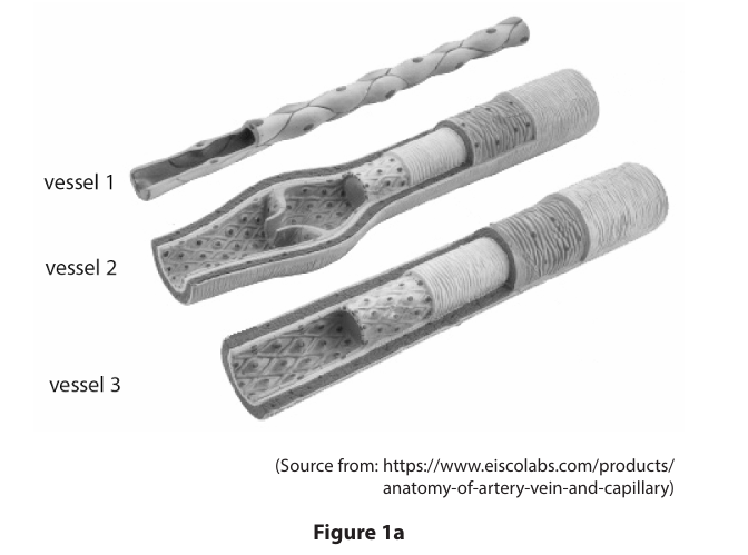
Figure 1a shows the structures of three different types of blood vessel.
Which row correctly identifies each type of blood vessel in Figure 1a? (1)
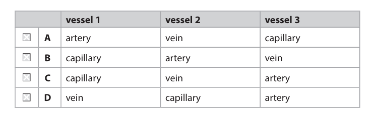
C capillary, vein, artery
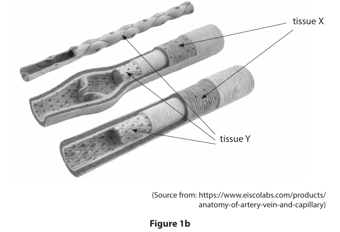
Figure 1b shows tissue X and tissue Y in the blood vessels.
Draw one line from each tissue to its correct name. (2)
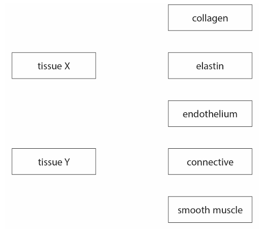
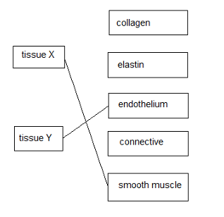
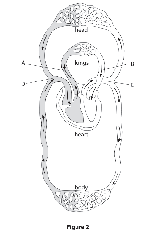
Figure 2 represents the human circulatory system.
Which letter, A, B, C or D, labels the pulmonary artery? (1)
A
B
C
D
A
Describe the function of the vena cava. (2)
1) carries deoxygenated blood (1)
2) (carries blood) from {body/head/veins} (1)
3) to {heart/ (right)atrium} (1)
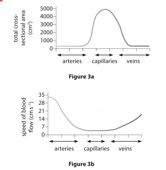
Figure 3a shows the total cross-sectional area of the three main types of blood vessel.
Figure 3b shows the speed of blood flow in the three main types of blood vessel.
State the relationship between the total cross-sectional area of the capillaries and the speed of blood flow in the capillaries, shown in Figures 3a and 3b. (1)
inverse proportion / negative correlation (1)
The total cross-sectional area of the arteries is 300 cm2.
The total cross-sectional area of the capillaries is 5 000 cm2.
Calculate the percentage increase in cross-sectional area when comparing arteries with capillaries.
Show working (3)
subtraction (1)
5000 - 300 division (1)
4700 ÷ 300 (15.67) multiplication (1)
X 100 (1567) = 1560-1570%
Give three functions of capillaries. (3)
1. carry {oxygen/oxygenated blood} (to cells/tissues) / absorb oxygen (at lungs) / gas exchange (1)
2. absorb nutrients (in gut) / carry nutrients (to cells) / absorb water (gut/kidney) (1)
3. allow (facilitated) diffusion / osmosis (in correct contest) / slow the blood flow (so time for exchange) (1)
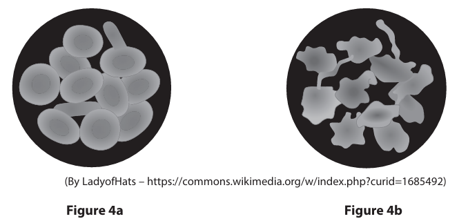
Figure 4a shows red blood cells in blood plasma.
Figure 4b shows red blood cells after being placed in a 5% salt solution for 10 minutes. Sentence 1 explains the changes that happened in the red blood cells in Figure 4b.
Identify the missing words, M and N, in Sentence 1. (2)
When red blood cells were placed in a 5% salt solution, … M… left the cells by the process of … N… .
M- water
N- osmosis
Cardiovascular disease (CVD) is a major cause of death in the UK and the rest of the world. CVD includes heart disease and strokes.
Diet and biological sex are two factors that may affect the risk of CVD.
Explain how diet and biological sex affect the risk of CVD. (6)
Diet- • Western diet with high amounts of calories, particularly sugar, leads to obesity, which is a major risk factor for heart attack and stokes.
• Obesity also leads to inflammation and type 2 diabetes - both of which are risk factors for CVD.
• High levels of saturated fat in the diet may lead to high blood levels of LDL/cholesterol, fatty plaques and atherosclerosis, which can lead to thrombosis in the coronary artery or in arteries in brain.
• High salt intake leads to high blood pressure – a risk factor for CVD.
Sex- More women than men die rather than recover from heart attack as their symptoms present differently and they are more often misdiagnosed.
• Women more likely to visit GP/have medical checks and thus have high blood pressure diagnosed, which may then be treated.
• There may be sex differences in other risk factors such as smoking and alcohol consumption, and in diet.
• Women store more fat/higher body fat

Figure 2 shows a section through a human heart.
Which symbol, in Figure 2, shows the location of the sinoatrial node (SAN)? (1)
A- star
B- diamond
C- pentagon
D- triangle
A- star
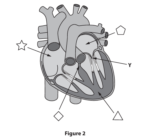
Name structure Y in Figure 2. (1)
AV/atrioventricular/bicuspid/ mitral} (valve) (1)
Heart muscle is myogenic.
Sentence 1 partially explains the meaning of the term myogenic.
Heart muscle .........................F..................................... on its own without any stimulation from .............................G................................. .
Identify the missing words, F and G, in Sentence 1. (2)
F = contracts / beats / pumps / pulses (1)
G = nerves / nerve impulses / nervous system / nerve signals / brain (1)
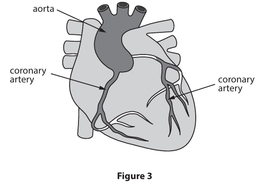
Figure 3 shows a heart, aorta and coronary arteries.
Describe the function of the coronary arteries. (2)
carry / provide / take /supply {oxygen/oxygenated blood} (1)
to heart {muscle / heart wall / wall of {ventricles/atria}/myocardium} (1)
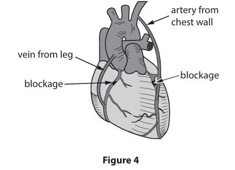
Figure 4 shows how bypass surgery can treat blockages in the coronary arteries. Bypass surgery involves a transplant operation.
Explain two advantages of using blood vessel tissue taken from the patient, rather than tissue taken from a donor. (4)
compatible / genetically identical / not foreign / has self antigens / same blood group (1) •
(so) no (risk of) rejection (1)
• (so) less risk of failure / less likely to need another operation (1)
• (so) no need for {immunosuppressants / drugs to prevent rejection} (1)
• (so) immune system will not be {weakened/compromised}/less risk of infections (1)
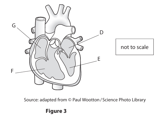
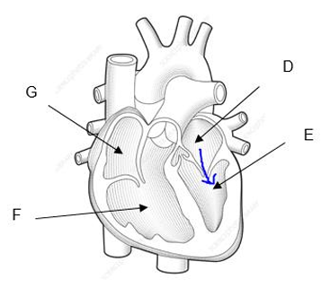
Figure 3 shows the structure of a human heart and associated blood vessels, seen from the front.
Draw an arrow on Figure 3 to show the direction of blood flow through the left side of the heart. (1)
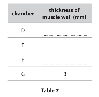
A scientist measured the thickness of the muscle wall of each of the four heart chambers, D, E, F and G, shown in Figure 3.
The measurements were:
8 mm 16 mm 3 mm 3 mm
Complete Table 2 for chambers D, E and F. Chamber G has been completed for you. (3)
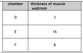
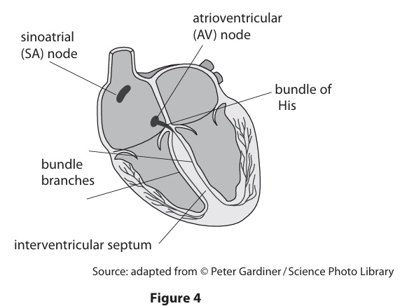
Figure 4 shows the positions of the nodes that regulate the rhythm of the heartbeat.
The impulse first spreads from the SA node to the AV node. Then the impulse travels through the bundle of His and the bundle branches. There is a time delay at the AV node as the impulse spreads.
Explain why this time delay is important. (3)
to allow atria to {finish contracting/to empty/finish systole/relax (1)
so blood {goes from atria into ventricles/goes through AV valves (1)
(to allow time) for valves to close/ to prevent backflow} (1)
before {ventricles start to contract/blood leaves heart/blood {pushed/pumped/forced} out of heart} (1)
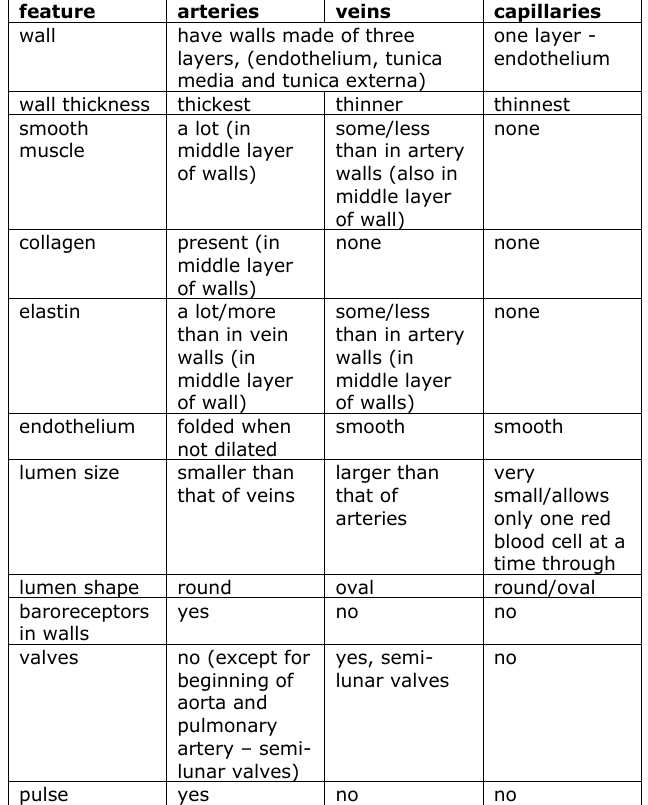
Compare the structures of arteries, veins and capillaries.
Your answer should refer to both similarities and differences.
You may use annotated diagrams and/or a table to support your answer. (6)