Histology- special senses
1/53
Earn XP
Description and Tags
Name | Mastery | Learn | Test | Matching | Spaced | Call with Kai |
|---|
No analytics yet
Send a link to your students to track their progress
54 Terms
tissue type
respiratory epithelium, aka pseudostratified columnar epithelium
tissue type
sero-mucous glands of the lungs
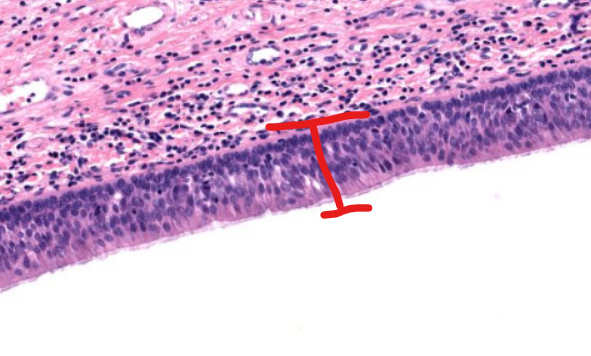
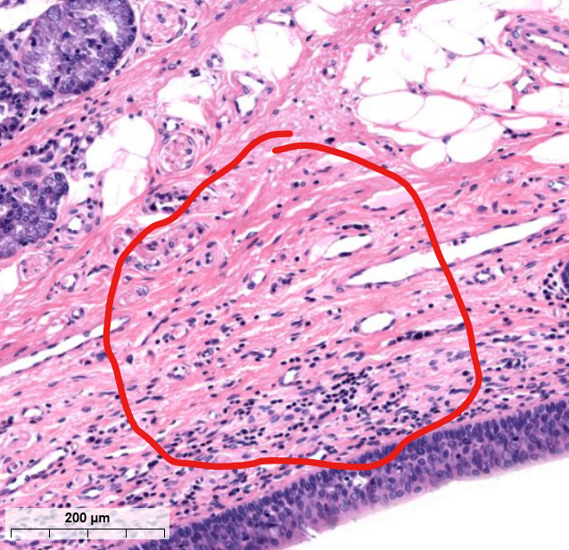
layer
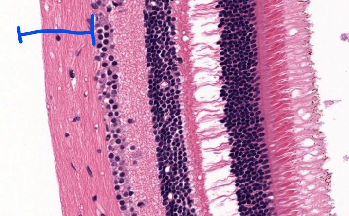
lamina propria
the layer of connective tissue that is under respiratory epithelium’s basement membrane
made of loose irregular CT
layer
basement membrane
supports respiratory epi
structure
terminal bronchiole-
conducting airway, has simple columnarepi
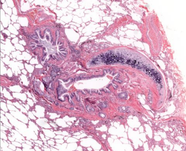
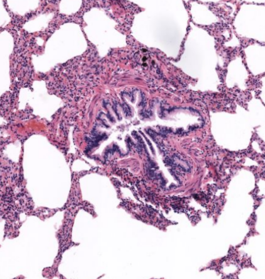
structure
primary bronchiole-
notice the bronchial cartilage
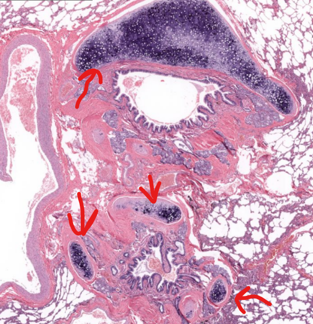
tissue type/structure
bronchiole cartilage-
found next to primary bronchi (just branched off of trachea)
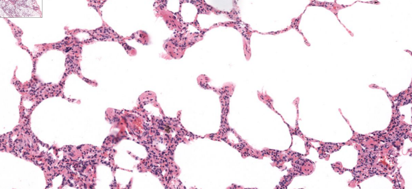
structure
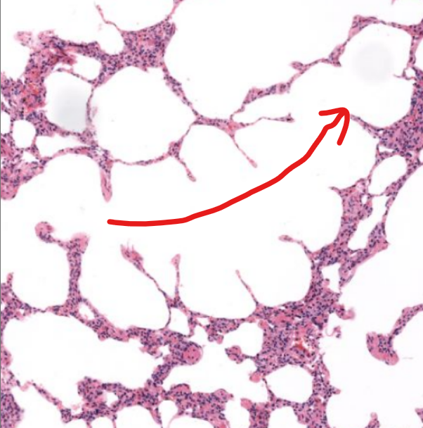
alveoli-
blind ending sacs, location of gas exchange within lungs

structure
alveolar sacs- spherical-like spaces where multiple alveoli connect to
structure
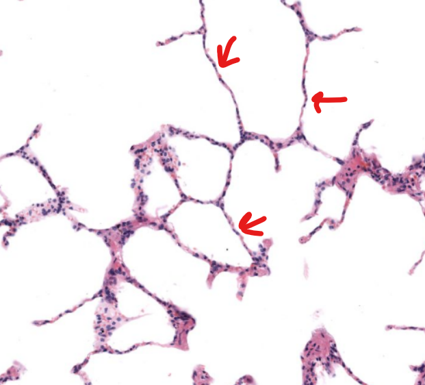
alveolar duct-
arise from bronchioles and are passageways lined with alveoli
tissue type
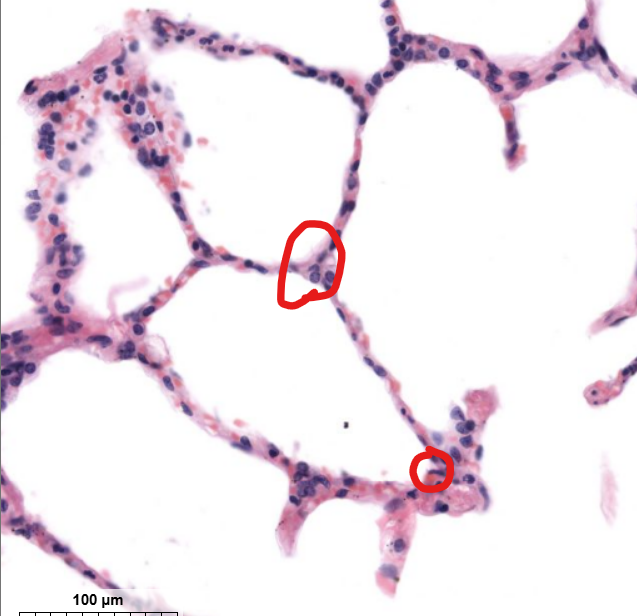
type 1- simple squamous epi
gas exchange occurs here
tissue type
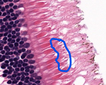
alveolar type 2 cells-
secrete surfactant
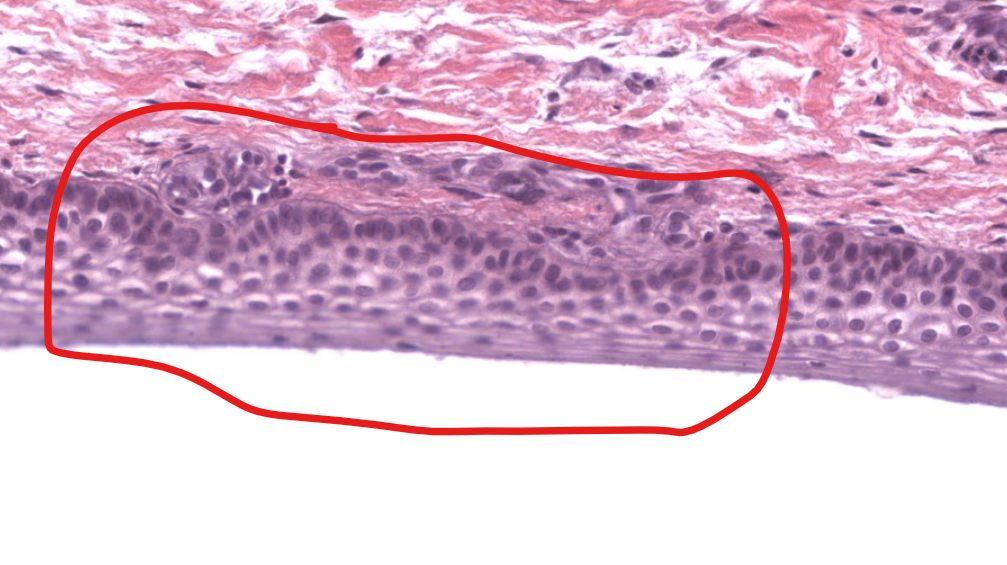
tissue type (on epiglottis)
respiratory epithelium
tissue type/layer?
lamina propria-
supporting respiratory epi
tissue type
elastic cartilage-
provides support to epiglottis
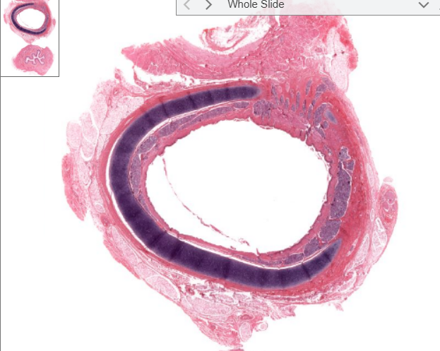
structure
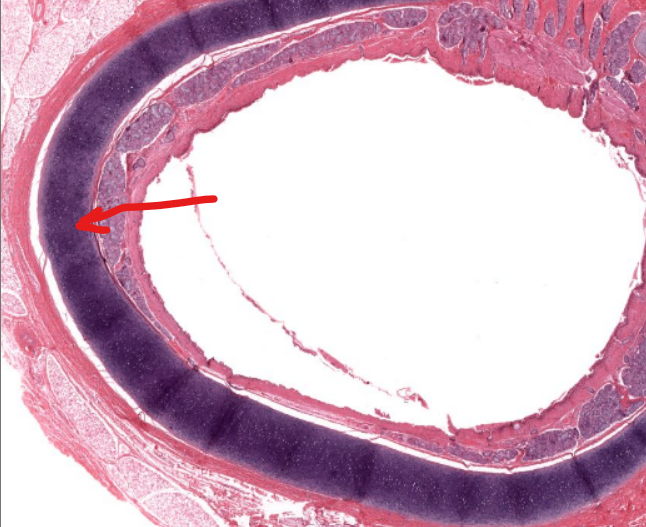
trachea
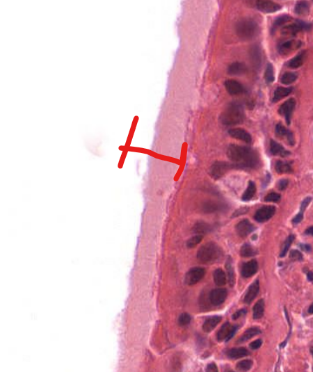
structure
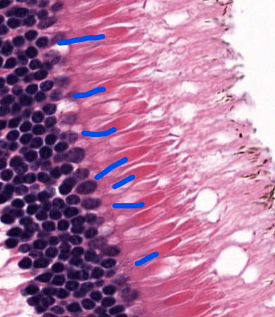
cilia of respiratory epithelium
structure
tracheal cartilage-
made of hyaline cartilage, is open on the posterior aspect to allow the esophagus to expand
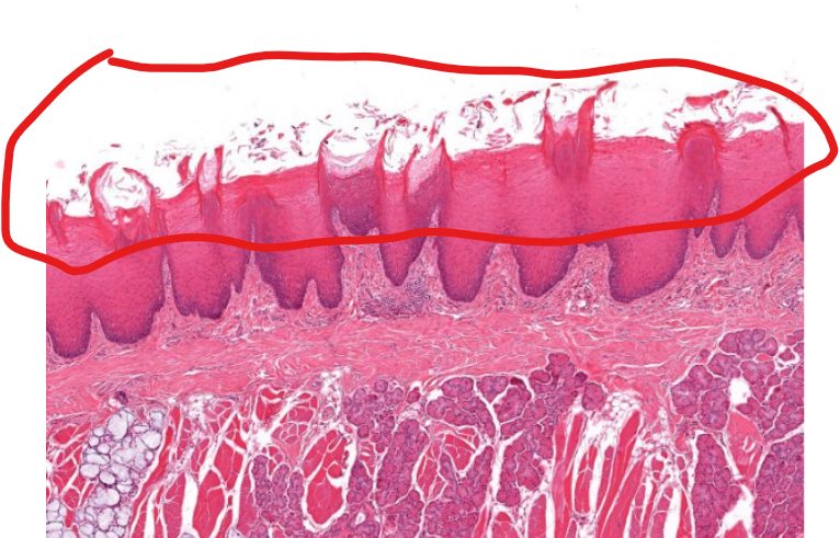
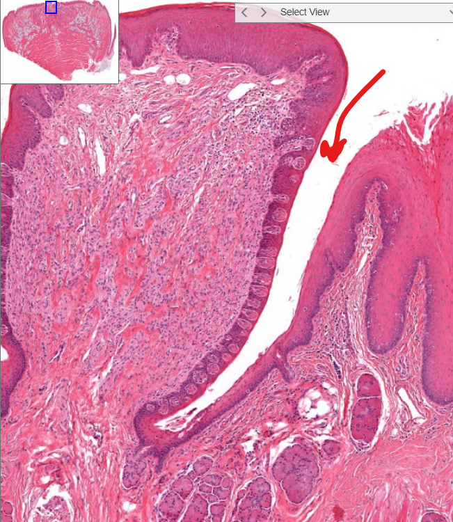
tissue type
stratified squamous epi, non-keratinized
structure
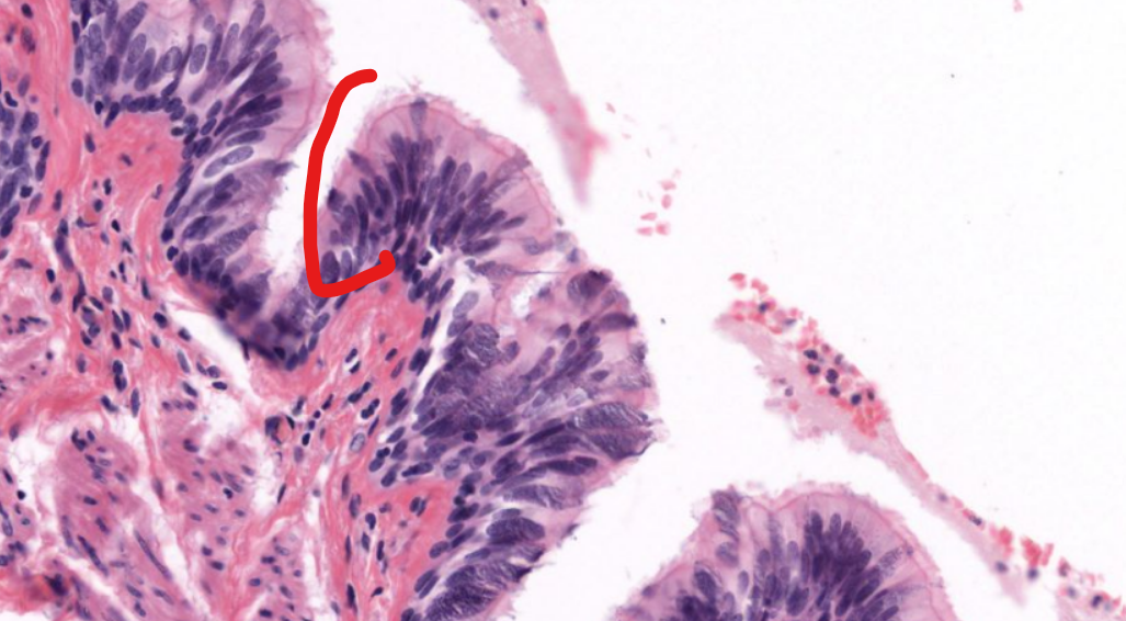
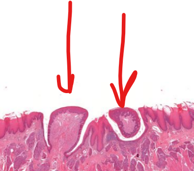
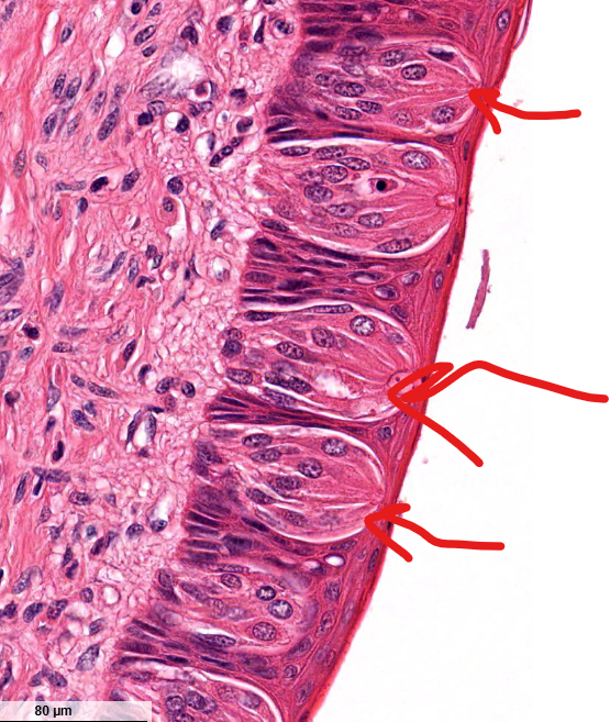
filiform papillae-
“flam-like”, serve to move food on the tongue
do not contain taste buds
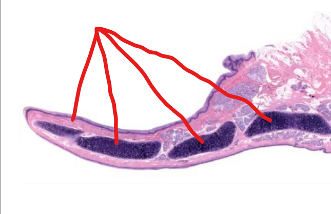
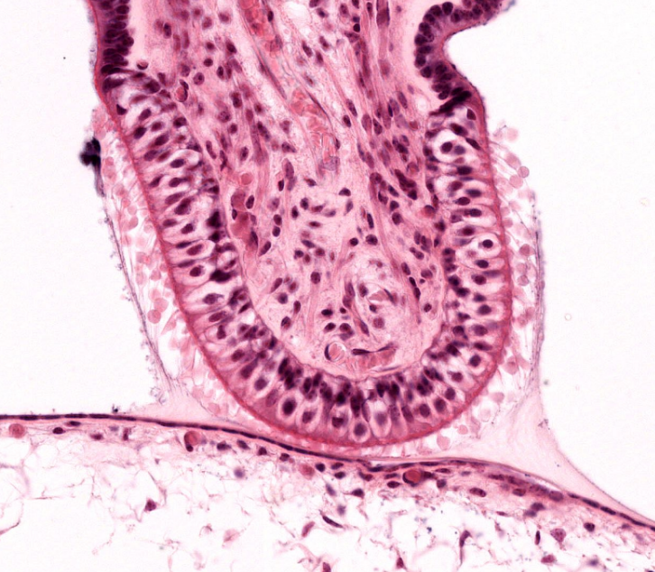
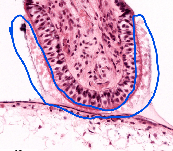
structure
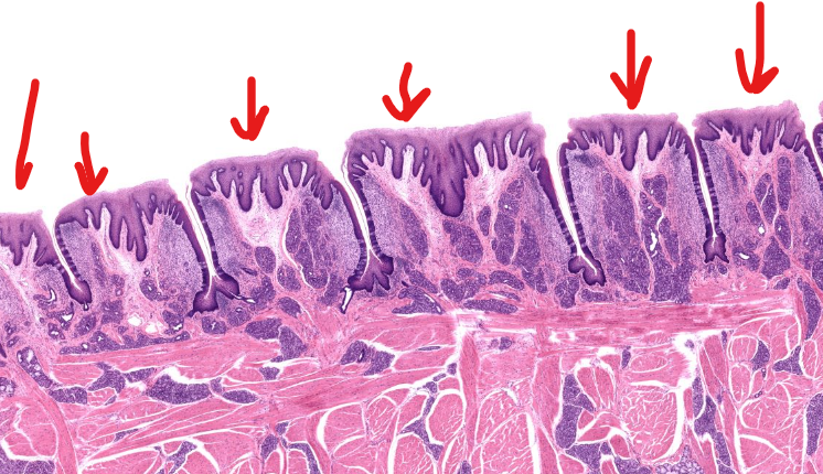
circumvallate papillae-
8-12 dome shaped structures in a V shape on tongue
largest in size and smallest in number papillae on the tongue
structure
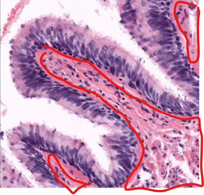
furrow-
“moat” around circumvallate papillae, which receives saliva from minor salivary glands
structure
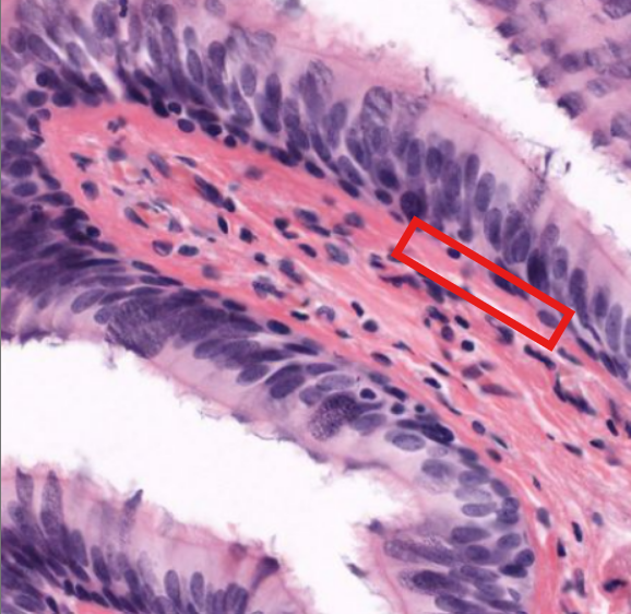
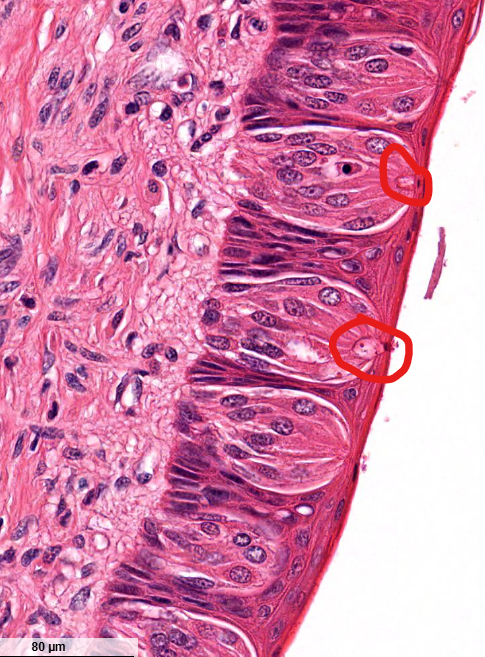
taste buds-
found on the “side” of the moats, contain cells with taste receptors
structure
taste pore
structure
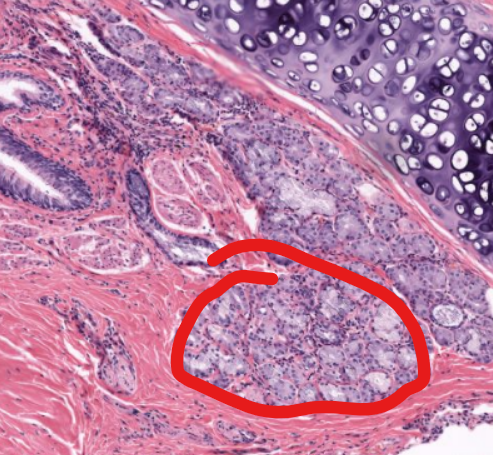
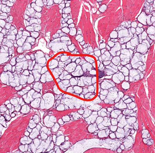
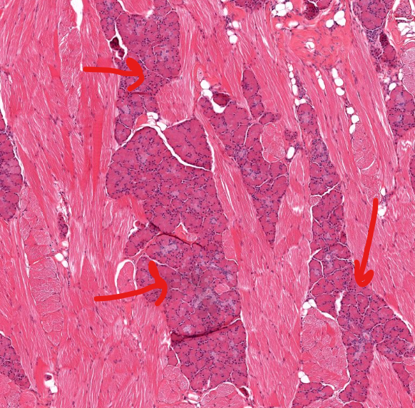
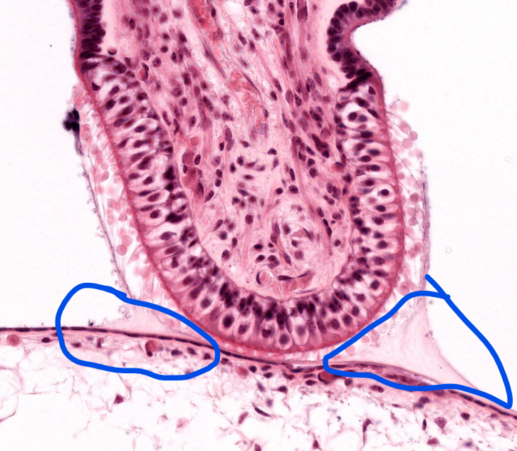
mucous salivary gland
structure
serous salivary gland
structure
foliate papillae-
parallel ridges on the lateral tongue, separated by deep moats

area
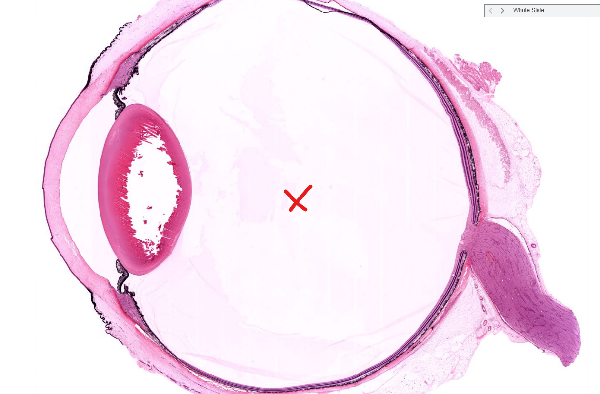
aqueous chamber of the eye
area
vitreous chamber of the eye
sclera
cornea
structure
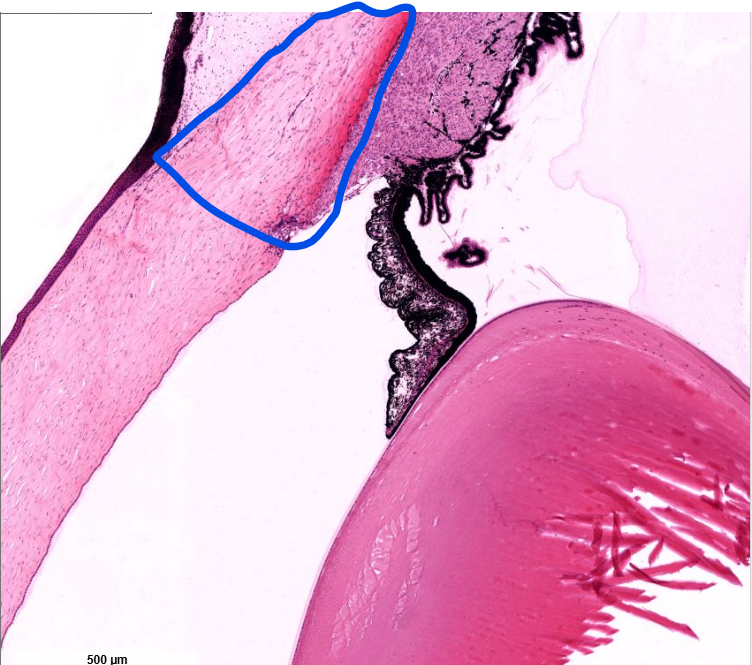
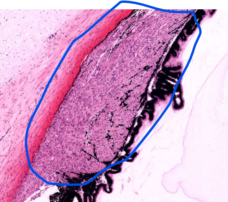
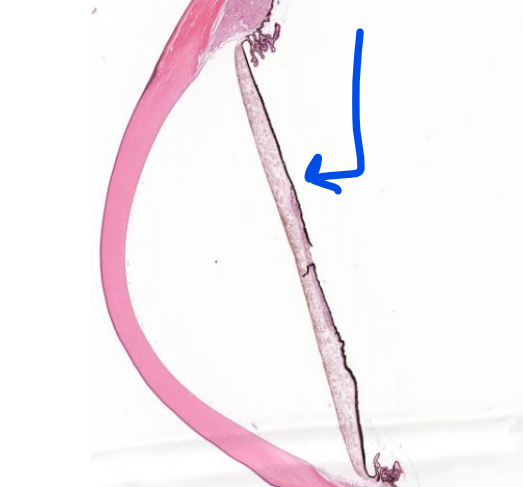
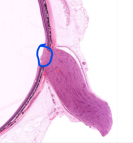
ciliary body
structure
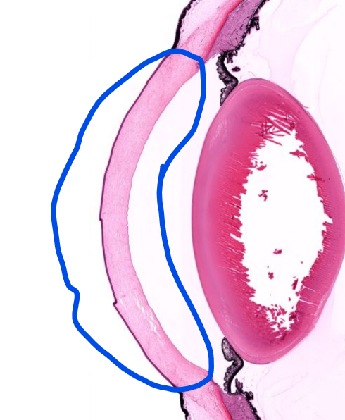
iris
layer
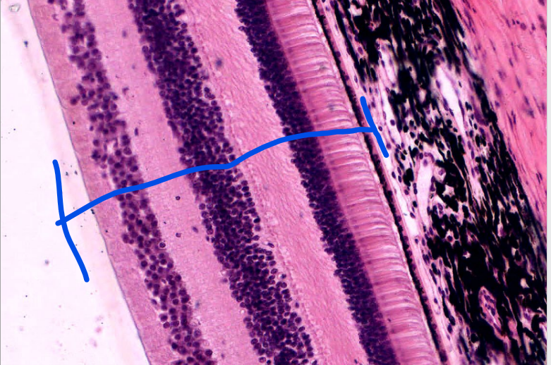
retina- (see the pigment epi and rods/cones?
structure
optic disc- where there are to photoreceptor cells, “blind spot” where the axons of the neurons become the optic nerve and leave the eye
structure
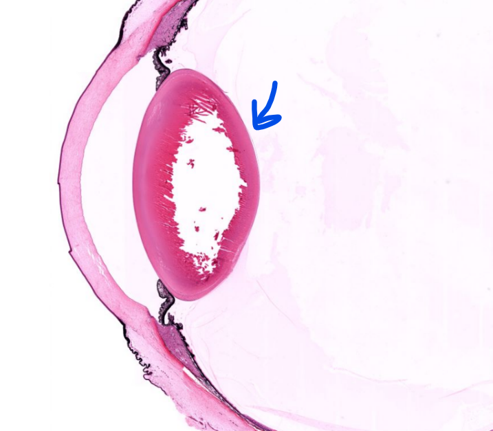
lens
structure
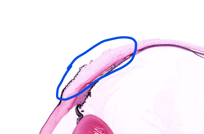
conjunctiva-
the mucous membrane covering the anterior sclera
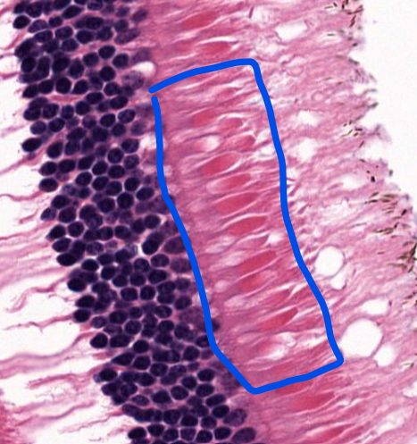
cell type
photoreceptor cells-
rods and cones, part of the retinal layer
cell type
cones
cell type
rods
structure
axons that receive signals from rods and cones, lead to the optic nerve
structure (look at photoreceptor cells
macula lutea-
area of the retina that has mostly cone cells

structure
cochlea of the inner ear- detects sound vibration
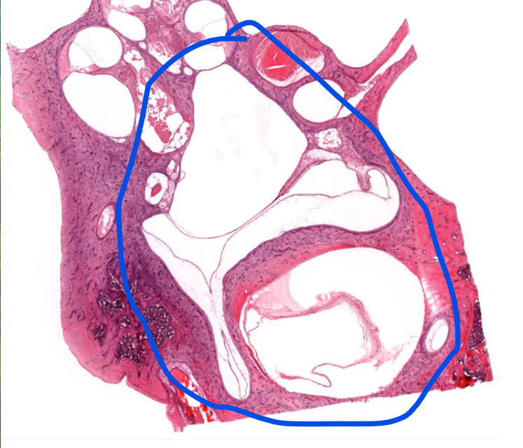
structure
vestibular apparatus- detects gravity/static position, and acceleration
structure
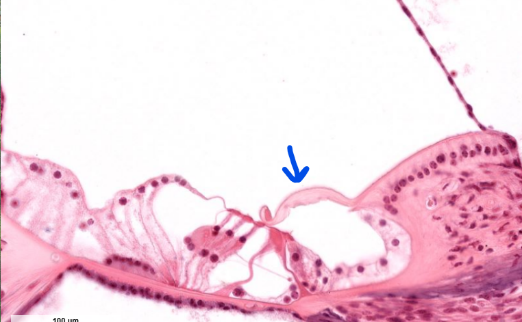
crista ampullaris- the sensory region of the semi-circular canals
structure
cupula- of the inner ear
structure
hair cells
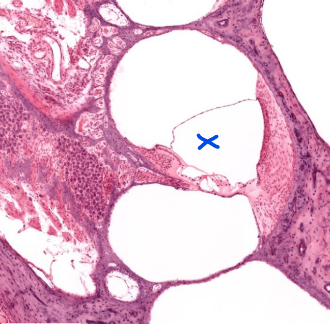
structure
scala media
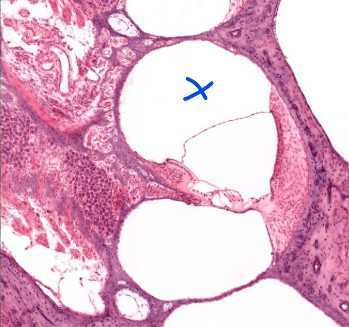
structure
scala vestibuli- the “first” part of the cochliea→ (vestibule)
notice the lame skinny layer of tissue dividing this and the scala media
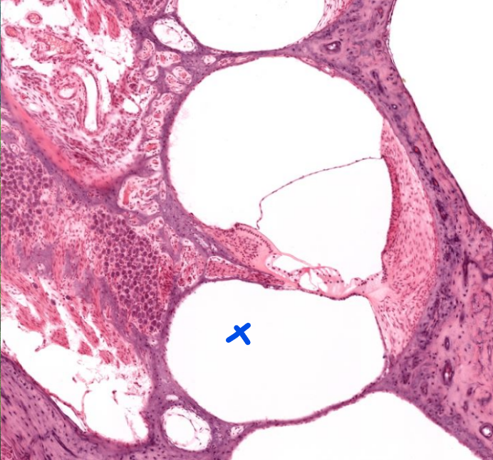
structure
scala tympani- the lower compartment- notice the complicated membrane between this and the scala media→ this is used to transmit hearing info
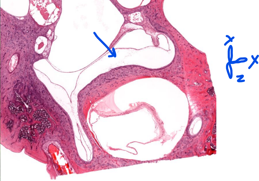
structure
semi-circular canals-
sense linear/directional acceleration
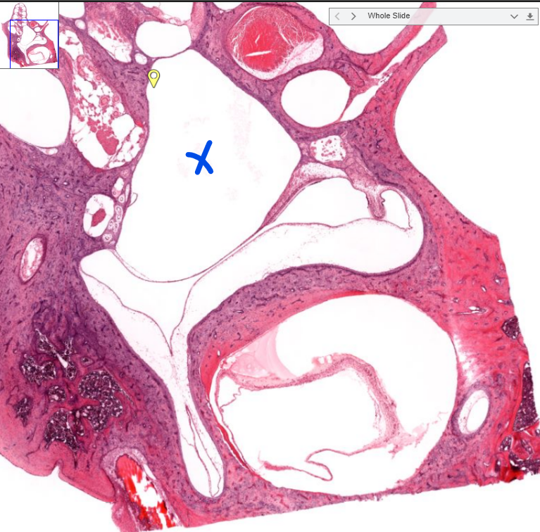
structure
utricule/saccule- impossible to say in this histology slide/one dimention
deal with sensing static position/gravity
structure
techtorial membrane