Muscles of the Neck and Back
1/21
There's no tags or description
Looks like no tags are added yet.
Name | Mastery | Learn | Test | Matching | Spaced |
|---|
No study sessions yet.
22 Terms
epaxial and hypaxial, transverse process
Both neck and back are provided with set of extrinsic and a set of intrinsic muscles. The intrinsic muscles of vertebral column are divided into two major groups, the __________ muscle. The axis of reference is the vertebral column itself at the level of the __________.

epaxial muscles
positioned dorsal to the transverse process of the vertebra. Their function is to support the spine, extend the vertebral column and allow lateral flexion.
Hypaxial muscles
positioned ventral to the transverse process of the vertebrae. They flex the neck and tail and contribute to the flexion of the vertebral column.
Extensors of the Neck
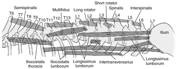
Epaxial muscles serve to extend the vertebral column and neck. They are arranged in three adjacent longitudinal groups: transversospinalis, longissimus and iliocostalis systems.
Transversospinalis system
most medial, lies directly to the vertebral bodies and spinous processes. It extends from the sacrum to the head.
spinalis et semispinalis cervicis
semispinalis capitis
multifidus cervicis
interspinales
muscles from the transversospinalis system
Spinalis et semispinalis cervicis
originates along the thoracic vertebral bodies and inserts on the spinous process of vertebrae C5 - C2
Semispinalis capitis, biventer cervicis (more dorsal) and complexus (more ventral)
lies adjacent to the nuchal ligament and has two parts which are
Multifidus cervicis
series of small, deeply placed muscle. It pass cranially from the midregion of one vertebra to the spinous process of the vertebra which they insert.
Interspinales
tiny muscle that extend between adjacent spinous process, are distinct in the cervical region
Longissimus system
Longissimus cervicis
Longissimus capitis
Longissimus atlantis
Intertransversarius
(intermediate system), is the longest and strongest of the three. It extends from the wing of the ilium to the skull.
give the muscles that forms the system
Longissimus cervicis
the direct continuation of the thoracic part
Longissimus capitis
flattened yet relatively thick and strong muscle positioned fairly deeply
Longissimus atlantis
an inconstant muscle, when present this muscle is a deep slip of the longissimus capitis
Intertransversarius
set of muscles derived from deeper portions of the longissimus system
Iliocostalis system
the most lateral, extends from the ilium to C7. It can be easily identified in most region by the length of its flat, shiny tendons. This system is minimally represented in the neck; only its cranial-most belly extends so far
Sternocephalicus
Sternohyoideus
Sternothyroideus
Scalenus
Hypaxial muscles
Flexors of the Neck
Sternocephalicus
- sternomastoideus (medial)
- Sternooccipitalis (lateral)
flex the neck and joints of the head and draws the head and neck laterally. There are two divisions of this muscle which both arise from the sternum and insert on the head and neck. In their diverging paths, these two embrace the ventral and lateral surface of the neck in the form of letter “V”.
What are the two divisions?
Sternohyoideus (strap muscle)
flat muscle that draws the basihyoid bone caudally.
Sternothyroideus (strap muscle)
draws the larynx and tongue caudally
Scalenus
wedge-shaped muscle ventral to the serratus ventralis muscle. It flexes the neck ventrally and draws the neck laterally
Hypaxial muscles
- longus capitis (lateral-most)
- longus coli (cervical and thoracic portions)
- rectus capitis ventralis (flexes the altantooccipitalis joint)
- rectus capitis lateralis (lateral to the rectus capitis ventralis)
act as ventral flexors of the neck and occipitoatlantal joint
what are the muscle parts that comprised this muscle?