CHP 14 Brain and Brainstem
1/82
There's no tags or description
Looks like no tags are added yet.
Name | Mastery | Learn | Test | Matching | Spaced |
|---|
No study sessions yet.
83 Terms
Gray matter
-collection of neurosomas, dendrites, and synapses
-forms the surface layer (cortex) over cerebrum and cerebellum
-forms nuclei deep within the brain
-dull color due to little myelin
White Matter
-Bundles of Axons
-white color from myelin around nerve fibers
-composed of tracts (bundles of axons) that connect one part of brain to another and to the spinal chord
Ventricles
-four internal chambers within the brain; two lateral ventricles, third ventricle (superior), fourth ventricle (inferior) that connects to the central canal
-the cerebral aqueduct connects 3rd and 4th
-lines by Ependymal
-contains the choroid plexus that supplies capillaries
Choroid Plexus
spongy mass of blood capillaries on the floor of each ventricle
-covered by ependymal
Ependymal
type of neuroglia that lines ventricles and covers choroid plexus
produces cerebrospinal fluid
Cerebrospinal fluid
clear, colorless liquid that fills the ventricles and canals of CNS
production of cerebrospinal fluid
-begins with filtration of blood plasma through capillaries of the brain
-ependymal cells modify the filtrate
-now there is more sodium and chloride than plasma, less calcium+glucose+ and VERY LITTLE protein
Functions of Cerebrospinal fluid
1) Buoyancy =. floats so brain doesn’t rest of cranial floor
2) Protection = protects brain from striking the cranium when head is jolted
3) Chemical stability = flow of CSF rinses away metabolic wastes from nervous tissue, chemical homeostasis
Flow of cerebrospinal fluid
start in lateral ventricle
through the third ventricle
through the cerebral aqueduct
through the fourth ventricle
through the central canal
into the subarachnoid space
extra fluid leaves through the arachnoid villi (“holes”) where it is filtered back into the dura mater sinuses
Blood Supply to brain
brain is only 2% of body weight but gets 15% of blood
neurons have a high demand for ATP so needs oxygen + glucose supplied from blood
Brain Barrier System
regulates what substances can get from the bloodstream into the tissue fluid of the brain
two entries protected: blood capillaries of brain tissue + capillaries of choroid plexus
characteristics of Blood brain barrier system
-consists of tight junction between cells: everything must go through a cell and not between them
-highly permeable to water, glucose, and lipid soluble substances (oxygen, carbon dioxide, alcohol, caffeine, nicotine etc)
-can be an obstacle for letting medications through
-trauma + inflammation cna causę damage and allow pathogens to enter through
Reticular formation
-loose web of gray matter that runs vertically through all levels of the brainstem
-has connections with many areas of cerebrum
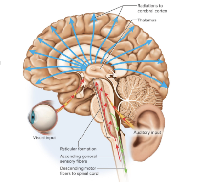
Functions of the Reticular Formation
1) somatic motor control= adjust muscle tension, gaze centers (allow fixation), central pattern generators (neural pools that produce rhythmic signals to muscles of breathing and swallowing)
2) cardiovascular control
3) pain modulation/blockage
4) sleep and consciousness (circadian rhythm)
5) habituation
Cerebellum
“little brain”
contains more than half of all brain neurons (because mostly gray matter), the branching white matter is the arbor vitae
function of cerebellum
motor coordination and locomotion
mostly subconscious
Parts of Forebrain
-Diencephalon= top of brain stem
-Telencephalon= cerebrum
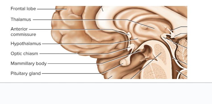
Diencephalon parts
-thalamus
-hypothalamus
-epithalamus
Thalamus
-different parts do different things, 23 different nuclei
-”the front desk/gateway to the cerebral cortex (gray matter covering on the cerebrum) = the thalamic nuclei filters information on the way to the cerebral cortex
-motor control = feedback loop between cerebral cortex and basal nuclei
-memory and emotional functions of limbic system
Hypothalamus
-control center of autonomic nervous system + endocrine system
-essential for homeostasis
Basic Functions:
Hormone secretion (pituitary gland)
Autonomic effects (the subconscious things; heart rate, blood pressure, etc)
What attaches the hypothalamus to the pituitary gland
Infundibulum (a stalk)
Main functions of the hypothalamus
-thermoregulation
-food and water intake (thirst/hunger feels)
-sleep + circadian rhythms
-memory (short term to long term)
-emotional behavior and sexual response (the big feelings)
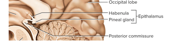
Epithalamus
-very small mass of tissue composed of the pineal gland, habenula (bridge from the limbic system to the midbrain)
Cerebrum (Telencephalon)
-largest part of the human brain
-sensory perception, memory, thought, judgement, voluntary motor actions
-five lobes named after the cranial bones overlying them
How are the left and right cerebral hemispheres connected
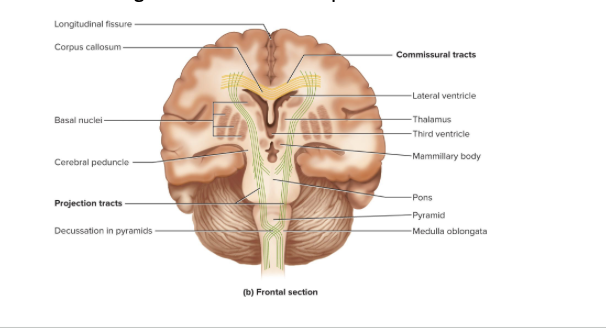
-the longitudinal fissure (superficial)
-corpus callosum = white matter tract (deep)
What feature determines amount of cranial activity/”smartness”
gyri (ridges)
sulci (indentations)
“more wrinkles = smarter brains”
What are the five lobes
frontal
parietal
occipital
temporal
insula (deepest one)
note: functions spread out across multiple lobes
Cerebral White Matter
most of the cerebrum is this
includes tracts (bundles of nerve fibers) in the central nervous system
Three types of Cerebral White Matter Tracts
1) Projection Tracts
2) commissural tracts
3) association tracts
Projection Tracts
-extend vertically between higher and lower brain and spinal cord centers

association tracts
connect different regions within the same cerebral hemisphere
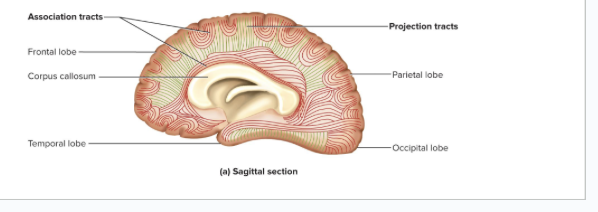
Commissural tracts
cross from one cerebral hemisphere to the other allowing for communication between the two sides of the cerebrum
*the corpus callosum
Gray Matter
-neural integration
located:
Cerebral Cortex (outer covering of the cerebrum)
Limbic System
Basal Nuclei
Cerebral Cortex
-covers the surface of the cerebrum
-contains billions of neurons
-gray matter found here
Neurons of the Cerebral Cortex
Stellate Cells = many dendrites projecting off the neurosomas, receive sensory input + process that information
Pyramidal Cells = thick dendrite w many branches that have dendritic spines, only neuron that leaves the cortex and connects with other parts of the CNS
Limbic System
-the center of emotion and learning
-circular feedback, can cause over activation “big feelings”
-have both gratification (pleasure or reward) + Aversion (fear or sorrow)
Cingulate Gyrus
of the limbic system
arches over corpus callosum in frontal ad partial lobes
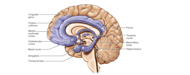
Hippocampus
of the limbic system
for memory functions (short term to long term)

Amygdala
of the limbic system
for emotional functions

Basal Nuclei
masses of cerebral gray matter deep within the white matter
involved in motor control
sends signals back and forth between itself and the midbrain + cortex
3 brain centers of the basal nuclei
-caudate nucleus
-putamen
-globus pallidus
Lentiform
the putamen and globus pallidus together
Integrative Functions of the Brain
higher brain functions such as:
sleep
memory
congnition
emotion
sensation
motor control
language
combining parts of brain working/integrated together
functions of the brain do not have easily defined anatomical boundaries
How do we perceive sensation?
1) Primary Sensory Cortex = where sensory input is first received and you become conscious of the stimuli
2) Association Areas process and interpret the sensory info
Primary Visual Cortex= make sense of what we see
Multimodal Association areas = take in multiple senses and combine them into an overall perception of our surroundings
Overall what are special senses?
limited to the head and employ relatively complex sense organs
can an earth worm do it?
What are the special senses? list
1) Vision = visual primary cortex in occipital lobe + visual association area
2) Hearing = Primary auditory cortex in temporal lobe & insula + auditory association area
3)Equilibrium = Cerebellum + several brainstem nuclei + association cortex
4) Taste = “Gustatory” primary gustatory cortex in the post central gyrus in the parietal lobe
5) Smell = “Olfactory” primary olfactory cortex in the temporal lobe/frontal lobe
What are general senses?
“somatic senses”
“Primary Somatosensory cortex”
distributed over the entire body and use simple receptors
touch, pressure, stretch, movement, heat; cold, pain
How do we perceive our general senses?
1) head = cranial nerves carry info, body= ascending tracts bring info
2) Thalamus processes the input from contralateral side
3) We become aware when it reaches the POST CENTRAL GYRUS DUE TO the Somesthetic association area
Sensory Homunculus
a diagram of the primary somesthetic cortex
resembles an upside down sensory map of the contralateral side of the body
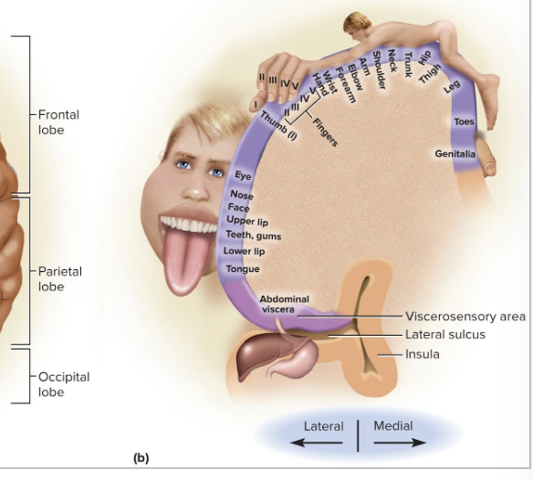
Somatotophy
point to point correspondence between an area of the body and an area of the CNS
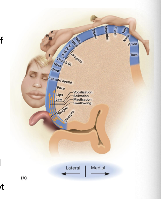
Steps of Motor Control
1) Association (Premotor) area in the frontal lobes is where we plan to move
2) Transmitted to neurons of PRECENTRAL GYRUS (primary motor area)
3) send to brainstem and spinal chord which leads to muscle contraction
Neurons of the Precentral Gyrus for Motor control
1) Pyramidal Cells = upper motor neurons
the fibers from upper motor neurons synapse w lower motor neurons whose axons then go to the skeletal muscles for contraction
Somatotopy of the pre central gyrus
motor homunculus is distorted because the amount of cortex devote to a given body region is the number of muscles/ motor unites and not the body region size
What are basal nuclei and how do they help motor control
control repetitive movements (ex: walking), start/stop of intentional movement, and learned behaviors/habits (ex: driving a car)
FEEDBACK LOOP between cerebrum to basal nuclei to thalamus back to cerebrum
Dyskinesias
Dys = abnormal
kines = movement
damage to basal nuclei causes this which causes abnormal movements
How does the cerebellum help with motor control
motor coordination
muscle tone and posture
joints working together coordination
Ataxia
damage to the cerebellum can cause this
clumsy, awkward gait
Language is caused by what areas
1) Wernicke Area
posterior to lateral sulcus usually in left hemisphere
RECOGNITION of spoken and written language
formulates phrases sends that “plan” to Broca area
2) Broca Area
inferior prefrontal cortex usually in left hemisphere
creates the MOVEMENT for speaking and signing
sends to primary motor cortex to lower motor neurons that grab the right muscles
3) Affective Language Area
usually right hemisphere
inflection in voice
Aprosody
damage to the affective language area
causes flat emotionless speech
Aphasia
a language deficit from damage to the hemispheres with Wernicke and Broca areas
Types of Aphasia
1) Nonfluent (broca) aphasia
slow speech, slurring, words sound wrong, and difficulty choosing words
2) Fluent (wernicke aphasia)
speech normal but it’s all gibberish
3) Anomic Aphasia
can speak and understand speech but cannot identify words or pictures
Cerebral Lateralization
= the difference in the structure and function of the cerebral hemispheres
Left = categorical, analytical, spoken/written language
Right = representational, artistic, imagination, patterns, sense comparisons
Cerebral Lateralization in correlation to handedness, age, sex
-Right Handed = left hemisphere categorical in 96%
-Left Handed = left hemisphere categorical in 70%, right hemisphere categorical for 15%, neither for other 15%
-males more lateralization than females
Cranial Nerves Usage
how the brain communicates with the rest of the body
12 pairs of cranial nerves
exit the cranium through foramina
mainly send to organs in head and neck
Pathways of Cranial Nerves
1) Motor neurons start in nuclei of brainstem and go to glands and muscles, sensory fibers start in receptors in head and neck and go to brainstem
2) most carry fibers between brainstem and ipsilateral (same side) receptors and effectors, EXCEPT optic nerve and trochlear nerve that sometimes do decussation
Classification of Cranial Nerves
Sensory = I, II, VIII
Motor = III, IV, VI, XI, XII
Mixed = V, VII, IX, X
Olfactory Nerve
(I)
send sense of smell to the brain
Optic Nerve
(II)
send visual signals to the brain by the optic chiasma
Oculomotor Nerve
(III)
control size of pupil and movement of the eye
Trochlear Nerve
(IV)
movement of the eye
Trigeminal Nerve
(V)
LARGEST cranial nerve
most important sensory nerve of the face
3 divisions:
Ophthalmic Division (sensory)
Maxillary Division (Sensory)
Mandibular Division (mixed)
Abducens Nerve
(VI)
movement of eye left and right
Facial Nerve
(VII)
Motor (facila expressioms, salviary glands, palatine glands ) + sensory (taste on the tongue0
What does damage to facial nerve (VII) cause
saggy facial muscles
no sweet or salty taste
Vestibulocochlear Nerve
(VIII)
sense of equilibrium and sound
Glossopharyngeal nerve
(IX)
control tongue, salivary glands, swallowing muscles
Vagus Nerve
(X)
“wandering” nerve
most extensive distribution of any cranial nerve
control cardiac, pulmonary, digestive, and urinary function
swallowing, speech, viscera regulation
damage to vagus nerve causes what
hoarseness and loss of voice
impaired swallowing
fatal if both are cut
Accessory nerve
(XI)
movement of the sternocleidomastoid and trapezius (move head and neck)
Hypoglossal Nerve
(XII)
movement of the tongue
Senescence of nervous system
peak development at age 30
brain weights 56% less by age of 75
cortex thinner, gyro narrower, sulci wider, fewer synapses and neurotransmitters/receptors
short term memory suffered most
autonomic nervous system less efficient at regulating body temp ad blood pressure
Alzheimers
nervous disability of old age
plaques of fibrillar proteins (amyloid) appear