Vascular midterm (1)
1/70
There's no tags or description
Looks like no tags are added yet.
Name | Mastery | Learn | Test | Matching | Spaced | Call with Kai | Chat |
|---|
No analytics yet
Send a link to your students to track their progress
71 Terms
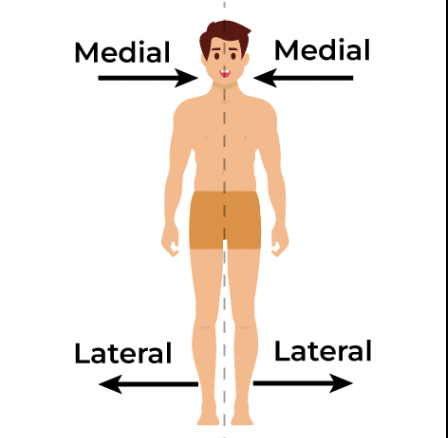
what is Medial and Lateral
Medial- towards the midline
Lateral- away from midline
What is Ipsilateral and Contralateral
Ipsilateral- on the same side of the body ( right hand right arm)
Contralateral- on the opposite side of the body ( right hand left hand)
What is Proximal and Distal
Proximal- closer to the heart
Distal- further from the heart
What is Superficial and Deep
Superficial is closer to the skin, deep is further into the skin
what is the difference between arteries and veins
arteries: carry blood (generally oxygenated) away from the heart, arteries don’t have valves.
Veins: carry blood ( generally deoxygenated) toward the heart, veins have valves to help them push blood toward the heart.
What are the three types of arteries
Elastic: Conducting arteries, Have thick walls with elastic fibers, the largest diameter, can uphold high blood pressure
Muscular: distributing arteries, contain more smooth muscle than elastic fibers. they are medium and small, they constrict or relax( vasodilate/vasoconstrict)
Arterioles: transports blood from small arteries to capillaries, have thin walls and high proportion of smooth muscle reltive to size ( the smallest in size but still have all 3 layers, just a small tunica media)
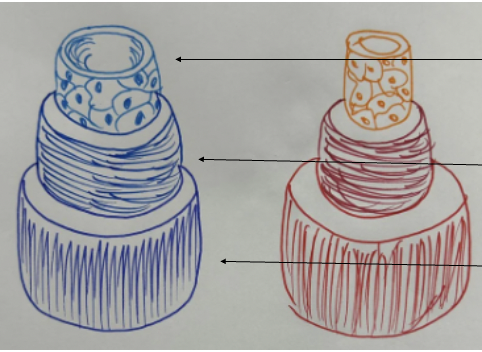
What are the 3 layers of the arteries and veins
Tunica intima: Innermost layer Consists of a single layer of Endothelial cells
Tunica media: Middle layer, smooth muscle cells arranged circularly around the vessel, Regulates blood flow with contraction or relaxation, comprised of 20-40 muscle cells in muscular arteries
Tunica adventitia: The outermost layer contains connective tissue and provides connection with the surrounding tissue. Has the vasa vasorum, which supplies nutrients to the vessels ( only for vessels that exceed 1mm in diameter) and has baroreceptors which Monitor stretch in blood vessel wall and detect changes in blood pressure
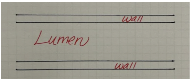
What is the lumen?
a channel within a vessel
What does hemodynamics mean
the study of blood flow through the circulatory system
What is pulsation?
the changes in the blood flow due to two phases of the cardiac cycle (systole and diastole )
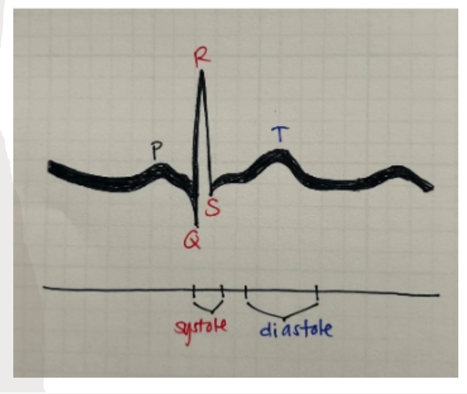
What is systole?
The first part of the cardiac cycle where the ventricles are contracting
What is diastole?
Portion of the cardiac cycle where the ventricles are relaxing
What is phasicity?
Changes in blood flow due to cardiac or pulmonary factors
What is a stenosis
Narrowing or obstruction in flow which causes the body to compensate in some way ( once you have this narrowing it will continue to build-ip )
What does Ischemic mean
Occurs when tissues of organs aren’t getting what they need, this is because they lack adequate oxygen.
Explain the pressure gradient
the difference in pressure between one area and another. Flow will naturally move from a higher gradient to a lower gradient
What does vasodilation and vasoconstriction mean
vasodilation: expansion of blood vessels
Vasoconstriction: contraction (constriction) of blood vessels
What are collaterals?
Make new routes of blood flow from the narrowing or closed-off vessels
What does heterogenous mean
When theres irregular composition of echo signals throughout
What does homogenous mean
composition of echo signals are uniform throughout ( the same)
What does heterogenous mean
When theres irregular composition of echo signals throughout
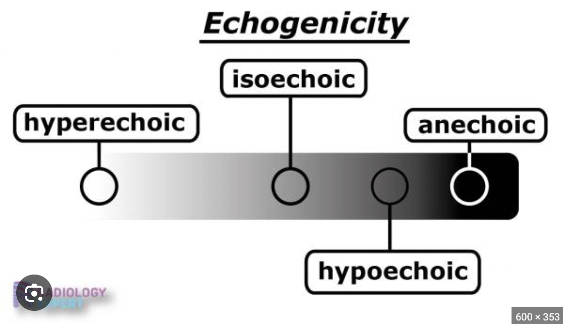
What is isochoeic and anechoic
Isoechoic means that the reflected echo density is the same ( shown in the image)
Anechoic: without any echo signal ( complete darkness this means it’s fluid-filled
Explain hypoechoic vs hyperechoic
hypoechoic: Echo signals are not as bright compared to other surrounding tissue or structures.
• Hyperechoic: Echo signals are brighter compared to other surrounding tissue /structures
What does Intra/Inter/Infra
Intra: within Inter: between Infra: below
What does Supra and Sub mean
Supra: above Sub: Below
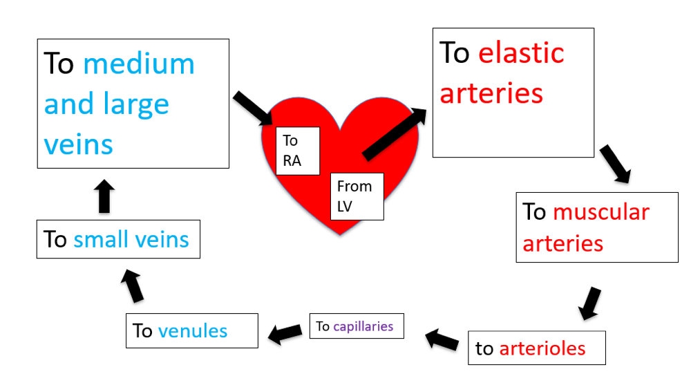
Explain pulmonary circulation
Moves blood between the heart and the lungs. more specifically, from the right ventricle to the lungs via the pulmonary artery, then back to the left atrium via the pulmonary veins
Explain systemic circulation
Moves blood between heart and body
From left ventricle to the aorta
What are the 3 main types of vessels in the body
arteries, capillaries and veins
Explain the relationship between the 3 main vessels
Arteries branch to arterioles and venules combine to become veins. Capillaries connect the arterioles to the venules, and this is where exchange happens

How does pressure change when we go from the elastic arteries to the veins?
BP is highest in the elastic arteries and muscular arteries, when we move down to the arterioles there is a sharp drop i pressure ( drops from 80mmhg to about 30-40mmhg)
The pressure decreases as blood flows through the capillaries usually around 20-40mmhg
As blood enters the venous side ( venules and veins) pressure drops further and is around 0-5mmHG
(Overview: biggest drop in P in the arterioles, moderate drop in capillaries, and very low pressure in veins)
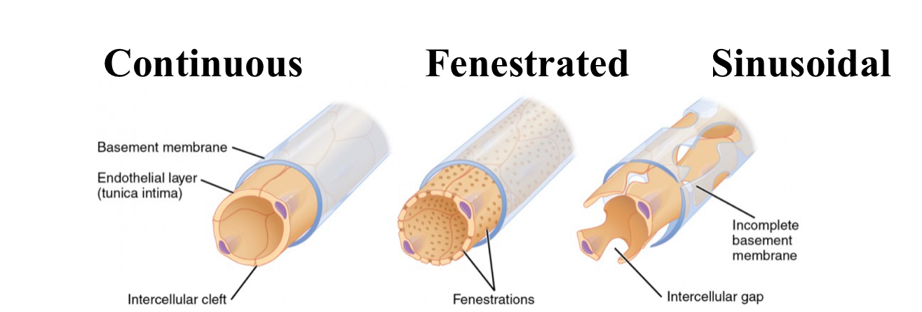
What are the different types of capillaries, how is each structures and how does that affect function
Continuous capillaries: in muscles, skin. Has endothelial cells form a continous lining with tight junctions, have small gaps. Allow exchange of small molecules like water and ions but restrict large molecules
Fenestrated capillaries: in kidneys and intestines. Has endothelial cells that contain small pores (fenestrations) permit rapid exchange of larger molecules, good for filtration and reabsorption
Sinusoidal capillaries: in liver and bone marrow. has large gaps between endothelial cells and a discontinuous basement membrane. Facilitates free exchange of large proteins and cells, allows filtration and cell migration
What are the different types of veins, how is each structures and how does that affect function
Venules and small veins: Venules have similar structure to capillaries but are larger. with connective tissue covering. Collect blood from capillaries and return flow to heart at low pressure.
Small veins: Form as venules increase in size ( have all 3 layers)
Medium veins: Thinner walls than arteries and have valves to prevent backflow. They transport blood back to the heart. (Collect blood from small veins and deliver to large veins)
Large veins: Have the thinnest walls relative to diameter and contain more smooth muscle. deliver blood to the heart
What are portal veins? how many are there? what do they do
Portal veins: a blood vessel that carries blood from the abdomen to the liver
hepatic portal vein: Carries blood from capillaries in the GI tract and spleen to the liver's sinusoids, allowing the liver to process nutrients and detoxify substances from digestion.
The hypophyseal portal vein: Connects the hypothalamus to the anterior pituitary gland, transporting hormones that regulate pituitary function.
What are venous valves? what types of veins have them
allow blood to keep moving forward to the heart, in veins greater than 2mm (medium and large veins) the number of valves are greater in lower limbs
What are the factors affecting blood flow
Elasticity of walls, distal vascular resistance, blood vessel structural pattern
Blood flows slower in larger vessels and faster in smaller vessels
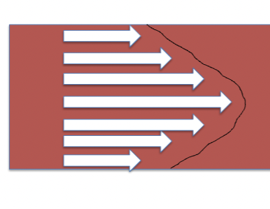
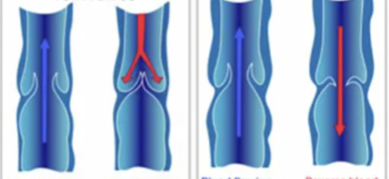
What is laminar flow
orderly blood flow where the layers of fluid move parallel to each other, the highest velocity is at the center of the vessel and slower near the vessel’s walls.
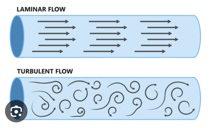
What is turbulent flow
more likely to occur in smaller vessels, EX: as blood moves from larger arteries into smaller arterioles or through narrowed segments
Chaotic, disordered flow of blood where the movement is irregular and multidirectional. often resulting in increased resistance to flow.
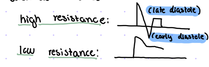
What has high resistence vs. low resistence, what arteries have high resistence vs low resistance
low resistance: organs such as the brain, liver and kidneys
arteries with LR: internal carotid artery and vertebral artery
High resistance: muscles
Arteries with HR:external carotid artery and subclavian
If a muscle needs constant blood flow it will go from high resistance to low resistance
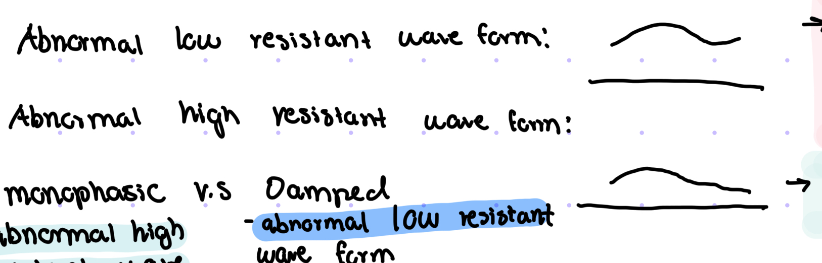
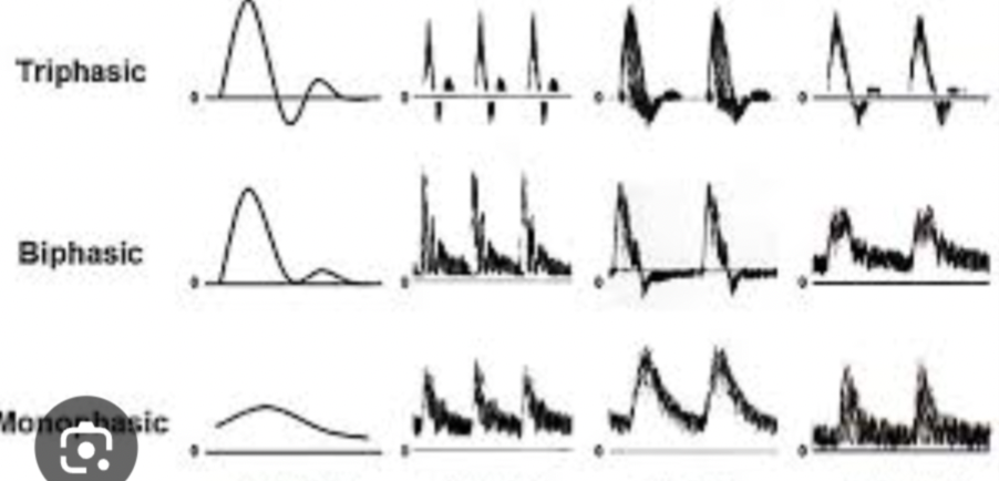
What are the abnormal wave forms, are they high resistance or low resistance
Abnormal low resistant waveform: Damped, the upstroke is more than 90 degrees caused by someone needing even more blood, making the wave change shape
Abnormal high resistant waveform: monophasic, when the wave more is greater than 90 degrees caused when the body isn’t getting what they need
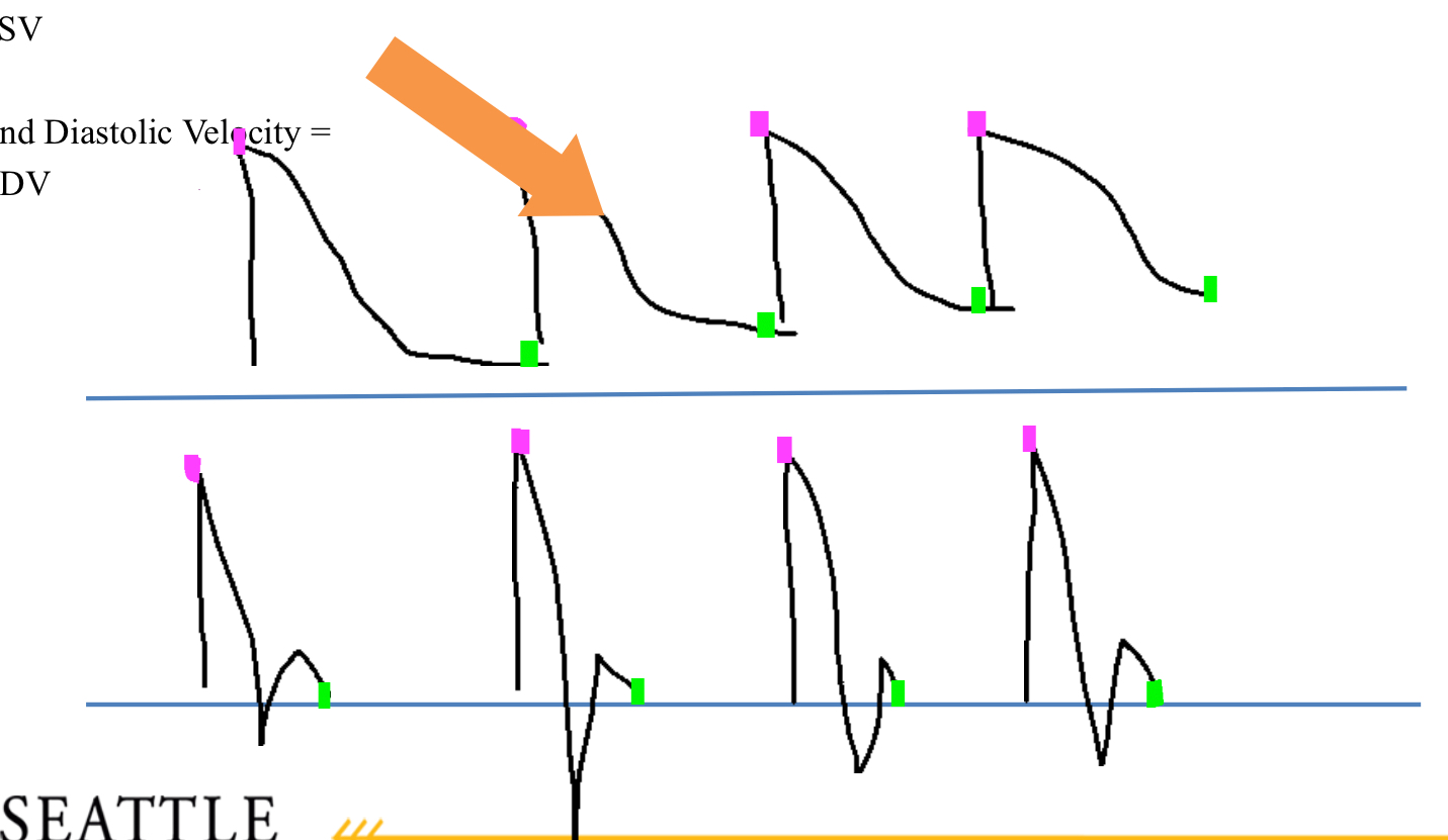
Which one is showing peak systolic velocity (PSV) and which point is showing EDV
Pink is the peak systolic velocity and green is the EDV
What is a triphasic waveform
Waveforms with 3 distinct points, like high resistant waveforms. These are found in the legs and arms
Legs- ( femoral artery, popliteal artery, dorsal pedis artery ( top of the foot), anterior tibial artery
Arms- radial and ulnar artery
What is the stenotic profile
describes the changes in blood flow dynamics that occur due to a stenosis
( can result from atherosclerosis or anything that restricts blood flow)
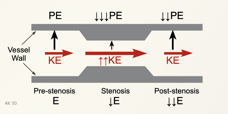
What is the relationship between Kinetic and potential energy in relation to a vessel
KE: Represents the energy of blood flow; it increases as blood passes through a stenosis As the diameter of the vessel decreases, blood velocity increases to maintain flow, which raises kinetic energy.
Potential Energy (PE): Relates to the pressure within the vessel; approaching the stenosis PE is at its highest as blood flows through a stenosis, pressure drops and makes PE at it lowest. Post stenosis PE increase and KE decreases but it’s still not as high as it was pre-stenosis
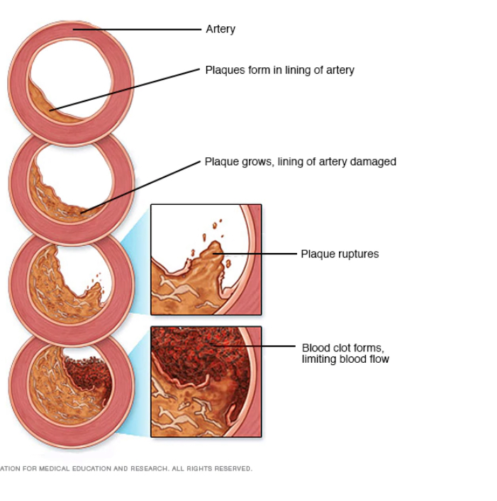
What is atherosclerosis, what are the types of plaque in the atherosclerosis process?
most common type of pathology in the arteries and veins, plaque formation ( develops over a long period of time)
Fatty streak, Fibrous Plaque, Complicated lesion, and Intraplaque hemorrhage
Explain what goes on in every type of plaque in atherosclerosis
Fatty streak- plaque forms in lining of artery
Fibrous plaque- accumulation of lipids covered by more lipid material ) collagen, elastic fiber deposits)
Complicated lesion- Fibrous plaque contains more fibrous tissue, more collagen, calcium and cellular debris
Ulcerative lesion- deterioration of the normally smooth surface of the fibrous cap. ( higher tendency to shed debris)
Intraplaque Hemorrhage- Plaque bleeding from within
What is being shown in this image, label the parts of this.
( also, draw out this image with all parts labeled)
thoracic aorta, the main artery from LV of heart, has 3 parts ascending arch and descending
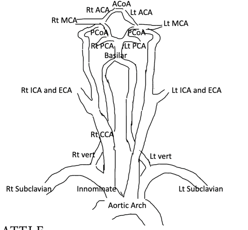
What vessels supply the brain with blood
RT and LT ICA, RT and LT vertebral
What is being shown, what does it supply?
this is the Rt and LT ICA, and it supplies blood to the brain, no branches typically posterior and lateral to the ECA branch
What is being shown, what does it supply
RT and LT ECA supply blood flow to the face and scalp, usually smaller than ECA, always more anterior and medial to ICA
What is being shown, and what does it perfuse? where does it branch off from
the RT and LT vertebral, perfuses posterior cerebral circulation of brain, branches from proximal subclavian artery.
What are the four lobes, what do they do?
Frontal lobe- critical thinking, memory, emotion
Temporal lobe- long-term memory, olfactory capabilities
Parietal lobe- Body sensations ( taste touch), calculations, spatial relationships
Occipital lobe: visual perception
How does the brain get blood flow?
2 intracranial circulation systems: anterior and posterior
Anterior Circulation: perfuses frontal, temporal, and anterior portions of the parietal lobe (FTAP)
Posterior circulation: perfuses brain stem, occipital lobe posterior portion of parietal lobe ( BOPP)
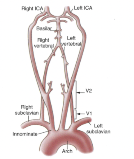
Based off this image, which arteries intracranially provide all of the cerebral blood supply
the ICA and vertebral arteries
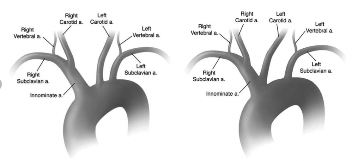
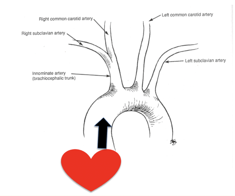
What is the Bovine arch?
Has a common origin for the innominate ( also known as brachiocephalic) and left common carotid arteries with a separate origin of the left subclavian artery.
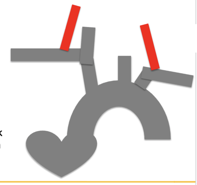
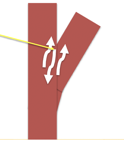
What are communicating arteries, how many do we have?
only used if necessary for collateralization to get blood flow to MCAs.
ACoA- joins bilateral ACAs
PCoA- ( we have 2 RT and LT) joins ipsilateral PCA to the origin of MCA
Draw this out 3 times and label it
give urself practice problems about what would happen if there was a stenosis
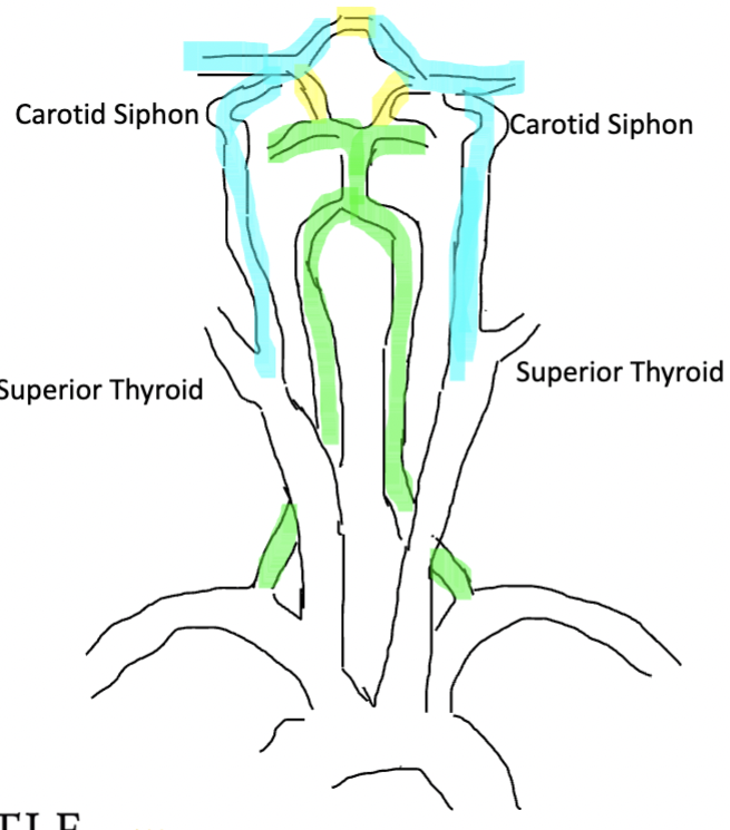
What types of circulation is being shown, when would you use them?
Anterior circulation (Blue)- Refers to the blood supply to the front part of the brain, primarily via the internal carotid arteries.
Posterior circulation (green)- blood supply to the back part of the brain, primarily via the vertebral arteries and basilar artery.
Collateral circulation (yellow)- the alternative or "backup" routes of blood flow that develop or are used when normal blood flow is obstructed
What are the segments of the ICA intracranially
carotid siphon- (Blue) resembles bottom of letter s
Terminal ICA (TICA)- (pink) Site of bifurcation into middle cerebral artery (MCA) and anterior cerebral artery (ACA)
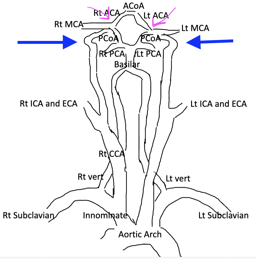
Fill in the blank:
At branch points blood flow can have both forward and backward velocity components called ____ the effective velocity is ___
( shown by arrow)
boundary layer separation, zero
Explain Parabolic flow
Refers specifically to laminar flow in a cylindrical or pipe-like geometry (e.g., blood vessels, pipes), where the velocity profile is parabolic.
( all parabolic flow is laminar but not all laminar flow is parabolic)
What factors affect turbulence
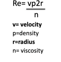
since viscosity and density of blood are fairly constant, turbulence depends on the size of the vessel and the velocity of blood flow ( as shown with Reynolds number)
A lumen diameter decrease of 70% corresponds to a cross sectional area decrease of 95 percent, explain why
A decrease in diameter significantly decreases the cross-sectional area because the area is calculated as the square of the diameter, meaning even a small change in diameter results in a much larger change in the area itself
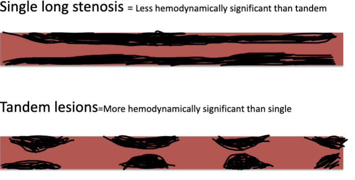
What is a tandem stenosis
Two or more stenotic lesions occurring in a
series, more hemodynamically significant than regular stenosis
The diastolic component of a Spectral Doppler arterial waveform is shaped by _____________. ( why?)
peripheral resistance of the end vascular bed
because this determines how easily blood can flow through the small vessels when the heart relaxes. If the small vessels are narrow, blood flows more slowly and makes the waveform rounder, while wider vessels allow blood to flow smoothly and create a clearer shape.
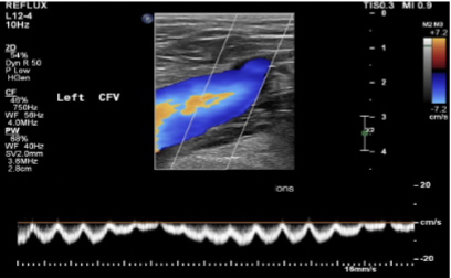
explain the calf pump
A mechanism in the lower leg that helps blood return to the heart. When you move your feet or walk, the calf muscles contract and squeeze the deep veins in the legs. This action pushes blood upward toward the heart, helping to prevent blood from pooling in the lower legs.
What factors influence venous waveforms but do not influence arterial waveforms
respiration, calf muscle contraction and relaxation, external compression, venous valves and gravity
How does respiration affect venous wave forms
in the thorax- during inspiration: venous volumes increase and pressure decreases. During expiration: venous volume decreases and pressure increases
This is reversed in the abdomen
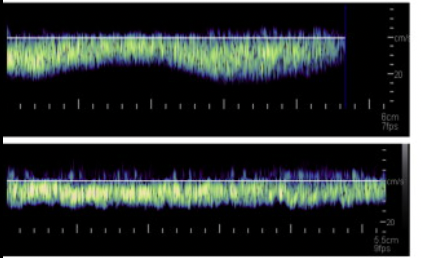
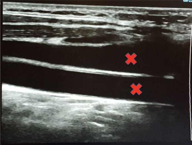
What is the normal venous spectral waveform called? What is an abnormal venous spectral waveform called
Respiro-phasic: ( no proximal obstruction)
shown on the top image, has some type of waveshape
continuous: (there is a proximal obstruction)
shown on bottom image, has no waveshape
What is another abnormal venous spectral waveform caused by rt atrial cardiac pressures
when Rt atrial cardiac pressures increase significantly, this can affect venous waveform, it loses variations from respiration and becomes pulsatile.
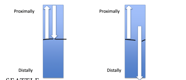
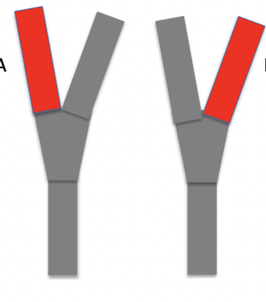
Which image shows a competent venous valve, how do you know?
the left image is a competent venous valve because it prevents bi-directional blood flow ( it only goes toward the heart)
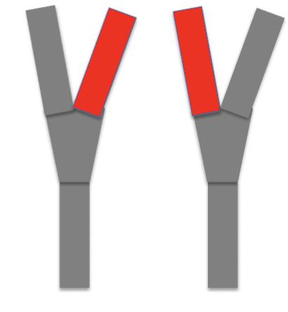
explain varicose vein valves
healthy valves restrict reverse blood flow but varicose valves reverse blood flow due to damaged valves, shown on the right image