2.1 Structure and Function of Eukaryotic and Prokaryotic Cells
1/60
There's no tags or description
Looks like no tags are added yet.
Name | Mastery | Learn | Test | Matching | Spaced | Call with Kai |
|---|
No analytics yet
Send a link to your students to track their progress
61 Terms
What is the function of the bacterial cell wall? (3)
- It exerts a pressure potential to prevent the cell from bursting due to osmosis.
- It maintains the structure of the cell and protects its internal contents.
- It is made of a substance called peptidoglycan.
What is the function of the slime capsule? (3)
- It protects the cell from being engulfed by white blood cells.
- It covers the antigens on the cell surface.
- This helps the bacterium to avoid being identified by the host's immune system.
What is the function of pili? (2)
- They are used for the attachment of the bacterium to a host cell.
- They are also used to transfer genetic material during sexual reproduction.
How do flagella move a bacterial cell? (2)
- Flagella are attached by a hook to a protein disc at their base.
- The disc rotates, spinning the flagellum like a propeller, which requires energy from ATP.
What is the function of prokaryotic ribosomes? (2)
- Prokaryotic cells have 70S ribosomes which are the site of protein synthesis.
- They are less dense and smaller than the 80S ribosomes found in eukaryotic cells.
What is the nucleoid in a prokaryotic cell? (3)
- It is the region within the cytoplasm where the genetic material is located.
- The genetic material consists of a single, often circular, length of DNA.
- Unlike a true nucleus, it is not contained within a membrane.
What is a plasmid? (3)
- A plasmid is a small, self-replicating loop of DNA found in some bacteria.
- They can code for specific traits, such as the production of certain metabolites.
- They can also be responsible for carrying genes for antibiotic resistance.
What is the function of mesosomes and photosynthetic membranes? (2)
- Mesosomes are infoldings of the plasma membrane that provide a surface for the attachment of enzymes for aerobic respiration.
- Photosynthetic membranes are infoldings of the plasma membrane where bacterial chlorophyll and the enzymes for photosynthesis are located.
What is the function of glycogen granules and lipid droplets? (2)
- Glycogen granules are for carbohydrate storage.
- Lipid droplets are for lipid storage.
What is Gram staining? (1)
It is a technique used to identify different types of bacteria and the factors affecting their cell wall vulnerability.
How is the cell wall of Gram-positive bacteria structured? (3)
- They have thicker layers of peptidoglycan in their cell walls.
- The peptidoglycan contains chemicals such as teichoic acid within its net-like structure.
- Gram-positive bacteria have a lower lipid content compared to Gram-negative bacteria.
How is the cell wall of Gram-negative bacteria structured? (3)
- They have thinner layers of peptidoglycan that do not contain teichoic acid.
- They have an outer membrane, which is a phospholipid bilayer.
- The outer membrane is made from lipopolysaccharides.
How is the staining process initiated to distinguish between Gram-positive and Gram-negative bacteria? (3)
- The primary stain, crystal violet dye, is applied and retained by the bacterial cell walls.
- Iodine is applied as a mordant, which forms a crystal violet-iodine complex that is difficult to remove.
- Decolourisation with propanone or ethanol dissolves the lipid bilayer of Gram-negative bacteria, enhancing the leaching of the primary stain.
How is the staining process completed to distinguish between Gram-positive and Gram-negative bacteria? (2)
- The solvent dehydrates the thicker Gram-positive cell walls, closing the pores and preventing the crystal violet-iodine complex from diffusing out.
- A counterstain, such as safranin, is applied to the decolorized Gram-negative bacteria, giving them a pink colour.
What are the expected results of a Gram stain test? (2)
- Gram-positive bacteria will appear violet or purple under the microscope.
- Gram-negative bacteria will appear red or pink under the microscope.
What are the different classifications of bacteria based on their shape? (3)
- Bacteria that are spherical are known as cocci.
- Bacteria that are rod-shaped are called bacilli.
- Bacteria that are twisted are known as spirilla.
How are bacteria classified based on their respiratory requirements? (3)
- Obligate aerobes are bacteria that require oxygen for respiration.
- Facultative anaerobes can respire aerobically but can also use anaerobic respiration.
- Obligate anaerobes are bacteria that can only respire anaerobically.
How can antibiotics interfere with bacterial processes? (2)
- One method is to interfere with protein synthesis by specifically targeting the 70S ribosomes.
- This process does not affect human cells because they contain 80S ribosomes.
What is another process targeted by antibiotics? (2)
- Another method is to inhibit the bacterial synthesis of the B vitamin, folate.
- Folate is required by the bacterium for the synthesis of DNA and RNA.
What are two differences between Gram-negative and Gram-positive bacteria? (2)
- Gram-positive bacteria have a thick layer of peptidoglycan, whereas Gram-negative bacteria have a much thinner layer.
- Gram-negative bacteria have an outer membrane, a structure which is absent in Gram-positive bacteria.
How do beta-lactam antibiotics affect Gram-positive bacteria? (2)
- Beta-lactams, such as penicillin, interfere with the synthesis of peptidoglycan.
- Peptidoglycan is the major component of bacterial cell walls, making it a significant target.
Why are glycopeptide antibiotics not effective against Gram-negative bacteria? (2)
- Glycopeptide antibiotics are large, polarised molecules.
- These molecules cannot penetrate the outer membrane layer of Gram-negative bacteria.
How do polypeptide antibiotics affect Gram-negative bacteria? (1)
Polypeptide antibiotics, such as polymyxins, interact with the phospholipids of the outer membrane.
How is the plasma membrane structured and what is its function? (3)
- The plasma membrane mainly consists of lipids and protein.
- It is differentially permeable, which means it regulates the movement of solutes between the cell and its environment.
- Receptor molecules are integrated across its surface, allowing it to respond to chemicals.
How is a plant cell wall structured? (3)
- It is a rigid structure composed of long cellulose molecules grouped in bundles called microfibrils.
- The microfibrils are embedded in a matrix of pectins and hemicelluloses.
- The middle lamella, which is part of the wall, contains calcium pectate.
What are the functions of lysosomes? (2)
- They are involved in the breakdown of imported food vacuoles.
- They break down old organelles and pathogens.
What is the role of lysosomes in programmed cell death? (1)
Lysosomes are involved in apoptosis, or programmed cell death.
How do lysosomes digest materials within a cell? (3)
- A lysosome fuses with a vacuole that was formed at the cell membrane.
- Digestion occurs, and the useful products are absorbed into the cytosol of the cell.
- Undigested remains are discharged from the cell by exocytosis.
How is a centriole structured? (3)
- Centrioles are small, hollow cylinders made of microtubules.
- The microtubules are made from a globular protein called tubulin.
- They are located in animal cells and occur in pairs, positioned at right angles to each other.
How are amyloplasts and centrioles involved in cell processes? (2)
- Centrioles are involved with the separation of chromosomes during cell division by producing a spindle.
- An amyloplast is a small organelle found in plant cells, enclosed by a membrane, which contains starch granules.
What is the function of an amyloplast? (2)
- Its main function is the storage of starch grains.
- It also converts starch back into glycose for release when the plant requires it.
How is a chloroplast structured? (3)
- It is surrounded by a double membrane as well as an internal thylakoid membrane.
- These membranes are stacked up to form grana.
- The grana are linked by lamella.
What are the functions of the plant cell wall? (3)
- Pressure from the cell protoplast against the wall maintains cell turgidity.
- It is permeable to water and most solutes.
- The secondary cell wall can be impregnated with lignin for mechanical strength or suberin for waterproofing.
How is the nucleus structured? (3)
- It contains the cell's DNA, which is bound to histone proteins to form chromatin.
- It is surrounded by a double nuclear membrane, known as the nuclear envelope, which contains nuclear pores.
- The nucleus contains one or more nucleoli, where ribosomal RNA and ribosome subunits are manufactured.
What are the functions of the nucleus? (3)
- It acts as the centre for the regulation of cell activities.
- It is the site of transcription and semi-conservative DNA replication.
- The nucleolus within the nucleus makes ribosomes.
What is the role and structure of ribosomes in eukaryotic cells? (3)
- They are the site of protein synthesis, a process called translation.
- Eukaryotic cells contain 80S ribosomes.
- These ribosomes consist of a 40S small subunit and a 60S large subunit.
How is the rough endoplasmic reticulum structured and what is its function? (3)
- It is a system of membranes that encloses a fluid-filled space called cisternae.
- Its surface is covered with ribosomes and the system is joined to the nuclear envelope.
- It transports proteins synthesised at the ribosomes towards the Golgi bodies for secretory packaging.
What is the structure and function of the smooth endoplasmic reticulum? (2)
- It is a system of membranes similar to the RER but it does not have encrusted ribosomes.
- It is the site of synthesis and processing of lipids and steroids.
How is a lysosome structured? (3)
- A lysosome is a spherical vesicle that is bound by a single membrane.
- It contains high concentrations of hydrolytic enzymes.
- These enzymes are produced by the rough endoplasmic reticulum and modified by the Golgi apparatus.
What are three similarities between chloroplasts and mitochondria? (3)
- They are both large organelles that are biconcave in shape and contain their own DNA.
- Both are surrounded by an outer membrane.
- Both have a folded inner membrane, which provides a greater surface area for reactions.
What are three differences between chloroplasts and mitochondria? (3)
- Chloroplasts are the site of photosynthesis, whereas mitochondria are the site of aerobic respiration.
- Chloroplasts contain chlorophyll, the green pigment for trapping light energy.
- Chloroplasts are formed from a type of unspecialised plant organelle known as a leucoplast.
How are cells organised in multicellular organisms? (3)
- In multicellular organisms, cells are organised into tissues.
- Tissues are organised into organs.
- Organs are organised into organ systems.
What are two features of eukaryotic cells? (2)
- Eukaryotic cells contain a nucleus.
- They also contain membrane-bound organelles.
What is the structure and function of the Golgi apparatus? (3)
- It is a series of fluid-filled, flattened, and curved sacs called cisternae.
- The Golgi apparatus sorts, processes, and packages proteins and lipids.
- It is also responsible for producing lysosomes.
How is a mitochondrion structured? (3)
- It is usually oval-shaped and is bound by a double membrane called the envelope.
- The inner membrane is folded to form projections called cristae.
- The matrix contains the enzymes needed for cellular respiration.
How are 70S and 80S ribosomes structured and what is their function? (3)
- 70S ribosomes are composed of a large (50S) subunit and a small (30S) subunit.
- 80S ribosomes are composed of a large (60S) subunit and a small (40S) subunit.
- Both types of ribosomes are the site of protein synthesis.
How do the three main types of microscopes work? (3)
- An optical microscope uses a beam of light passed through the object.
- A scanning electron microscope scans a beam of electrons over the surface of the sample, producing a 3D image.
- A transmission electron microscope transmits a beam of electrons through a very thin sample, producing a 2D image.
Why is staining used in microscopy? (3)
- Staining of samples is required for both light and electron microscopes.
- It provides contrast between the organelles and the cytoplasm.
- This allows cellular structures to be observed more easily.
What is an advantage of electron microscopes? (1)
They have a significantly higher resolution and magnification compared to optical microscopes.
What are three disadvantages of electron microscopes? (3)
- The sample must be placed in a vacuum, so living things cannot be magnified.
- They are very expensive and are not portable.
- The images they produce are only in black and white.
How can rough endoplasmic reticulum be separated from smooth endoplasmic reticulum? (3)
- The rough endoplasmic reticulum has ribosomes, which makes it more dense than the smooth endoplasmic reticulum.
- During density gradient centrifugation, organelles settle at the point that matches their own density.
- This results in the denser rough endoplasmic reticulum forming a distinct band separate from the smooth endoplasmic reticulum.
Why do Gram-positive and Gram-negative bacteria react differently to some antibiotics? (3)
- Gram-positive bacteria have a thick layer of peptidoglycan, whereas Gram-negative bacteria have a thin layer.
- Some antibiotics, such as penicillin, work by inhibiting the enzymes involved in forming the peptidoglycan wall.
- These antibiotics are more effective against Gram-positive bacteria because their thick cell wall is susceptible to this type of disruption.
Why do antibiotics not affect human cells? (2)
- Human cells are eukaryotic and do not have a cell wall or peptidoglycan.
- Human cells also have different types of ribosomes and enzymes compared to bacteria.
Why can clumps of misfolded proteins be seen with an electron microscope but not a light microscope? (2)
- A light microscope has a limited resolution, meaning it cannot distinguish between two points that are very close together.
- This is because the wavelength of visible light is much longer than the wavelength of the beam of electrons used in an electron microscope.
Why are stains used when preparing tissue for a light microscope? (2)
- Stains are used to provide greater contrast, making different cellular components easier to see.
- This works because different stains are taken up by or attach to specific parts of cells, making them stand out.
How can a student accurately measure the size of a specimen under a microscope? (3)
- First, calibrate the eyepiece graticule by aligning its scale with the scale on a stage micrometer.
- Use the calibrated eyepiece graticule to measure the specimen in graticule units, then convert this measurement into an actual unit, such as micrometres.
- Repeat the measurement on several similar specimens and calculate a mean value to ensure the result is accurate.
What are two examples of Gram-negative bacteria that release endotoxins? (2)
- Salmonella
- Escherichia coli (E. coli)
Why do antibiotics that inhibit ribosome production prevent oxidative phosphorylation? (2)
- Ribosomes are responsible for synthesising the essential proteins and enzymes needed for oxidative phosphorylation to occur.
- If ribosome production is inhibited, key components such as electron transport molecules cannot be made, which stops the process.
Why may inhibiting glycolysis and mitochondrial respiration be an effective cancer treatment? (2)
- Inhibiting these pathways would prevent cancer cells from being able to produce any ATP.
- Without ATP, cancer cells would have no energy to carry out metabolic processes or the rapid cell division that allows them to grow.
What is the difference between a tissue and an organ? (2)
- A tissue is a group of similar, specialised cells that work together to carry out a particular function.
- An organ is a structure composed of different tissues that work together to perform one or more specific functions.
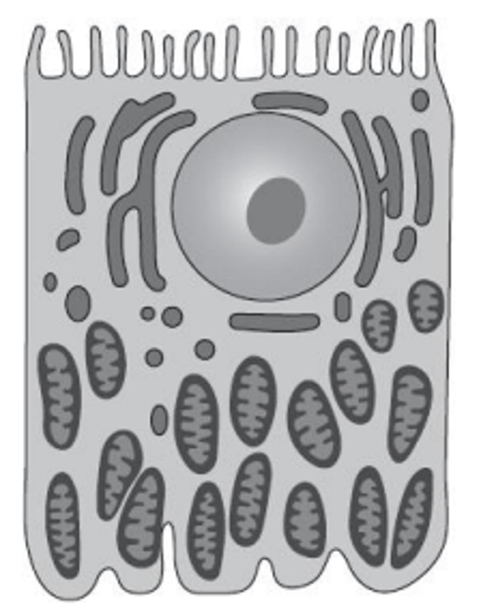
How do the features of a cell from the proximal convoluted tubule enable its function? (3)
- The cell contains a large number of mitochondria, which provide the ATP required for active transport.
- The cell surface has many microvilli, which create a large surface area for reabsorption.
- This surface area accommodates a high number of carrier proteins needed for transport from the filtrate back into the blood.