Exam #3 (Spinal column, somatic nervous system, & somatic reflexes, autonomic nervous system, and senses)
1/149
There's no tags or description
Looks like no tags are added yet.
Name | Mastery | Learn | Test | Matching | Spaced |
|---|
No study sessions yet.
150 Terms
white matter
large numbers of myelinated and unmyelinated axons
white matter is organized into columns and each column contains
tracts or bundles of axons
what do white matter tracts or bundles of axons do?
they carry info to and from the brain
they carry the same type, rate, and direction of information
ascending tracts
spinal cord to brain
sensory info
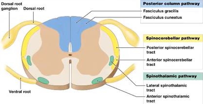
descending tracts
brain to spine
motor info
gray matter
neurons, neuroglia, unmyelinated axons
horns
cell bodies of gray matter organized into functional groups called
nuclei
sensory nuclei
receives information from PNS
motor nuclei
sends motor commands to effectors
sensory nuclei are _____ from motor nuclei
separated
posterior gray horns
somatic and visceral sensory nuclei
anterior gray horns
somatic motor nuclei
lateral gray horns
only located in the thoracic and lumbar areas
visceral motor
autonomic N.S.
what is the pathway of a dorsal root (posterior)
axons + cell body of sensory neurons
unipolar neurons
what is the pathway of a ventral root
axon of motor neurons
somatic motor neurons cover
skeletal muscles
visceral motor axons cover
smooth muscles
cardiac muscles
adipose tissue glands
dermatome
a specific strip of skin that is innervated by a specific spinal nerve
Peripheral neuropathy
nerve damage that causes weakness, numbness, and pain, typically in the hands and feet
examples:
- Trauma
- Diabetes – peripheral axon compression
- Sciatica
- Shingles
- Paraplegia
- Quadriplegia
Paraplegia
nerve damage in the lower extremities
Quadriplegia
nerve damage in all 4 extremities
decussation
crossing over of white matter tracks in the brainstem or the spinal cord to the opposite side
- Most motor or somatic motor does cross sides, usually around the brain stem area
somatic sensory pathway (ascending (afferent) tracts)
first-order neuron —> second-order neuron —> third-order neuron

first-order neuron
where the cell body is in the dorsal root ganglion
what’s detecting the stimuli
axon travels through that posterior or dorsal root and into the posterior horn
the body, entering the spinal cord

second-order neuron
- Between two neurons, typically found in between the brain or the spinal cord
- Takes the info up the spinal cord and take it to the thalamus
spinal cord, entering the brain
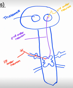
third-order neuron
- In the cell bodies in the thalamus
- Takes the signal out of the appropriate area of cortex, goes wherever that info needs to go in the cortex to be processed
- Shorter in length
thalamus and sending info where it needs to be

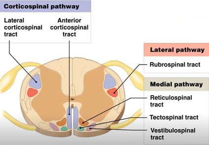
somatic motor pathway (descending (efferent) tracts)
thalamus isn’t needed
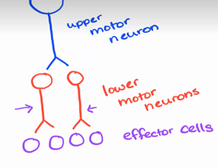
upper motor neuron —> lower motor neuron
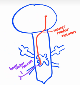
upper motor neuron
- Goes from brain down to the spinal cord
- Cell body is in the brain

lower motor neuron
axons travel out the anterior route and goes out to the effector

somatic motor
innervates skeletal muscle
types of neural pools
diverging circuit
converging circuit
serial
parallel
reverberating circuit
diverging circuit
- Motor neurons (upper)
- Upper motor neurons excite lower motor neurons which excite effector cells
- If the upper motor neuron is about to stimulate, it doesn’t mean it will give the same effect to each lower motor neuron
- Meaning the left lower motor neuron could be excited while the right lower motor neuron is inhibited
- Example of this is when you’re doing a bicep curl, the bicep is getting excited (used/contracted)) while the tricep is being inhibited (relaxed)
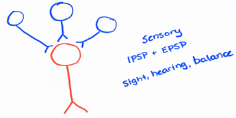
converging circuit
- A lot of neurons going to one / receiving signals from multiple neurons (spatial summation)
- Mostly sensory (IPSP and EPSP)
- Sight, hearing, balance
serial circuit
- One presynaptic neuron, one postsynaptic neuron, and maybe another neuron
- The middle one (postsynaptic neuron) would be the interneuron
- First order, second order, third order neurons
- Touch and pain, sensory

parallel circuit
- Looks like diverging
- Signal is going down neurons at the same exact time (parallel)
- Seen when both sides of the body want to be activated (walking, putting one foot down and raising the other)
reverberating circuit
- Positive type feedback
- Second neuron has a collateral that comes back to the first neuron and innervates the first neuron
- In more detail, first neuron sends a signal down that will transmit to the second neuron. The signal gets sent down through the second neuron but then the signal gets sent back up and reactivate the first neuron
- Seen with consciousness, keeping you awake
- This can’t happen forever
- Two ways to turn this off is synaptic fatigue and inhibitory stimulus
- Synaptic fatigue: can happen in either synapse and there isn’t enough neurotransmitter anymore or can’t make anymore to where the signal can’t be sent anymore
- Inhibitory stimulus: neuron inhibits the neuron to turn off the pathway
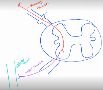
reflexes
rapid, automatic or involuntary responses to specific stimuli; always results in the same response
reflex arc
negative feedback loop
order:
sensory receptor —> sensory neuron —> integration center (interneurons) —> motor neuron —> effector
sensory receptor
receiving information
sensory neuron
taking information to the CNS
integration center (interneurons)
spinal cord or CNS
interneuron may or may not be present
motor neuron
carries information to the effector
effector
any muscle or gland that responds to motor neuron signals
innate reflex
born with this reflex
feeding
pain reflex
acquired reflex
reflexes learned throughout life
driving
walking
location of processing
information being processed in either the spinal cord or the brain
spinal (location of processing)
skeletal muscle
cranial (location of processing)
brain
response
somatic or visceral
somatic response
skeletal muscle
visceral response
smooth, cardiac muscle, adipose, and glands
complexity
simple or complex
simple complexity
monosynaptic
Involving only one synapse in the reflex arc
complex complexity
polysynaptic
Involving multiple synapses and neural pathways
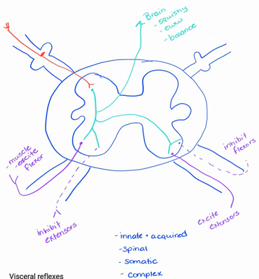
stretch reflex - essential for posture control and coordination of movement
Patellar reflex (knee-jerk)
Patellar tendon is attached to the tibia and to the quad muscles
Tendon pulls on the quadricep and stretches the muscle
This then causes the tibia to jerk up
withdrawal reflex
- Example: Touching something hot and removing your hand from it
- Only affects one side of the body
- Have info be sent down using a sensory neuron to a interneuron going down to two motor neurons
- The first motor neuron is excited and create an EPSP, this will cause the muscle to contract
- The second motor neuron inhibits the other side and relaxes it
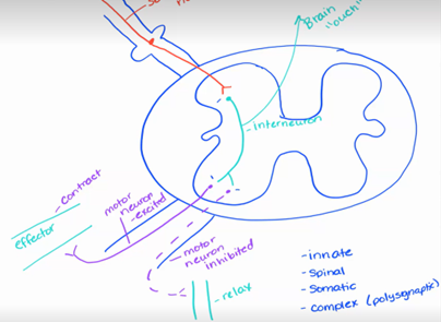
why does the info get processed and sent back out to the body in the spinal cord instead of the brain?
Because it’s more quicker compared to sending the info to the brain and receiving it back
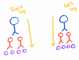
cross-extensor reflex
Example: you’re walking in the middle of the night and your right foot steps on something cold, wet, and disgusting. Your first move is to lift up your right foot but at the same time your left foot registers that it’s now standing on it’s own and has to balance the body. This happens at the same time.
- Signals are sent to one side of the body to do one task and at the same time signals get sent to the other side of the body to perform another task
- Right side = lifting your right foot
- Left side = balancing your body with your left leg
lateral horn is in which sections
T1 —> L2
in the posterior horn, it goes
somatic sensory then the visceral sensory
What is the clinical importance of dermatomes?
- To know what nerves or part of the limb or body is affected
What is a peripheral neuropathy?
- Diabetes causes peripheral neuropathy, usually in the feet to where they can’t feel their legs or arms
- When the limbs “fall asleep”, usually from compression
Someone with a spinal cord injury, are their spinal cord reflexes still intact?
The reflexes are still active (sensory to motor), but the brain process doesn’t exist because the signals don’t get sent to the brain
autonomic nervous system
is concerned with maintaining homeostasis within the body by increasing or decreasing the activity of various organs in response to changing physiological conditions
- Regulates homeostasis
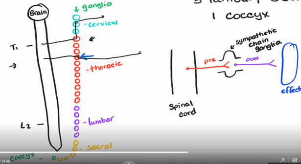
Sympathetic
prepares body for physical activity (arousal, competition, stress, danger, anger or fear)
“Fight or Flight” or Thoracolumbar
what happens to your body with sympathetic
- Increased heart rate
- Increased blood pressure
- Increased respiratory rate
- Increased blood sugar glucose
- Mental awareness increases
- Pain signals decrease
- Pupils dilate, increasing visual acuity
- Decreased digestion and urinary
lateral horns are only found in which sections
T1-L2
motor neurons only come out of which sections
T1-L2
are motor neurons found in the sympathetic nervous system
no
it doesn’t come out of anywhere else in the spinal cord besides T1-L2
Parasympathetic
calming of body
”Rest and Digest” or Craniosacral
what happens to your body with parasympathetic
- Lower heart rate
- Lower blood pressure
- Lower respiratory rate
- Increased digestion, nutrient absorption, storage of nutrients
- Increasing gland secretion
- Increase urination and defecation
- Pupils are constricted
where do motor neurons come out of for parasympathetic
either cranial nerves or the sacral region
motor neurons that come out of the cranial area/nerves are
parasympathetic
motor neurons that come out of the cervical are
neither sympathetic or parasympathetic
motor neurons that come out of the thoracic and T1-L2 of the lumbar are
sympathetic
motor neurons that come out of sections L3-I4-I5 are
neither sympathetic or parasympathetic
motor neurons that come out of the sacral region are
parasympathetic
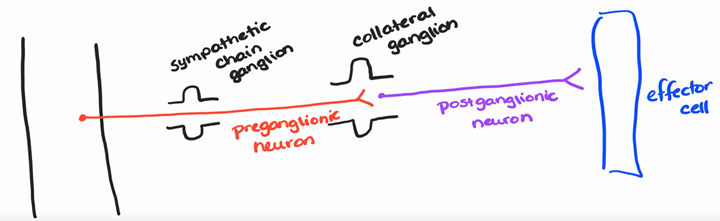
ganglion
area where two neurons synapse
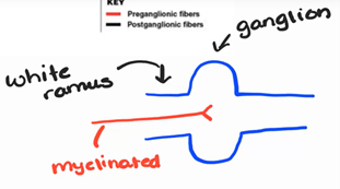
preganglionic neuron
cell bodies in the lateral gray horns of spinal segments T1-T12 and L1, L2; axons are myelinated
enters into the ganglion through a white ramus (myelinated)
autonomic ganglia
sympathetic chain ganglia (paired (meaning both sides) – 3 cervical, 11 thoracic, 5 lumbar, 5 sacral, 1 coccyx)
preganglionic nerve fibers enter the chain ganglia via white rami (ramus)
collateral ganglia - celiac, superior mesenteric, inferior mesenteric
- They are not paired
- They are found anterior to the spinal cord
- they go to organs and tissues that are in the abdominal cavity
- they come out of the spinal cord between T1-L2, coming out of the lateral column
- travels through without stopping through a sympathetic chain ganglia
- synapses inside of a collateral ganglia
adrenal medullae
- travels through a sympathetic chain ganglia and through collateral ganglia and synapses directly on the adrenal medulla with multiple different neurons
- releases both epinephrine and norepinephrine (neurotransmitters)
postganglionic neurons
Axons unmyelinated; fibers long
Leaving the ganglion through a gray ramus (unmyelinated) and going to the effector
pathways of ANS nerve fibers
Chain ganglia
Collateral ganglia (celiac, superior mesenteric and inferior mesenteric)
Adrenal medullae
§ A single sympathetic preganglionic fiber has many axon collaterals and may synapse with 20 or more postganglionic fibers; then postganglionic fibers may innervate several visceral effectors (Widespread response)
what does this mean?
- This allows one neuron to have a widespread effect on multiple effector cells
- One message goes to multiple post ganglions neurons which goes to multiple effector cells
- Widespread effect
- This also activates the adrenal medullae since this is releasing epinephrine or norepinephrine into the bloodstream
preganglionic neuron
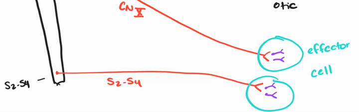
cell bodies in the nuclei of the four cranial nerves that initiate in the brain stem (III, VII, IX, X) and segments S2-S4
cranial nerve III
oculomotor – pupil diameter - For parasympathetic, it will decrease the size of pupils
cranial nerve VII
facial – controls submandibular salivary glands
cranial nerve IX
glossopharyngeal – controls parotid salivary gland
cranial nerve X
vagus – controls cardiovascular, respiratory, and digestive
S2-S4
goes to reproductive, bladder, and colon
types of ganglionic neurons
terminal ganglia
intramural ganglia
terminal ganglia
they go near target organs but they’re not on it (ciliary (III), pterygopalatine (III), submandibular (VII), and otic (IX))
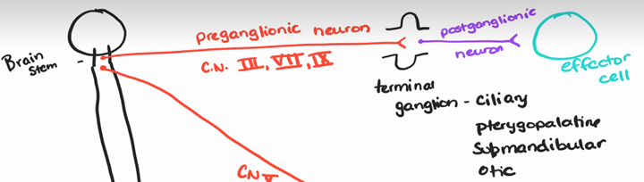
intramural ganglia
embedded in the wall of the target organ (they go to the target organ)
postganglionic neurons
Postganglionic fibers are short passing between the terminal and intramural ganglia and the target organ; - synapses with only 4 to 5 postsynaptic neurons from one effector (Localized response)
- Example being: it can increase your digestive system without having to lower your heart rate at the same time
neurotransmitters
what sends signals from the neuron to whatever the next cell (chemical signal)
all preganglionic sympathetic neurons release
ACh (cholinergic)
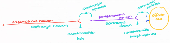
postganglionic neurons have what receptors for ACh
nicotinic receptors for ACh
varicosities
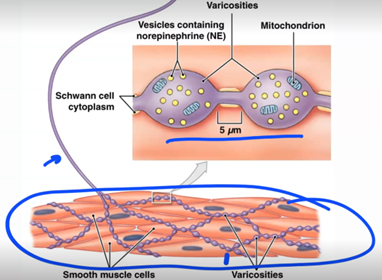
adrenergic neurons can release what
norepinephrine (NE): released by sympathetic postganglionic