2. ascending sensory pathways
1/42
Earn XP
Description and Tags
longitudinal organization
Name | Mastery | Learn | Test | Matching | Spaced | Call with Kai |
|---|
No analytics yet
Send a link to your students to track their progress
43 Terms
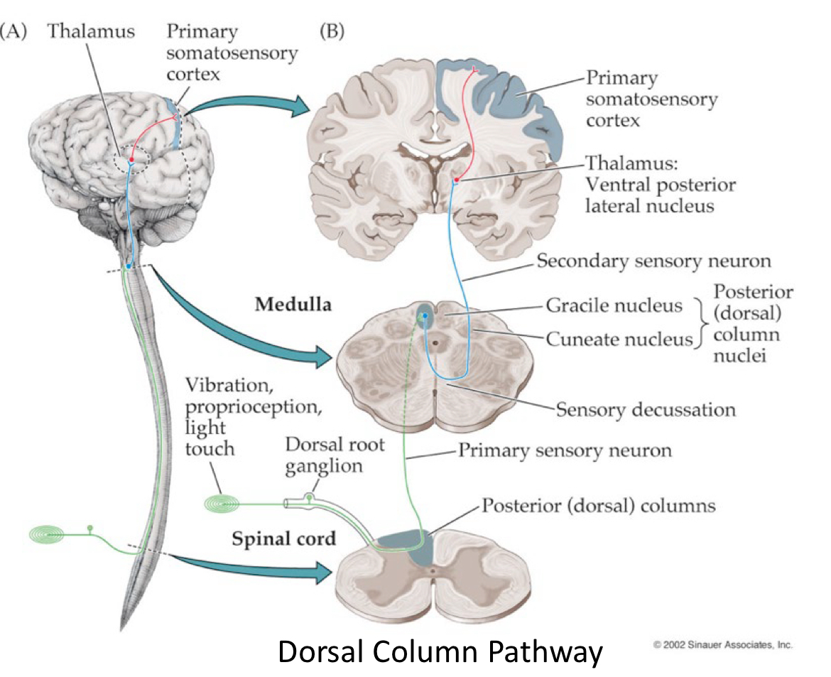
walk through this graphic
dorsal column-medial lemniscus pathway
how many and which ascending pathways are testable?
2
dorsal column/medial lemniscus pathway and
spinothalamic pathway
ascending tracts {organization of white matter (funiculi)} are segregated by?
sensory modality
dorsal column/medial lemniscus pathway and
spinothalmic pathway
there are two clinically testable conscious ascending somatosensory pathways that convey info from periphery to cerebral cortex
relays sense vibration sense and discriminatory touch (2 point) from periphery → cortex
function of dorsal column/medial lemniscus pathway
relays pain, temp, crude touch from periphery → cortex
function of spinothalamic pathway
ascending pathway conveys unconscious sensory info muscles → cerebellum
spinocerebellar pathway
fasciculus gracilis and cuneatus
dorsal column pathway
anterior spinothalamic tract- crude (non-discriminative) touch and pressure
lateral spinothalamic tract (pain and temp)
spinothalamic pathways
posterior/dorsal (blank) tract
anterior/ventral (blank) tract
cuneocerebellar tract
spinocerebellar pathways
why do long tracts have to decussate during ascent or descent?
left cortex deals w right side of body
right cortex deals w left side of body
longitudinal organization
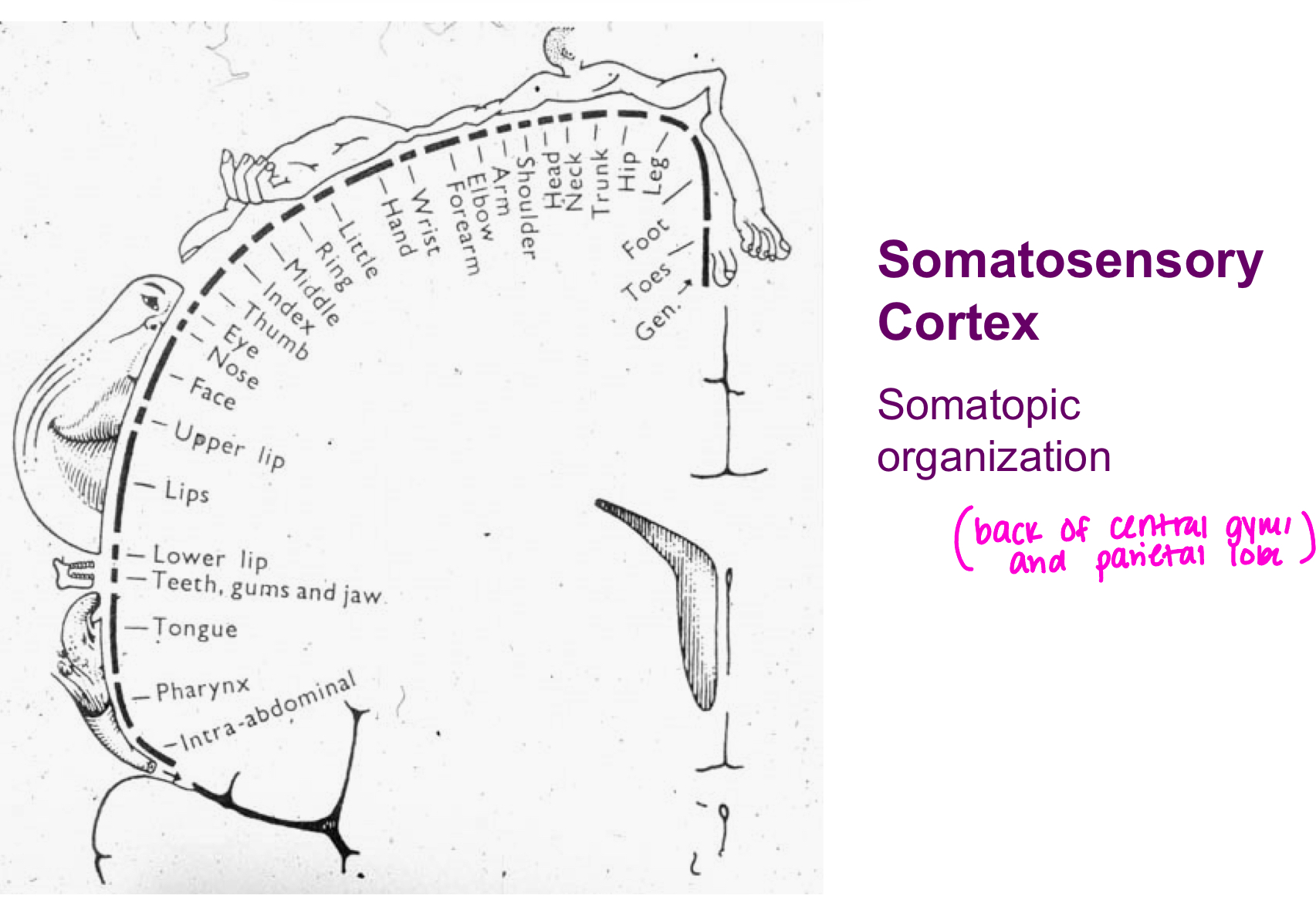
somatotopic organization
the two sensory modalities (sensory endings/structures sensitive to specific stimuli) relative to dorsal column:
proprioception
muscle spindles
golgi tendon organ
fine/discriminatory touch
the three sensory modalities (sensory endings/structures sensitive to specific stimuli) relative to spinothalamic pathway:
crude touch
pain
temp
describe the 3 neuron pathway:
1: first order neuron cell body in DRG and terminates in brainstem or spinal cord synapse w 2
2: second order neuron axon crosses the midline of brainstem or spinal cord, terminates in thalamus
3: third order neuron cell body in thalamus and terminates in cerebral cortex/post central gyrus, somatosensory
all info heading to cerebral cortex must stop at (blank) except olfaction
thalamus-relay for cortex
*thalamus also good for higher level cortical function and cortical activation
cortical activation
he process of increased neuronal activity in the cerebral cortex
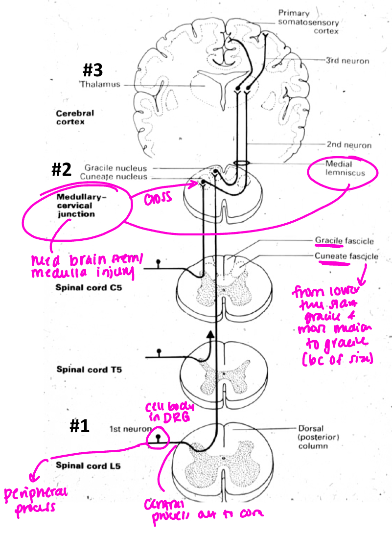
what pathway is this? talk through graphic
dorsal column/posterior
where does the second neuron of posterior/dorsal pathway cross?
at medulla and form medial lemniscus; injury consistent with same side
where does posterior/dorsal column first order neuron enter?
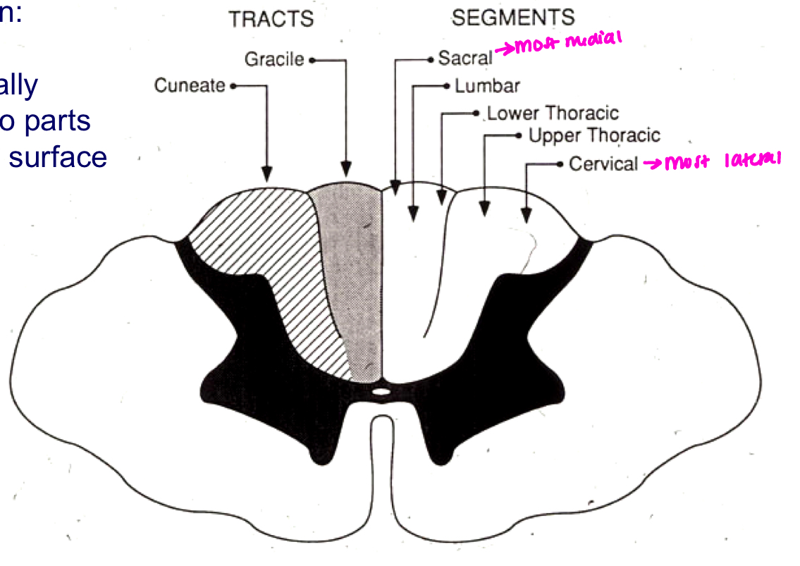
for T5 and above receives input from cuneate
T6 and below receives input from gracilis
is gracile or cuneate more medial in posterior funiculus?
gracile
describe somatosensory cortex somatotopic organization
proprioception
sense of where body is in space
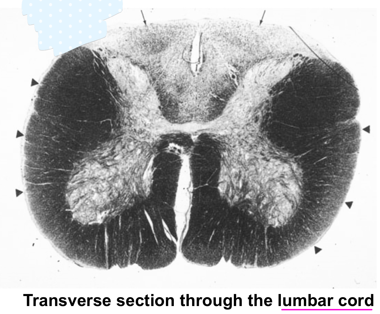
what is wrong in this picture? what would you expect to observe upon examination?
there is demyelination of the dorsal column pathway (posterior funiculi); loss of proprioception: would not know where low extremities are without seeing them; loss of vibration
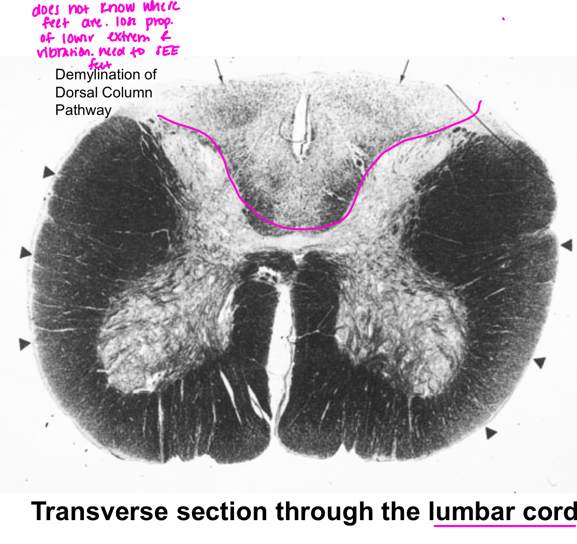
what about demyelination of just fasciculus gracilis?
no proprioception and vibration too BUT gracilis is medial so that means lower part of body is affected
what about demyelination of just fasciculus cuneatus?
no proprioception and vibration too BUT cuneatus is lateral so that means upper part of body is affected
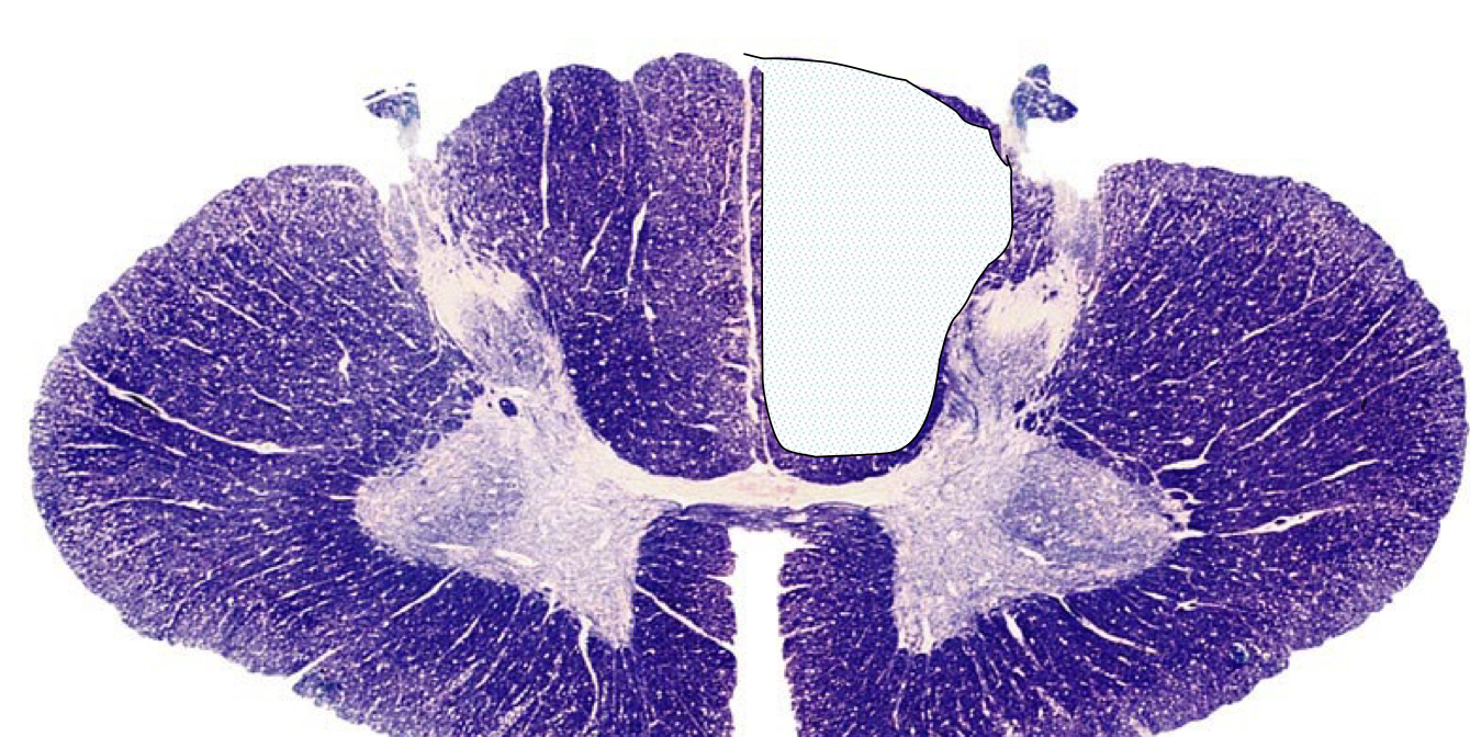
what findings would you expect to see in an individual with this lesion?
lesion to L posterior funiculus so both L cuneatus and gracilis; second neuron crosses all the way up in medulla so injury affects the same side
what separates the L and R fasciculus gracili?
posterior median sulcus
what separates Cun vs Grac?
posterior intermediate sulcus
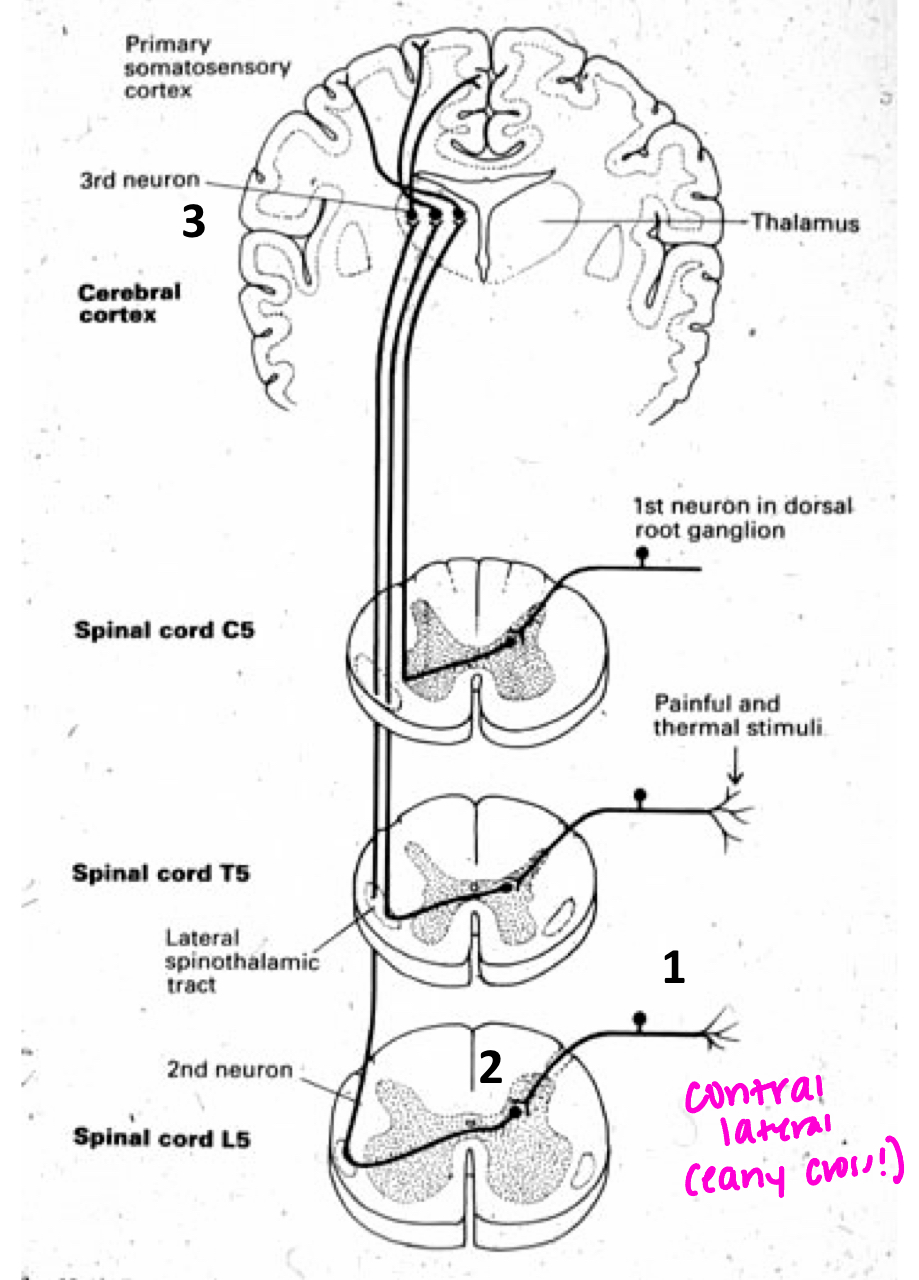
what pathway is this?
spinothalamic
spinothalamic neuron location and desiccates
in dorsal horn and crosses immediately at ventral white commissure
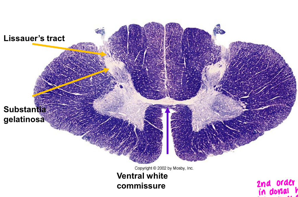
where do poorly myelinated pain and temp neurons enter
dorsal horn and follow path to ventral white commissure to desiccate (gray matter)
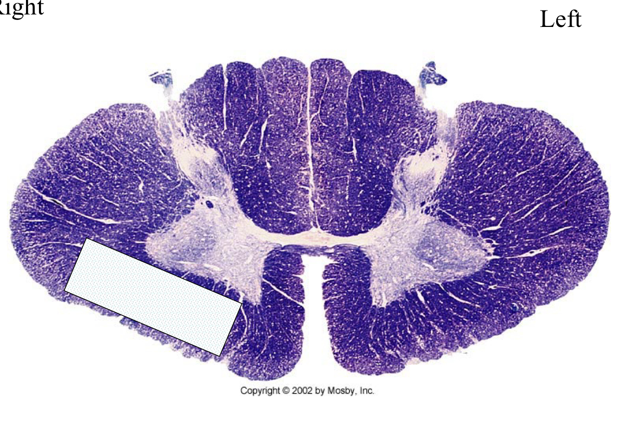
what would you expect to see on examination of this individual?
spinothalamic loss of pain and temp on L side of body
dorsalateral pathway vs spinothalamic tract
dorso: ipsilateral deficits and good myelination
spinothalamic: contralateral deficits and no or poorly myelinated
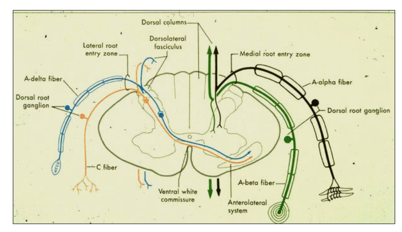
what is suspended sensory loss?
suspended sensory loss: pain and temperature sensation is lost at a particular spinal cord level, band-like effect
long tract lesion damage to any of the major sensory pathways in the spinal cord, causing sensory loss that extends below the lesion level and can affect multiple sensory modalities depending on which tract is involved
lesion at white commissure?
spinothalamic neurons could not desiccate so bilateral loss of pain and temp suspended sensory loss!
where do all pathways to the cerebellum end?
they (dorsal spinocerebellar, ventral spinocerebellar, and cuneocerebellar pathways) end ipsilaterally in cerebellum
what is the DSC pathway?
dorsal spinocerebellar/cuneocerebellar
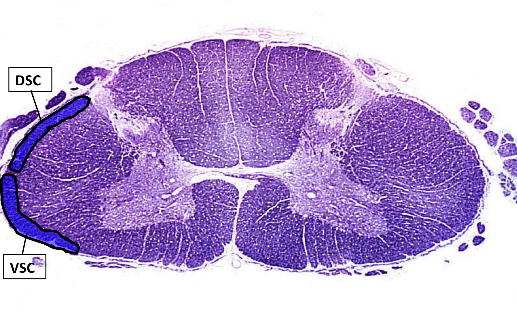
spinocerebellar pathways; not testable; unconscious to limbs
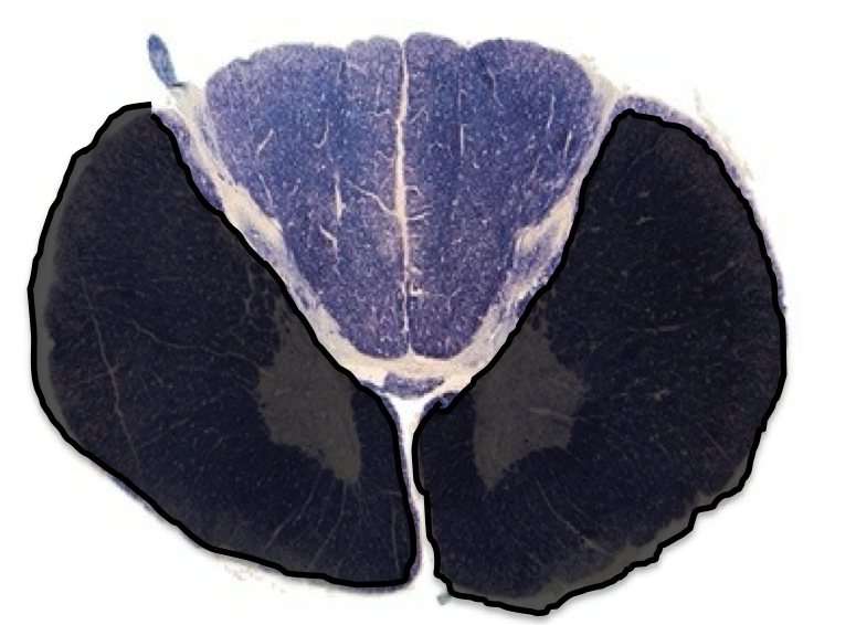
rare, after occlusion of anterior spinal artery
motor: bilateral paralysis or weakness
fine touch: normal
pain/temp: bilateral loss
anterior cord syndrome (spinothalamic; @ and below)
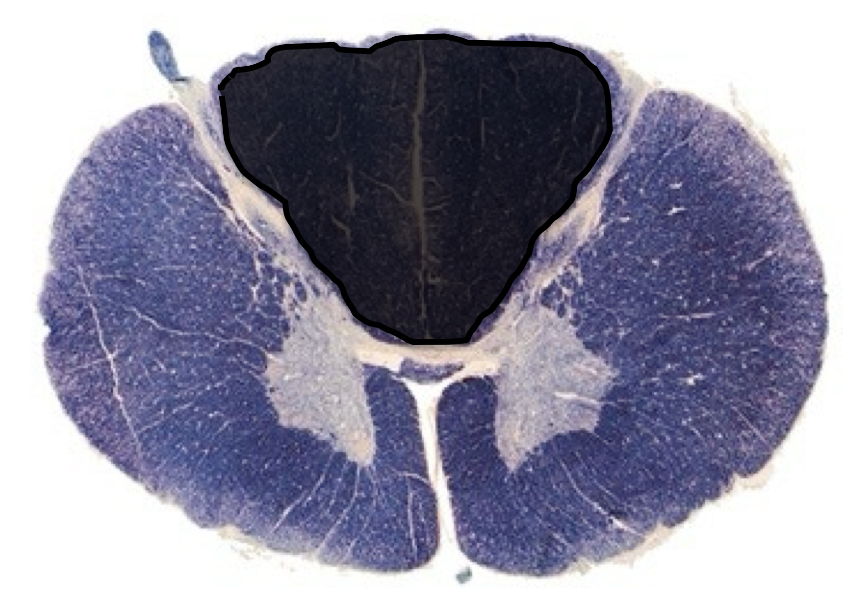
rare, after occlusion of posterior spinal artery
motor: normal
fine touch vibration: bilateral loss
pain/temp: normal
posterior cord syndrome (dorsal column @ and below)
damage to central area of spinal cord, damages crossing fibers first
motor: immediate bilateral loss
fine touch: normal
pain/temp: progressive bilateral paralysis by direct damage on motor neurons
central cord syndrome
ventral white commissure-spinothalamic
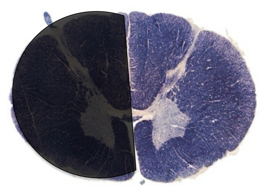
aka lateral spinal hemisection
motor: ipsilesional paralysis (same side lesion)
fine touch: ispilesional loss
pain/temp: contralesional loss
(symptoms show loss of both pathways)
Brown-sequard syndrome
(motor and touch = dorsal column and pain/temp is spinothalamic)
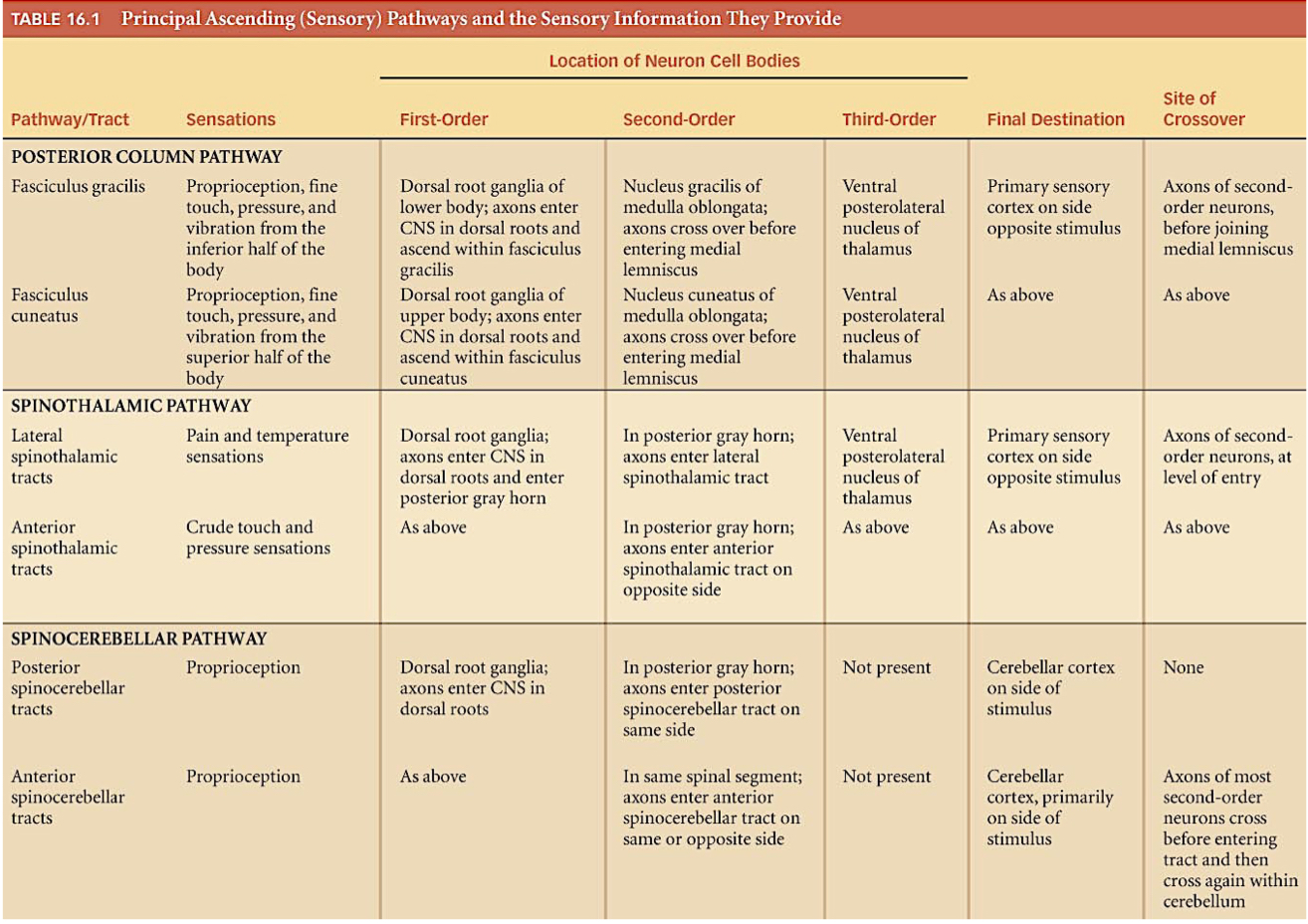
memorize! summary!