Biology, Membranes
1/90
Earn XP
Name | Mastery | Learn | Test | Matching | Spaced | Call with Kai |
|---|
No analytics yet
Send a link to your students to track their progress
91 Terms
Membrane Strucuture
Phospholipid Bi-layers
Globular protines
fluid Mosaic model
Phospholipid Bi-layer
Structure of Membranes
Include Glycerol Phosoplipids and Sphingolipids
Fluid Mosaic Model
Structure of membranes
Mosaic of proteins float in or on the fluid lipid bi-layer
EX: Boats on a pond
Boats on a pond are a example of what?
Fluid Mosaic model
Fluid Mosaic model of Cell membranes
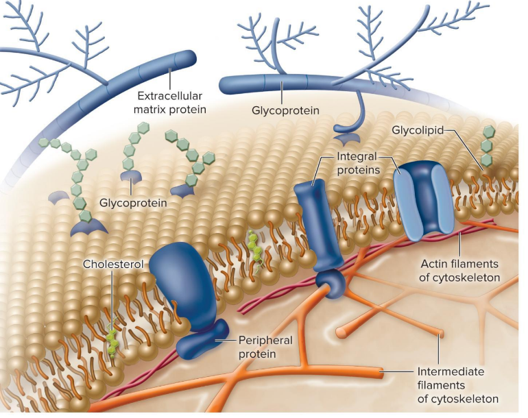
Components of Cellular membranes
Phospholipid bilayer
Transmembrane Proteins
Interior Protein Network
Cell Surface Markers
Cellular membranes- Phospholipid Bilayer
Flexible Matrix, barrier to permeability
Cellular membranes- Transmembrane Proteins
Integral membrane proteins
Cellular membranes- Interior Protein Network
Peripheral or intracellular membrane proteins
Cellular membranes- Cell Surface Markers
Glycoproteins and Glycolipids
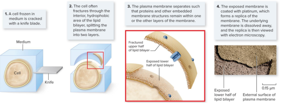
Studying the cell membrane- TEM
Transmission electron microscope (TEM), one method of TEM is to embed specimen in epoxy
less than 1 micro meter thick of of speciman can be imaged
Studying the cell membrane- SEM
Scanning electron microscope (SME)
Freeze Fracture Microscopy
Membrane lipids
Lipidomics
Glycerol phospholipids
sphingolipids
sterols
3 Classes of the 1000 distinct lipids in cells
Glycerol phospholipids
sphingolipids
sterols
Lipidomics
Defines the number and biological function of lipids
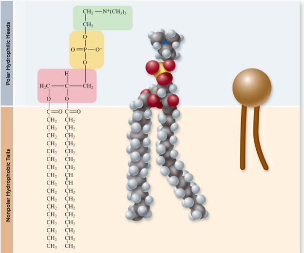
Sphingolipid Structure
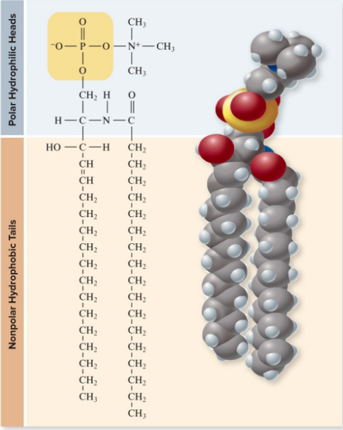
Phospholipids
Amphipathic structure ( bi-layer forms spontaneously)
Head is Polar, Hydrophilic, and has Phospate group attached
2 fatty acids, are non polar and hydrophobic
Phospholipid structure
Physiology of Phospholipid Bilayer
Bilayers are fluid
Hydrogen bonding holds layers together
unanchored proteins and individual phospholipids move through membrane
Bilayers are
fluid
what holds phospholipid bilayers together
Hydrogen bonds of: H2O -H2O and H2O-polar heads
Hydrophillic and Hydrophobic bonds
Saturated Fatty Acids affect on fluidity
Cause membrane to be less fluid compared to unsaturated fatty acids
How dose temperatures affect membranes fluidity
Warm temps make the membrane more fluid, cold makes them more viscous
Cold tolerant bacteria are possible by fatty acid desaturases
what compositions of lipids affect fluidity, thickness, and shape of membrane?
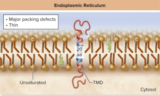
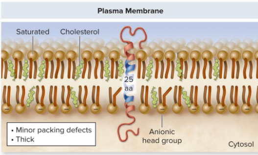
lipid composition of the ER membrane, Golgi stack and plasma membrane
Endolpasmic reticulum (ER)
Plasma Membrane (PM)
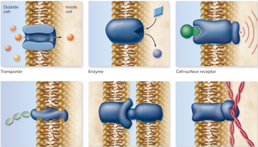
Functions of Membrane Proteins
Transport
Enzymes
Cell-surface receptors
Cell-surface ID Markers
Cell-Cell adhesion Proteins
Attachment to Cytoskeleton
Affect on membrane structure
Membrane Protein Function
Membrane Structure relates to Function
Diverse Function form Diverse structure of membrane proteins
Common Structural features relate to role of membrane Protein
Diverse Function form Diverse structure
Membrane Proteins
Anchor Molecules
Attach to membrane protein, to the membrane surface
modified by lipids
anchor molecules moded by lipids
by nonpolar intersecting regions of the internal portion of the lipid bilayer
Chemical bonding domains, linking directly to proteins
Transmembrane proteins
Span Lipid bilayer
Non polar regions of proteins are embedded interior of bilayer
Alpha Helices and Beta sheets
Polar regions of proteins protrude form both sides of bilayer
Affect of non polar and polar regions on Transmembrane Proteins
Non Polar embed inside bilayer
Polar protrude form both sides of bilayer
Transmembrane Domain
membrane spanning region
Hydrophobic amino acids form Alpha helices
one domain need to be anchored in Membrane for proteins
may have more than one domain
Class of receptors is based on # of Domains present
how are hydrophobic amino acids arranged in Transmembrane domains
Alpha Helices
how many trans membrane domains needed in proteins to anchor in Membrane?
A single TM domain needed, but may have more than one of the same domain with the amount domain based on class of receptor type.
Pores
Created by extensive non polar regions in a TM Protein
Beta barrel are secondary protein structure
polar interior allows water and small polar molecules to pass through membrane
polar interior that allows water and small polar molecules to pass through membrane
Pore
Passive transport
movement of molecules through membrane
no energy required, move in response to concentration gradient
diffusion
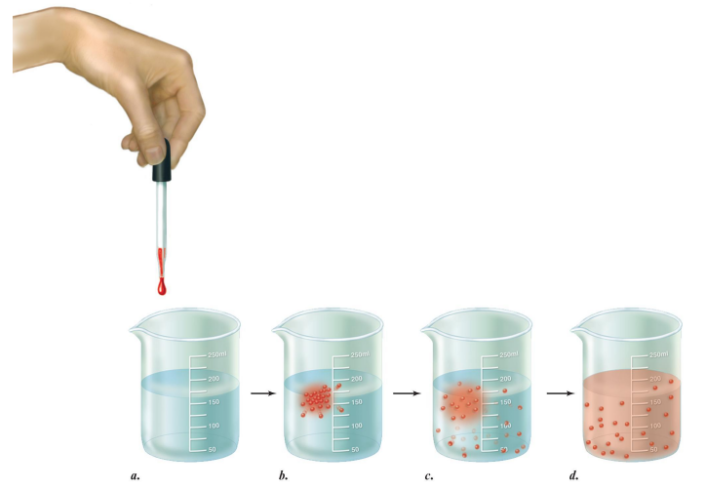
Diffusion
movement of molecules high to low concentration
continues till conc. is same in all regions
Diffusion
transport Across membranes
major barrier is hydrophobic interior, only repels polar molecules
nonpolar molecules function like diffusion
only small polar molecules
limited permeability to larger polar molecules & ions
Protein affect on membrane diffusion
-Facilitate diffusion:
Molecules can use proteins as gateways
Channel proteins are hydrophillic when open
Carrier proteins assist membranes when binded
-Membrane is selectively permeable due to channels and carriers
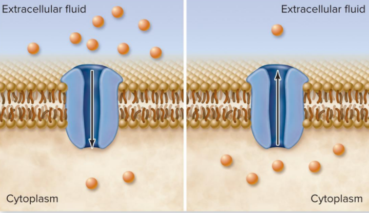
Channel Proteins
-Ion channels: passage of ions through non polar interior of plasma membrane
Gated channels, open or close to stimuli
direction dependent on concentration, voltage difference, Gate channels open or closed
direction in protein channels are dependent on
Relative Concentration on eather side of membrane
Voltage Difference across membrane
Gated Channels opened or closed
Facilitated Diffusion of Ions
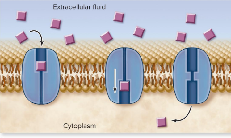
Carrier Proteins
Required to bind to molecule they transport
Transport both Ions and other solutes
Move by diffusion
Saturation in Carrier Proteins
Rate of transport limited by # of transporters
Facilitated diffusion by carrier proteins
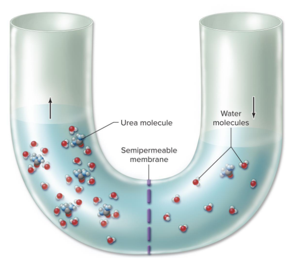
Osmosis
Net diffusion of water across membrane to higher solute conc.
Cytoplams of cell is an Aqueous solution
Cytoplasm of cell
Aqueous solution
Water
is a solvent
solute
dissolved substances
Osmosis Across a Semipermeable Membrane
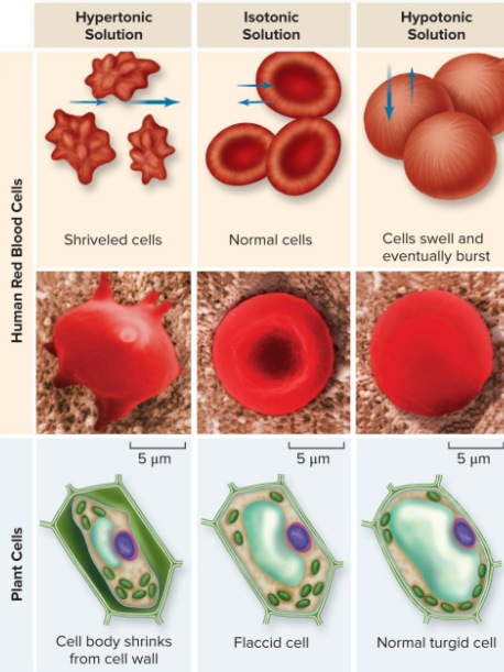
Osmotic concentration
Hypertonic solution- higher solute conc.
|Hypotonic solution- lower solute conc.
Isotonic solution- equal conc.
Aquaproins facilitate osmosis
Aquaporins
Channles for water in cells membrane
Facilitate Osmosis
Osmotic pressure
Force to stop osmotic flow
Cells swelling can create pressure
Hydrostatic pressure, balances pressure by driving water out
Plasma membrane are weak and may burst
Cell Swelling
In a hypotonic solution cells gain water causing swelling, that creates pressure
Hydrostatic Pressure
aids in reaching osmotic pressure, by driving water out
used by Prokarytoes, Fungi, Plants, and many Protists
Isotonic Environments
Animal cells must be in these environments to avoid bursting plasma membranes
Solutes and Osmotic Pressure in cells
Maintaining Osmotic Balance
Extrusion- injects water through contractive vacuoles
Isosmotic regulation- used by marine organisms and terrestrial animals
Turgor pressure- used by plants
Active Transport
Req. Energy ATP directly or indirectly used to fuel
Moves substance low to high concentration
Req. use of Highly Selective carrier proteins
Carrier Proteins used in Active Transport
Uniporters- move one molecule
Symporters- move 2 molecules, same direction
Antiporters- move 2 molecules, opposite direction
Uniporters
carrier proteins that move one molecule at a time
Symporters
carrier proteins, move 2 molecules in same direction
Antiporters
Move 2 molecules in opposite directions
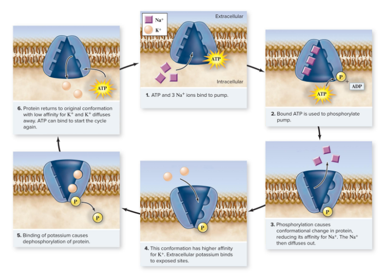
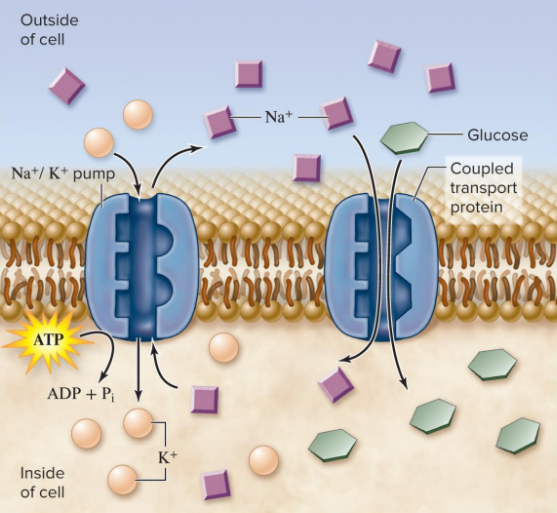
Na+-K+ Pump
Sodium-Potassium Pump
What type of transport is the Na+-K+ Pump
Active transport, uses ATP to change the conformation of carrier protein
what type of carrier protein is used for the Na+-K+ pump
Antiporter’s to move 3 Na+ out of cell and 2K+ in to cell
they move against the concentration gradient
How dose the Na+-K+ function
To change ions so they can be carried across the membrane, by moving 3 Na+ out of cell and 2K+ into cell using antiporter’s that have a affinity for Na+ or 2 K+
What is the Na+-K+ Pump ratio
3 Na+ out of cell and 2 K+ into cell
NA+-K+ Pump Function (Diagram)
How Coupled Transport Use ATP
Indirectly
How Coupled Transport Use Energy
Uses energy released when a molecule moves by diffusion to supply energy to activate transport of different molecule
Carrier Proteins in Coupled Transport
Symporter or Aniporter
Coupled Transport: Glucose-Na+ symporter
Captures energy form Na+ diffusion to move Glucose against a concentration gradient
Coupled Transport Via Membrane Proteins
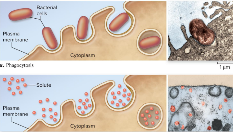
Bulk Transport
Endocytosis & Exocytosis
Active transport
Endocytosis
Movement of Sub into cell
Requires Energy
Phagocytosis
Pinocytosis
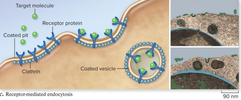
Receptor’s
Phagocytosis
Cell takes in Particulate Matter
Pinocytosis
Cell only takes in FLUID
Endocytosis Receptor
Mediate endocytosis
specific molecules are taken in after binding to receptor
Exocytosis
Movement of substance OUT of cell
Requires energy
Endocytosis (Diagram)
Example of Receptor mediate Endocytosis
In The disease Hypercholertolemia, The LDL receptors lack tails, therefor never fastened in the clathrin-coated pits and as a result do not trigger vesicle formation. The cholesterol stays in the blood stream of affected individuals, accumulating as plaques inside arteries, leading to heart attacks.
Example of Receptor mediate Endocytosis (Diagram)
Exocytosis (Diagram)
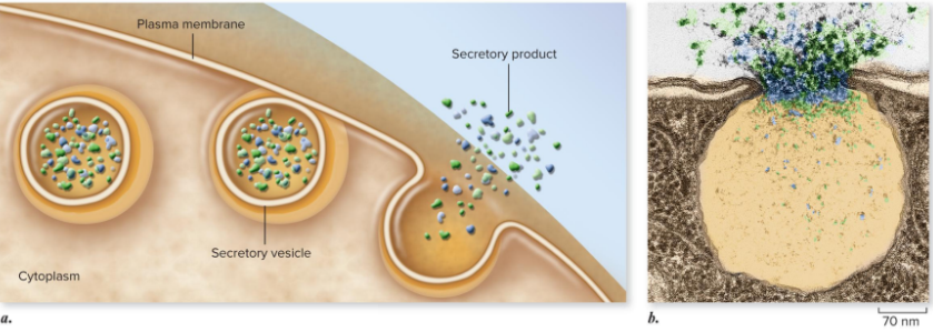
Exocytosis Function
Discharge of materials out of cell
Plants use to export cell wall material
Animals use to secrete Hormones, neurotransmitters, Digestive enzymes