IMED1001 - Renal System (Week 10)
1/25
There's no tags or description
Looks like no tags are added yet.
Name | Mastery | Learn | Test | Matching | Spaced |
|---|
No study sessions yet.
26 Terms

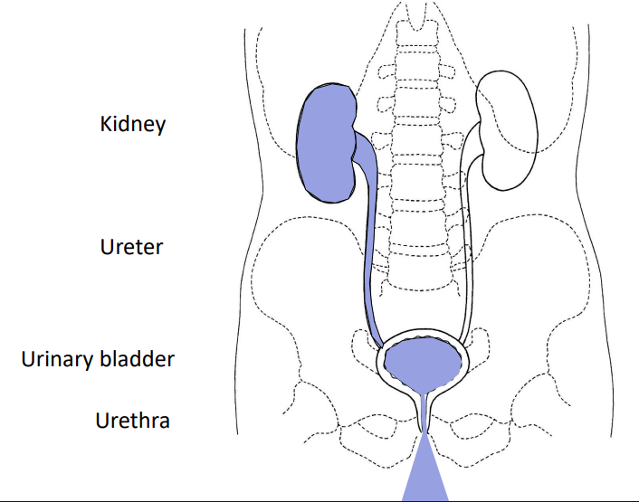
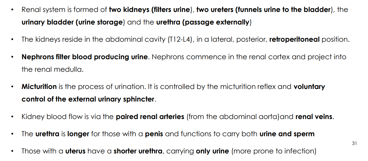
Gross Anatomy of The Renal System (Kidneys, Ureters, Urinary Bladder, Micturition)
- kidneys filter excess water, salts, and metabolic wastes from blood
- ureters carry urine to urinary bladder
- Urinary bladder stores urine
- micturition releases urine from urinary bladder into the urethra and out to the external environment (micturition is the decision making process for when we release urine)

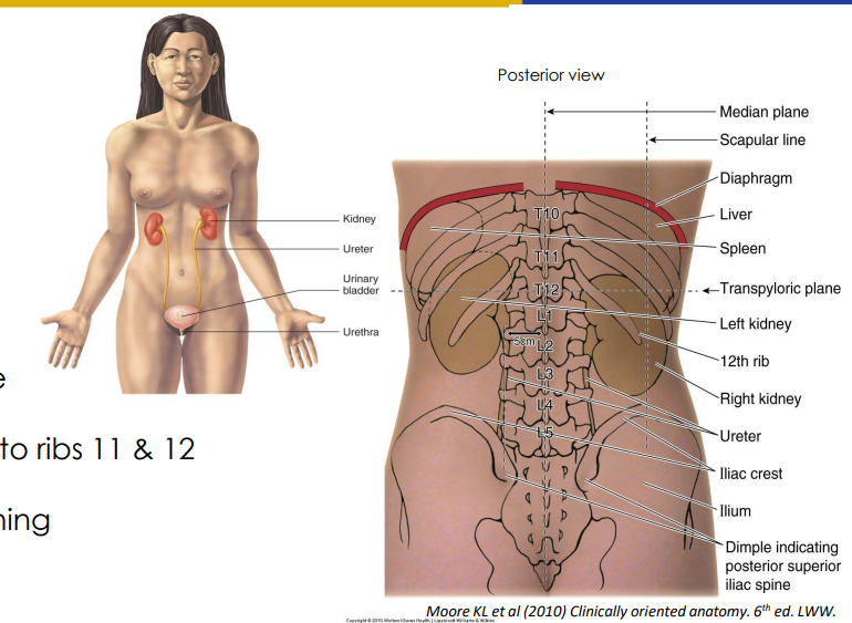
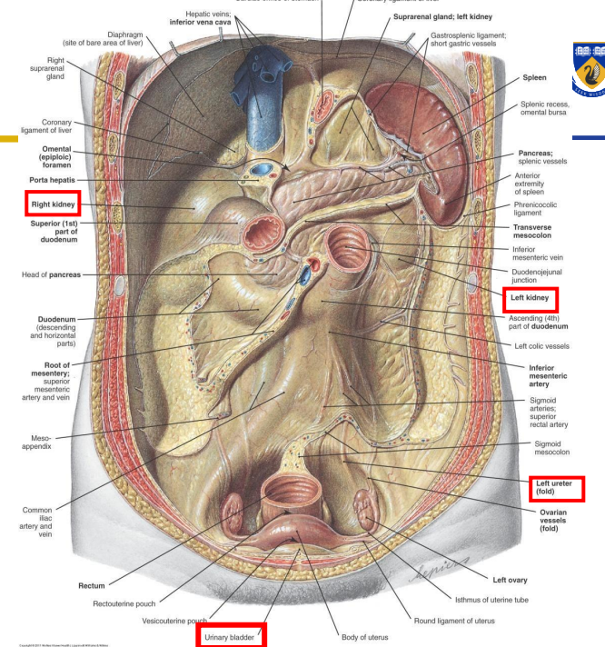
Topographic Location of the Kidneys
- left kidney more superior (liver displaces right kidney so it sits inferior)
- Left T12-L3
- Right: L1-L4
- Hilum approx. 5cm from midline (hilum is the place on side of kidneys where blood vessels enter and exit)
- Superior poles of kidneys deep to ribs 11 and 12
- kidneys move vertically with breathing

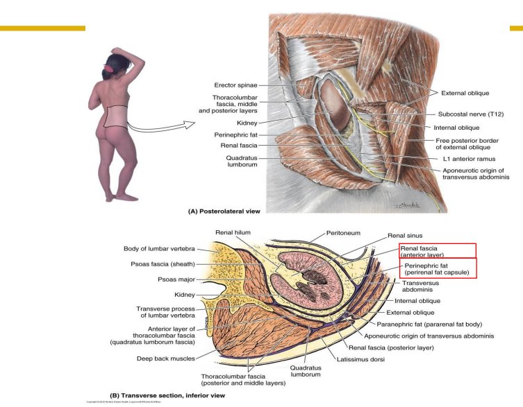
Location of Kidneys and Diaphragm
- diaphragm protects posterior aspect of superior poles of kidneys from ribs
- Kidneys are deep to the posterior abdominal wall muscles in hypochondrium regions
- fascia holds the kidneys in place
- fat protects the kidneys (perinephric fat)
Cadaveric Image of Kidneys
DIAGRAM ON SLIDE 8


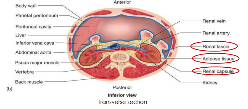
Transverse Section of Kidneys (Position of Kidneys, Renal Capsule, Adipose Tissue and Renal Fascia)
- kidneys and ureters are primary retroperitoneal (retro means behind, peritoneal means the peritoneal lining (covering abdominal organs)
- Bladder and urethra are subperitoneal
- Renal Capsule: fibrous connective tissue surrounding each kidney
- Adipose Tissue: engulfs renal capsule and acts as cushioning
- Renal Fascia: thin layer loose connective tissue, which anchors kidneys to posterior abdominal wall

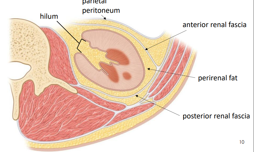
Attachment of Renal Fascia
RENAL FASCIA ATTACHES TO:
- diaphragmatic fascia superiorly
- attachment is to the diaphragm
- Kidneys move when diaphragm moves
- around 4cm forced respiration and 1cm during quiet respiration
Kidney Position Diagram
DIAGRAM ON SLIDE 11
kidneys and ureters are retroperitoneal
urinary bladder is subperitoneal

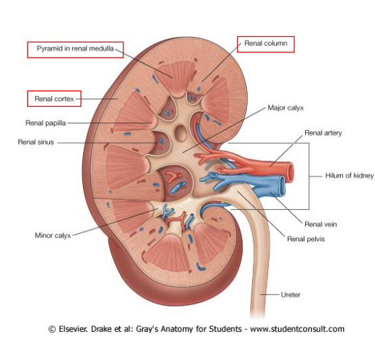
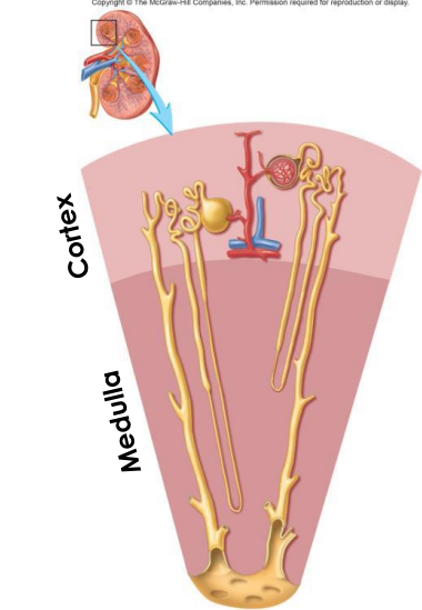
The Cortex and Medulla
- Cortex: Outer tissue with renal column extensions (between pyramids)
- Medulla: inner tissue containing renal pyramids


The Nephron
- functional and histological unit of kidney
- 1.3 million nephrons in each kidney
- 50-55mm in length
- blood enters the nephron for filtration producing urine
- Cortex contains renal corpuscle + convoluted tubules (proximal/distal) + collecting ducts (medullary rays)
- Medulla contains nephron loops (loops of Henle) + collecting ducts + papilla
- loop of Henle is not really the correct word, use nephron loop)
Location of the Nephron
DIAGRAM ON SLIDE 14


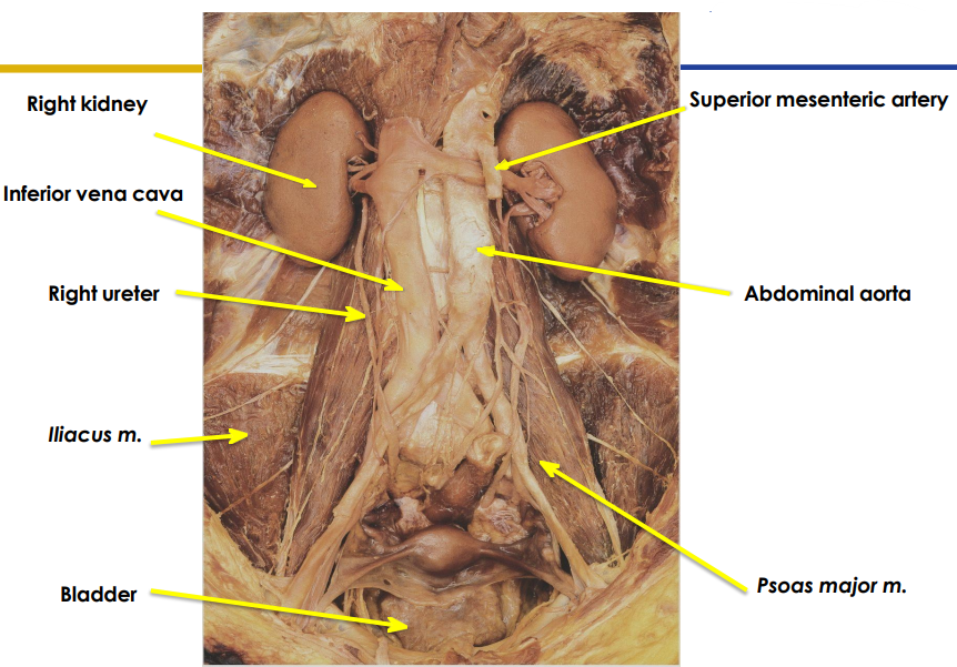
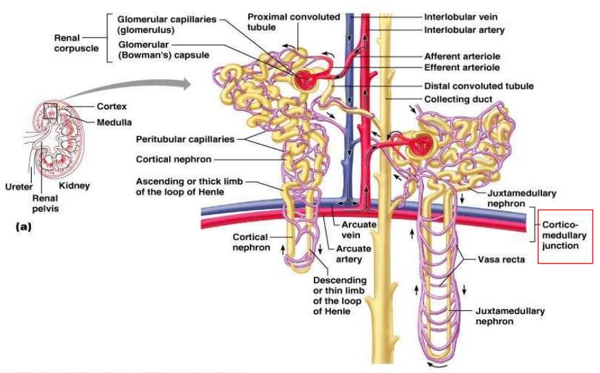
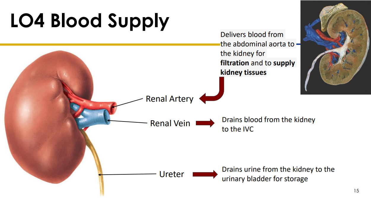
Blood Supply of Kidneys
- Renal Artery: Delivers blood from the abdominal aorta to the kidney for filtration and to supply kidney tissues
- Renal Vein: drains blood from the kidney to the Inferior vena cava
- Ureter: drains urine from the kidney to the urinary bladder for storage
Blood Supply of Kidney Diagram
DIAGRAM ON SLIDE 16


Arterial Supply and Venous Drainage
ARTERIAL SUPPLY:
- Renal a. -> segmental -> interlobar -> arcuate (to lobe) -> interlobular (to lobules) -> afferent arteriole (to individual nephrons)
VENOUS DRAINAGE:
- Mirrors arterial supply
accurate arteries demarcate junction between the renal cortex and renal medulla
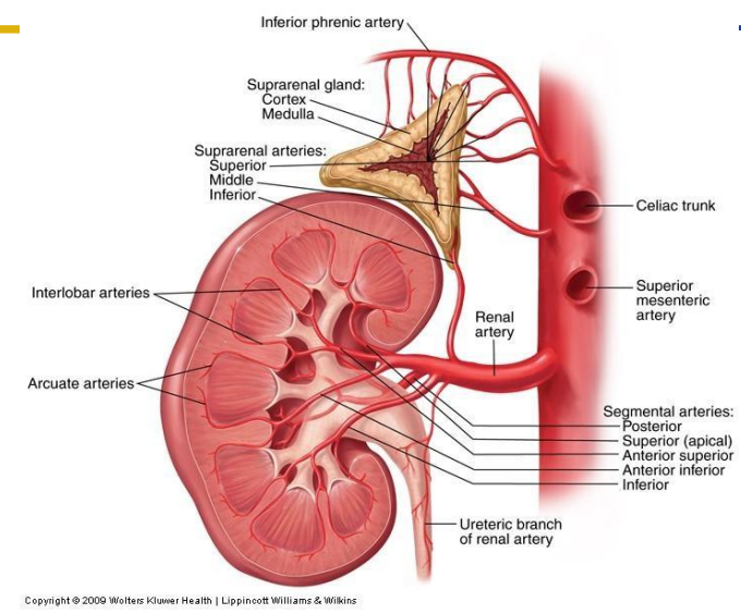
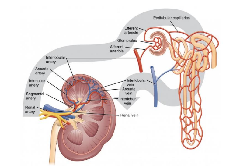
DIAGRAM OF KIDNEY BLOOD SUPPLY INSIDE KIDNEY
- Efferent is on the way out, afferent is on the way in
- efferent is smaller diameter then afferent
- efferent continues to be the peri-tubular capillaries (peri means around, so the capillaries around the tubules)

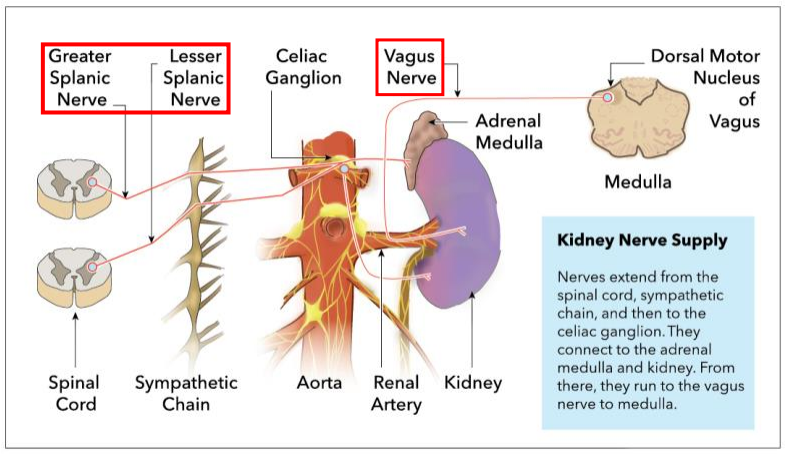
Kidney Innervation
NERVE SUPPLY:
- Vasomotor (control of vascular flow into kidneys)
- Parasympathetic: Vagus Nerve (increase renal artery flow which means increase glomerular filtration rate (GFR)
- Sympathetic: greater and lesser splanics (decrease renal artery flow means lower GFR)
- maintain a fairly constant GFR, creates around 180L of filtrate/day. Reabsorb most, only 1-2L of urine
- Vagus nerve is the main nerve involved in the parasympathetic nervous system


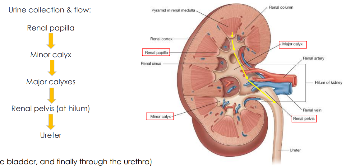
Passage Through Kidney
Urine Collection and flow:
- Renal Papilla -> Minor Calyx -> Major Calyxes -> Renal Pelvis (at hilum) -> Ureter (and then to bladder, and finally through urethra)
DIAGRAM ON SLIDE 20
- calyx means cup shape
- if its just collecting from one its called minor, once it collects from more then one its major
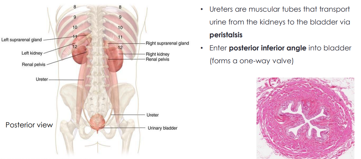
Ureters
- Ureters are muscular tubes that transport urine from the kidneys to the bladder via peristalsis (smooth muscle, two different orientations: longitudinal and circular, which allow for peristalsis movements)
- Enter posterior inferior angle into bladder (forms a one-way valve)


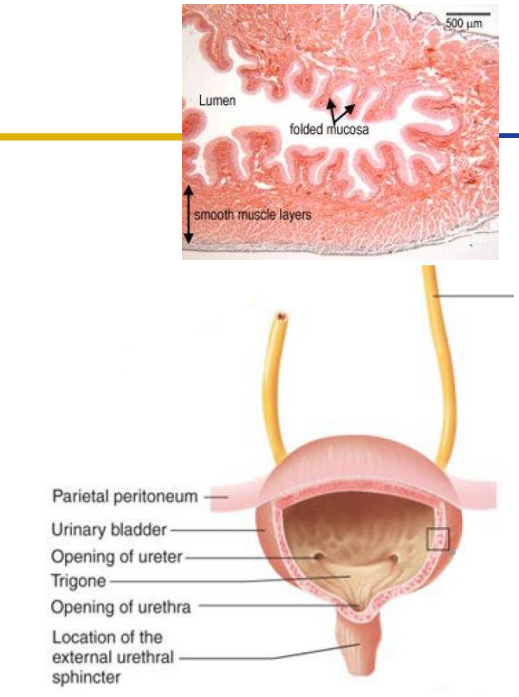
Urinary Bladder
- stores urine prior to micturition. Capacity (around 500mL) effected by gender, age etc.
- Detrusor muscle: muscularis externa smooth muscle
- Trigone: internal feature formed by openings of ureters and urethra
- the ureters come from behind because it means they get smaller when the bladder expands, preventing things from going up the urinary tract (UTI)
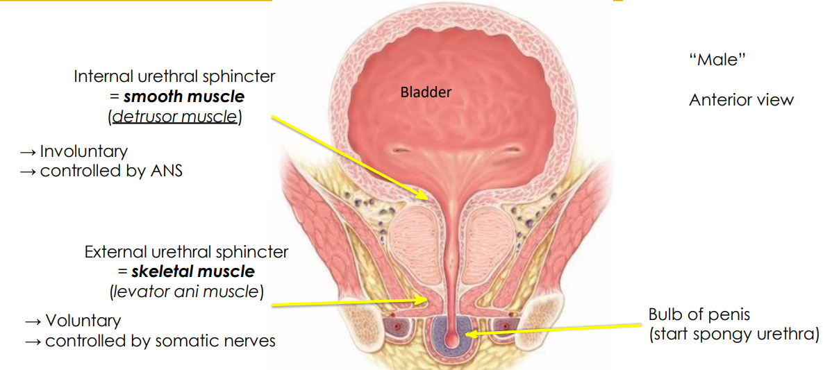
Urinary Bladder Diagram (Male)
- internal urethral sphincter (smooth muscle = detrusor muscle). This is involuntary and controlled by ANS
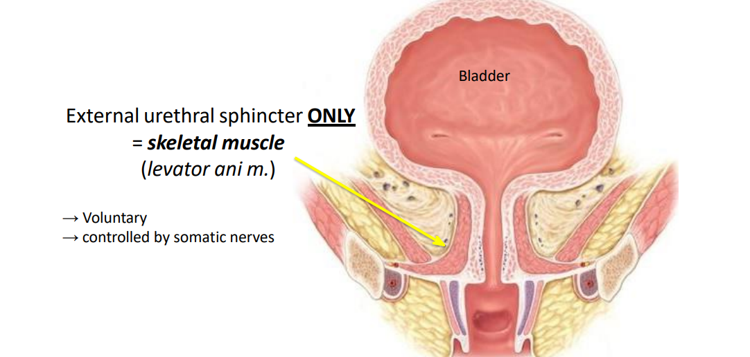
- external urethral sphincter (skeletal muscle = levator ani muscle). Voluntary and controlled by somatic nerves


What we control in Urinary Bladder
DIAGRAM ON SLIDE 24

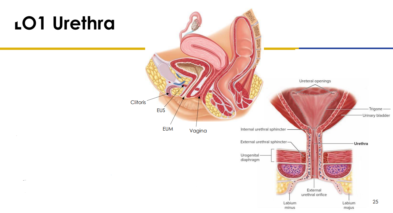
Urethra (for those with uterus)
- External Urinary Sphincter (EUS) within the pelvic floor is skeletal muscle (therefore voluntary control)
- Urine is forcefully expelled by detrusor muscle
- People with a uterus (3-4cm) (from inferior bladder to external urethral meatus (EUM) in vulva
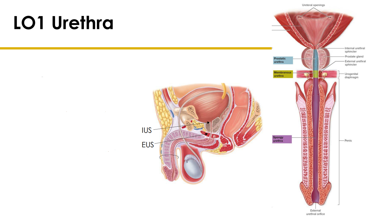
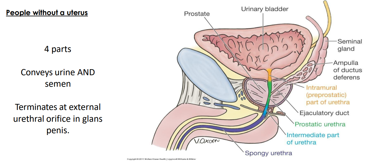
Urethra (for those without uterus)
- Passes through the prostate and the length of the penis
- Common passage for urine and sperm
- Internal urinary sphincter (involuntary) also present which contracts during ejaculations to prevent semen entering bladder


Urethra Structure in Males
- prostatic urethra (part of prostate)
- intermediate part is sometimes written as bulbourethral part
- spongy urethra

Innervation of Urinary Bladder
- bladder is innervated by the parasympathetic pelvic splanchnics (from S2-S4) and sympathetics originating from T10 to L2
- Pudendal nerve is somatic innervation for the EUS
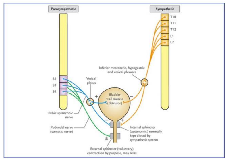
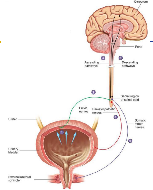
Micturition Reflex
1. Urine stretches the bladder walls
2. Excite parasympathetics which signals to the detrusor muscle to contract (and relax the IUS)
3. Ascending pathways let your brain know you need to urinate
4. Pudendal nerve (somatic motor) consciously control external urinary sphincter (contract to hold it in, relax to let it go)

Take Home Messages
DIAGRAM ON SLIDE 31
