Lab #7 Histology || —Muscle tissue, nervous tissue, membranes, and the integumentary system
1/29
There's no tags or description
Looks like no tags are added yet.
Name | Mastery | Learn | Test | Matching | Spaced | Call with Kai |
|---|
No analytics yet
Send a link to your students to track their progress
30 Terms
What are muscle cells called?
muscle fibers
What is the cytoplasm of the muscle cell called?
Sarcoplasm
What is the cell membrane of the muscle cell called?
Sarcolemma
What are the 3 kind of muscle tissue found in the human body?
skeletal, smooth, cardiac
What are the similarities between the 3 muscle types (skeletal, smooth, and cardiac muscle)
Similarities:
Basic function: all three muscle types generate force and enable movement
Excitability: all muscles respond to stimuli by generating action potentials
Contractility: All muscles contract (their fibres shorten to generate force)
Elasticity and extensibility: Muscles can stretch and return back to their original shape
Actin and myosin filaments: contraction in all muscle relies on these interactions; but their composition differs
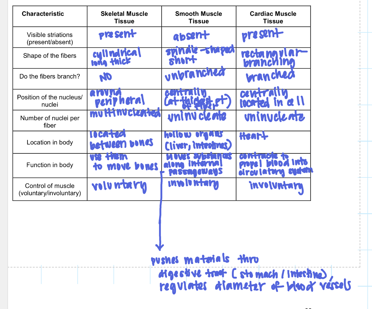
What are the differences between the 3 muscle types (skeletal, smooth, and cardiac)
Can you identify this muscle type, a function, location, and a characteristic?
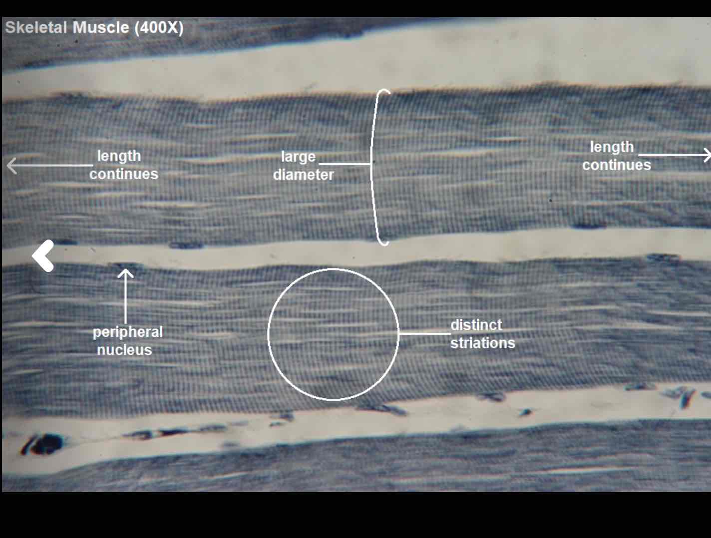
Skeletal muscle:
Function: When these muscles contract, it causes the movement of the bone. — this is the only voluntary movement
Location: Found throughout the body between bones by tendons
Characteristic: Striations (due to the composition of the actin and myosin filaments) they are arranged in compartments called sacromeres.; multinucleate (located peripherally), cylindrical
Can you identify this muscle type, provide a function, location, and characteristic.
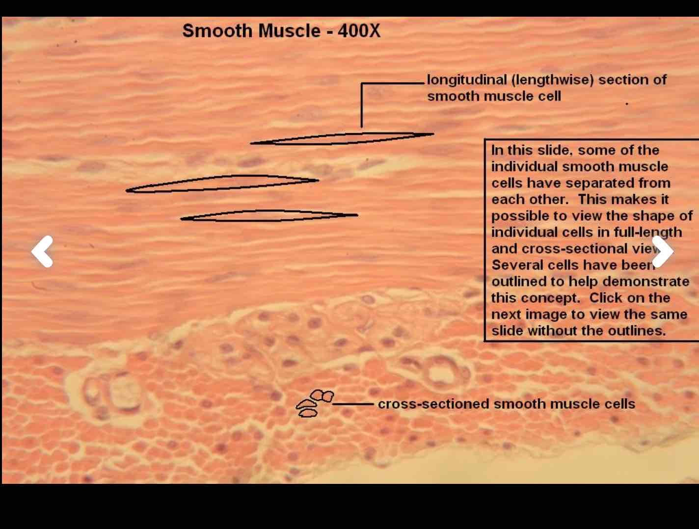
Smooth Muscle type:
Location: found in hollow organs — intestines, esophagus, bronchi (and blood vessels)
function:
Involuntary movement; helps regulate processes in body like the smooth muscle in the stomach and intestine helps with digestion and moving waste through the body (helps move materials along internal passageways), and regulates the diameter of blood vessels + respiratory tracts.
Characteristics:
No striations (because their actin and myosin filaments are arranged in a disorganized pattern); they are uninucleate (located centrally); shorter spindle shaped cells; there are cross-sectioned and longitudinal sections.
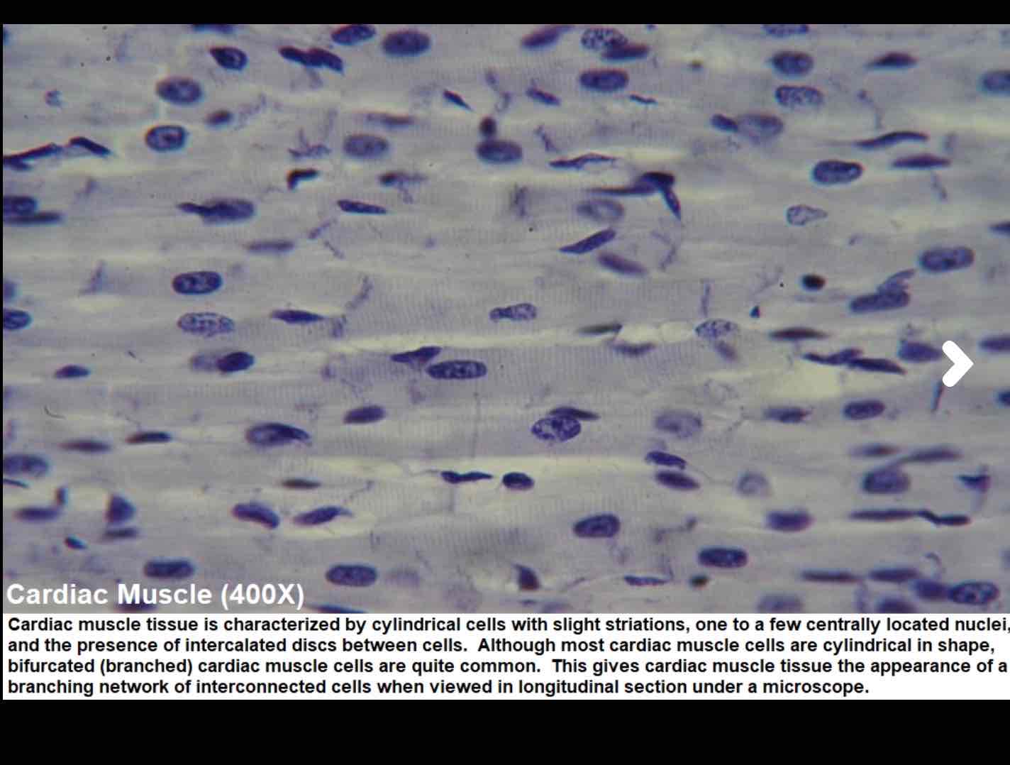
Can you identify this muscle, provide a function, location and characteristic
Location: Found in the heart
Function:
Involuntary muscle and is primarily responsible to pump blood throughout the body by contraction rhythmically and continuously
Characteristic:
Uninucleate (centrally located), striations (because of the arrangement of sacromeres), cells branch in irregular pattern and cells join end-to-end by regions called intercalated discs (these are where the cell membranes of adjacent cells contact each other and contain desmosomes and gap junctions; which allow for cohesion and rapid electrical conduction)
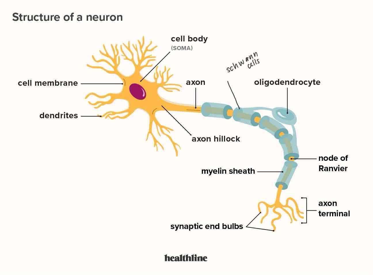
Can you identify parts of the neuron?
Dendrites, soma (cell body) - consists nucleus and cytoplasm (contains mitochondria, nissl bodies (rough er) and ribosomes, neurofibrils (cytoskeleton), axon (in central nervous system myelin sheath = from oligodendrocytes and peripheral (myelin from schwann cells), Nodes of ranvier (gaps between myelin), telodendrites (axon terminals)
The cell of neurons in the CNS are mostly found in GREY MATTER; grey matter is found in the thin outer layer of the brain and as butterfly shaped structure in the centre of the spinal cord.
Outside of the CNS, neuron cell bodies are clustered within structures called GANGLIA (located in PNS)
Ganglia play a role in processing sensory input or regulating autonomic motor functions.
Can you give the functions of the structures in the neuron and the function & location of nervous tissue
function of the structures in the neuron:
Dendrites:
Structure: dendrites are branching, tree-like extensions that extend from the cell body
Function: Receive signals (electrical impulses) from other neurons and transmit those signals to the cell body (soma) of the neuron.
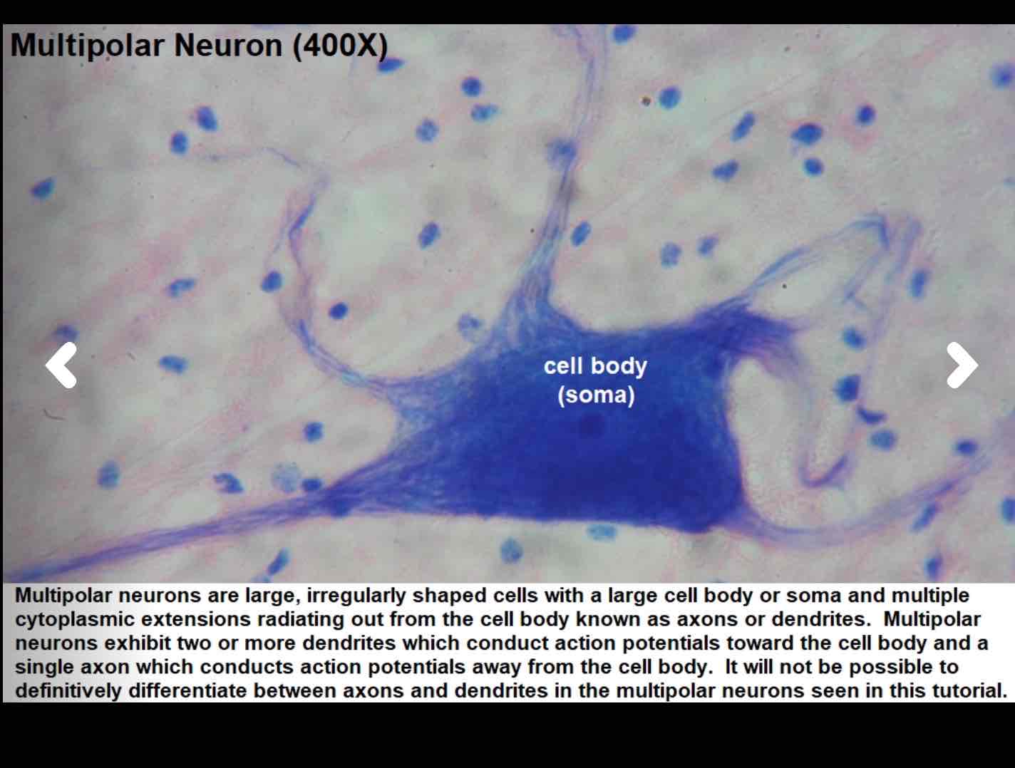
Cell body (soma):
Structure: Contains the nucleus, mitochondria, ribosomes, nissl bodies (rough er; plays a part in protein synthesis and carries ribosomes) and neurofibrils (cytoskeleton).
Function: It’s the control center; makes decisions and processes the neuron’s actions/what the neuron should do; soma also produces proteins and energy needed for the neuron’s functions
Axon:
Structure: A long, thin, tube-like structure that extends from the cell body.
Function: Carries electrical impulses/signals away from the cell body and delivers them to other neurons, muscles, or glands (the highway for electrical impulses)
Myelin sheath:
Structure: Fatty-protein coating wrapped around the axon; in the CNS it is made from oligodendrocytes; in the PNS it is made from Schwann Cells
Function: Responsible for speeding the transmission of electrical signals along the axon by allowing them to jump between the gaps in sheath (gaps = nodes of ranvier; That allow for saltatory conduction where electrical signals/action potentials to jump from one node to the next)
Axon terminals/synaptic terminals (telodendrites):
Structure: Small branches at the end of the axon.
Function: Allow neurons to communicate with neurons, muscles, or glands; they transmit messages to other cells using neurotransmitters that travel across the synapse (tiny gaps between neurons)
Dumb down the relationship between CNS, PNS, Sensory (afferent) division, Motor (efferent) division, somatic nervous system, autonomic nervous system, parasympathetic nervous system, and sympathetic nervous system
CNS: Is the command/control centre of our body; processing info and sending commands (it is our brain and spinal cord)
PNS: It connects our body to the CNS (the wires connecting a computer to the screen); it is the nerves outside of our brain and spinal cord
Afferent (sensory) division: Nerves that carry sensory information (like touch, temperature, pain) from our body to our CNS (brain) so we know what’s happening.
Efferent (motor) division: Nerves carry information from the CNS (brain) to our muscles and organs (effectors), telling them what to do (move or contract)
Somatic Nervous System: It’s part of the motor division; controls voluntary (skeletal muscles) actions (like moving our muscles to walk, talk or wave)
Autonomic Nervous System: Another part of the motor division; controls involuntary (cardiac and smooth muscle) actions (like heart beating, digestion, and breathing).
Parasympathetic Nervous System: Part of the autonomic nervous system; it helps your body relax and conserve your energy; helps slow your heart rate and aids w digestion (rest and digest system)
Sympathetic Nervous System: Part of the autonomic nervous system; “fight or flight system”; gets our body ready for action (like during stress or excitement); speeds up our heart rate and gets muscles ready to move.
What are the three types of membranes?
Mucous, cutaneous, and serous
Can you describe the three types of membranes?
Subcutaneous (hypodermis) layer:
Structure: made up of fat cells, collagen, blood vessels, and nerves; sits below the epidermis (where keratinocytes are found — cells that produce keratin which makes the skin waterproof and tough), melanocytes (cells that make pigment) and Langerhans cells (involved in immune defense); and epidermis (contains connective tissue with collagen and elastin fibres, blood vessels, nerves, hair follicles and glands — sebaceous and sweat glands).
Location:
Beneath the epidermis and dermis
function:
Acts as a barrier against UV radiation, pathogens, and physical injury; provide insulation, regulate temperature, and store fat, act as shock absorber.
Mucous Membrane:
Structure: Surface layer made of epithelial (stratified squamous epithelium); loose connective tissue (lamina propria) that consists of nerves, veins, and structural proteins — attaches the epithelium to the smooth muscle; deepest layer (muscularis mucosae) is the smooth muscle that keeps the mucosa moving — most active in the stomach.
Location: Found in areas that are exposed to the external environment; places like the respiratory tract (nose, mouth, throat), urinary and genital tracts (vagina, urethra, ureter, bladder), Ears, anus, and digestive tract (stomach and intestines)
function: Protect organs from chemicals and physical damage; help food move; keep the tissues in the area moist.
Serous Membrane:
Location: Thin layers of tissue that line organs and body cavities; found in pleura (surrounds the lungs in the pleural cavity; reduces friction), peritoneum (lines the abdominal cavity and organs; reduces friction between the abdominal and pelvic organs and the body wall); pericardium (surrounds the heart)
Structure: Have two layers; parietal layer (lines the walls of cavity) and visceral layer (surrounds the organs); and then it has serous space (fluid filled cavity between the parietal and visceral layer)
Also made of simple squamous epithelium, connective tissue, and serous fluid (lubricating substance from blood vessels and lymph vessels — that helps reduce friction)
function:
Reduce friction in the body.
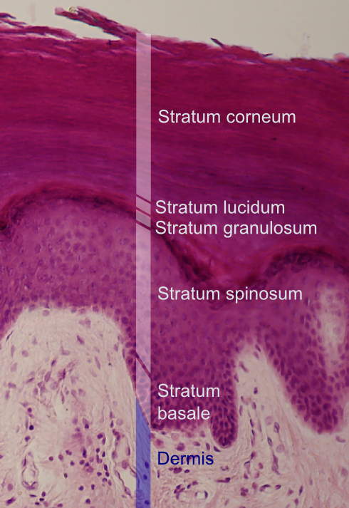
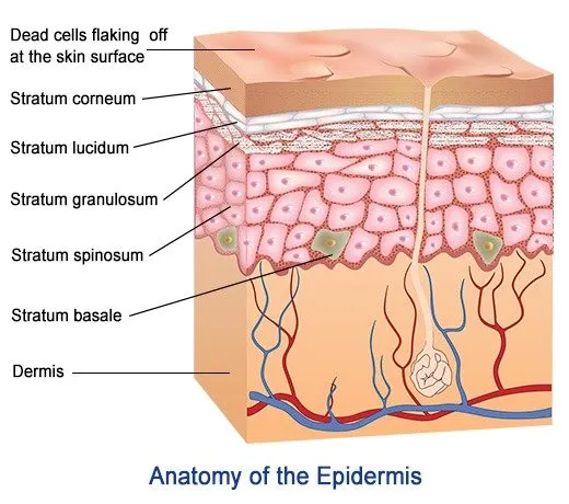
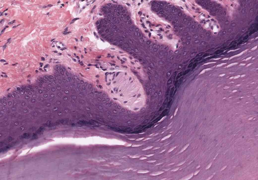
identify the layers of the epidermis the associated structures
Epidermis:
Outermost layer (composed primarily of keratinized stratified squamous epithelium); this layer is avascular and relies on diffusion from the dermis for nutrients
Stratum Corneum (horny layer):
Outermost layer (top layer of skin); made of many layers of dead, flattened skin cells that are filled with keratin; form a tough, protective layer that shields the body from physical damage, pathogens, and water loss; over time the dead skin cells are shed and replaced by newer cells from below.
Stratum Lucidum (clear layer):
Only found on thick (hairless skin) — like palms and soles of your feet; composed of thin, translucent, layers of dead cells (keratinocytes) and packed with eleidin (protects the skin from friction and stress); protects skin from friction and damage (extra protection for areas that experience a lot of wear and tear).
Stratum granulosum (granular layer):
Found below the stratum lucidum and above the stratum spinosum; makes the furthest limit that cells can receive nutrients; the cells in this layer begin to undergo keratinization (where the cells start to flatten out and fill up with keratin — making the cells tougher and helps protect the skin); how keratohyalin granules play a part (these granules contain protein that help the keratin fibres clump together and form a stronger structure); and then the lamellar granules inside the cells release lipid (fat) content into the space between the cells (which form a waterproof protective barrier — keeps moisture in and prevents harmful substances out); and then finally, as the cells continue to move upwards, they lose their nuclei and other organelles (the process of keratinization continues until they reach the stratum corneum)
Stratum Spinosum (spiky/spiny layer):
This layer is below the stratum granulosum and above stratum basale; the cells in this layer are starting to flatten out and begin to form connections with each other called desmosomes (which gives this layer a spiky layer); this layer has langerhans cells (help fight infections).
Stratum Basale (Basal Layer) or stratum germinativum:
The deepest layer of the epidermis; made up of columnar or cuboidal epithelial cells, 10-25% of melanocytes, a single row of keratinocytes, and stem cells; rests on basement membrane; separates dermis from the epidermis;
Here the single row of keratinocytes are mitotically active (constantly dividing to produce new cells, and these cells begin to produce keratin); the melanocytes produce melanin for skin pigmentation and UV protection.
What is the structure and function of the skin (epidermis, dermis, and hypodermis)
*Hypodermis (subcutaneous) is not considered part of the skin*
Epidermis:
Structures:
Keratinocytes: Produce keratin, a protein that strengthens the skin and creates a barrier
Melanocytes: Produce melanin; the pigment responsible for skin colour and gives protection against UV radiation
Langerhans Cells: found in the squamous cell layer of the epidermis; and is responsible for detecting and fighting off pathogens; tissue resident macrophages; (they surround pathogens to kill infectious cells).
Markel Cells: found in stratum basale; they touch a sensory structure called tactile (merkel) disc; they function in the sensation of touch.
**also the areas with thick skin, have 5 sub layers — stratum basale, stratum spinosum, stratum granulosum, stratum lucidum, stratum corneum**
Dermis:
Structures:
Contains numerous blood vessels, specialized sensory nerve endings, glands, hair follicles and their associated structures.
Divided into two layers: (a) Papillary (areolar loose connective tissue) - which is highly vascularized; this connective tissue produces projections called dermal papillae that extend toward the epidermis (has many functions, but is responsible for strengthening the skin’s structural integrity and also the dermal papilla makes epidermal ridges — on thick skin areas — which are often called fingerprints on fingers); dermal papillae also contains pain receptors (free nerve endings), capillary loops, and meissner’s corpuscle
Also houses the meissner’s corpuscle; which is responsible for detecting light touch (low frequency vibrations) and sits on the upper part of the dermis; it is primarily found in eyelids, lips, and fingertips
(B) Reticular layer; it is composed of dense irregular connective tissue (elastin fibres); houses sweat glands (eccrine glands — involved in thermoregulation by sweating to cool the body; apocrine glands; found in the armpits and groin area — emit thicker sweat; often response from stress); houses the sebaceous (oil) gland (secrete oily substance, sebum, that moisturizes skin and hair); houses hair follicles (which anchors the hair to the skin — hair grows from follicles in this layer); houses arrector pill muscles (contract to give appearance of goosebumps); houses blood vessels; houses nerve endings (Pacinian Corpuscle; detects deep pressure and vibrations — usually found in the skin of the fingers, in the pancreas, and the walls of the urinary bladder)
Hypodermis (subcutaneous) — not considered part of the skin:
Is a well-vascularized, loose, areolar connective tissue and adipose tissue; it functions as a mode of fat and storage — provides insulation.
Do you know the mode of secretion for sebaceous glands and sweat glands?
Sebaceous glands: Use the mode of secretion called HOLOCRINE secretion; where the glandular cell ruptures and dies to release it’s contents — in this case the cell produces sebum and then disintegrates to release the oily substance into the hair follicle or the skin.
Sweat glands: use the mode of secretion called Merocrine or apocrine secretion;
For merocrine; the sweat glands release their secretions through exocytosis (their products are packaged into vesicles that move to the cell membrane and release into the duct or the surface of the skin) and this process works without damaging the glandular cell; most concentrated on the palms and soles of the feet
for apocrine; the cell snaps and pinches a part of itself off (where the product, like milk, sweat, is contained) and breaks away — then the product is also released into a duct; most concentrated in the armpits, groin, around the nipples
explain the role of sweat glands in body temperature
Eccrine/merocrine sweat glands: release sweat (mostly water with some salt) which evaporates; taking the heat away from your body; making you feel cooler — natural AC
Apocrine sweat glands: Are not for regulating or meant for cooling; they release thicker sweat more for stress or emotion
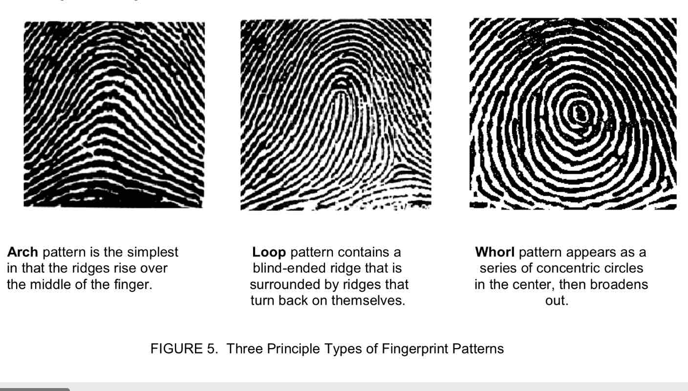
Identify the 3 principal types of fingerprint patterns
arch: Simplest; the ridges flow up and down like a hill; no cores or loops; least common
Loop: most common; curves back on itself; ulnar loop (opens toward the pinky) or radial (opens toward the thumb).
Whorl: Appears as a series of concentric circles in the center and then broadens out
What was the point of the tactile discrimination in various skin locations experiment?
To measure how well different parts of the skin can detect and distinguish between two points of contact.
Helps us understand the sensitivity of various areas of the body and how the nervous system processes touch.
results:
high sensitivity; areas like the fingertips, lips, and face (esp the tongue) have a high receptor density and can detect two points very close together
Low sensitivity; areas like the back, forearm, and thighs have a low receptor density and need the points to be father apart to distinguish them.
what result did The Princess and the Pea test reveal?
It revealed that the sensitivity to pressure decreased from the fingertips to the chin (so fingertips have more a higher density receptor compared to chins)
What did the experiment “adaptation to pressure” yield?
Revealed how the skin’s sensory receptors become less sensitive to continuous pressure over time; some receptors like pacinian corpuscle adapt more quickly to pressure and vibration, where as merkel discs may adapt more slowly.
What does the experiment “relative temperature sense” indicate about our sensory receptors?
Demonstrates how well our receptors distinguish between cold and hot sensations; and also after awhile the sensation might not feel as intense.
What does the experiment “liquid crystal thermography” demonstrate?
How temperature varies across the skin by using colour changing liquid crystal patches; can also demonstrate vascular health, circulatory issues, or inflammatory conditions — (places like the face, hands, and feet are usually the warmest)
What does the experiment of “the effects of temperature changes on blood flow” demonstrate?
Demonstrates how changes in temperature can influence the circulatory system and the flow of blood in the body
Summarize the effects of heating and cooling on peripheral blood flow (refers to the movement of blood through blood vessels in the outer regions of the body) regulation:
Heating increases peripheral blood flow via vasodilation (heat causes blood vessels in the skin to dilate) — vasodilation increases blood flow to skin (allowing more heat to disappear from body into environment) this increase blood to skin helps the body cool down by releasing excess heat (maintaining thermal regulation)
vasoconstriction (cold temperature causes blood cells to constrict); which reduces blood flow to conserve heat and reduce heat loss; reducing peripheral blood flow = body retains more warmth; helps keep internal organs at optimal temperature
Summary: Heat into peripheral blood flow (via vasodilation) to release heat; while cooling decreases peripheral blood flow and retains heat (helps regulate stable internal temp).
How is skin colour influenced?
Melanin (the primary pigment responsible for skin colour); produced by cells called melanocytes
types of melanin: Eumelanin (brown/black): gives skin darker colour
Phenomena (red/yellow): gives the skin a lighter or reddish tint
Sun exposure (UV radiation):
UV radiation stimulates the melanocytes to produce more melanin as a protective response (that’s why people tend to get darker when they spend more time in the sun)
Freckles can form as localized clusters of a lot of melanin.
Concentration of carotene: a yellow pigment that accumulates in the surface layer of the epidermis
Blood flow: Can influence skin colour — when you’re cold; your blood vessels constrict, making your skin appear paler.
when you’re hot; your blood vessels dilate; leading to redness.
hemoglobin can give a reddish think - esp when blood vessels are close to the surface (e.g., lips, cheeks, and hands).
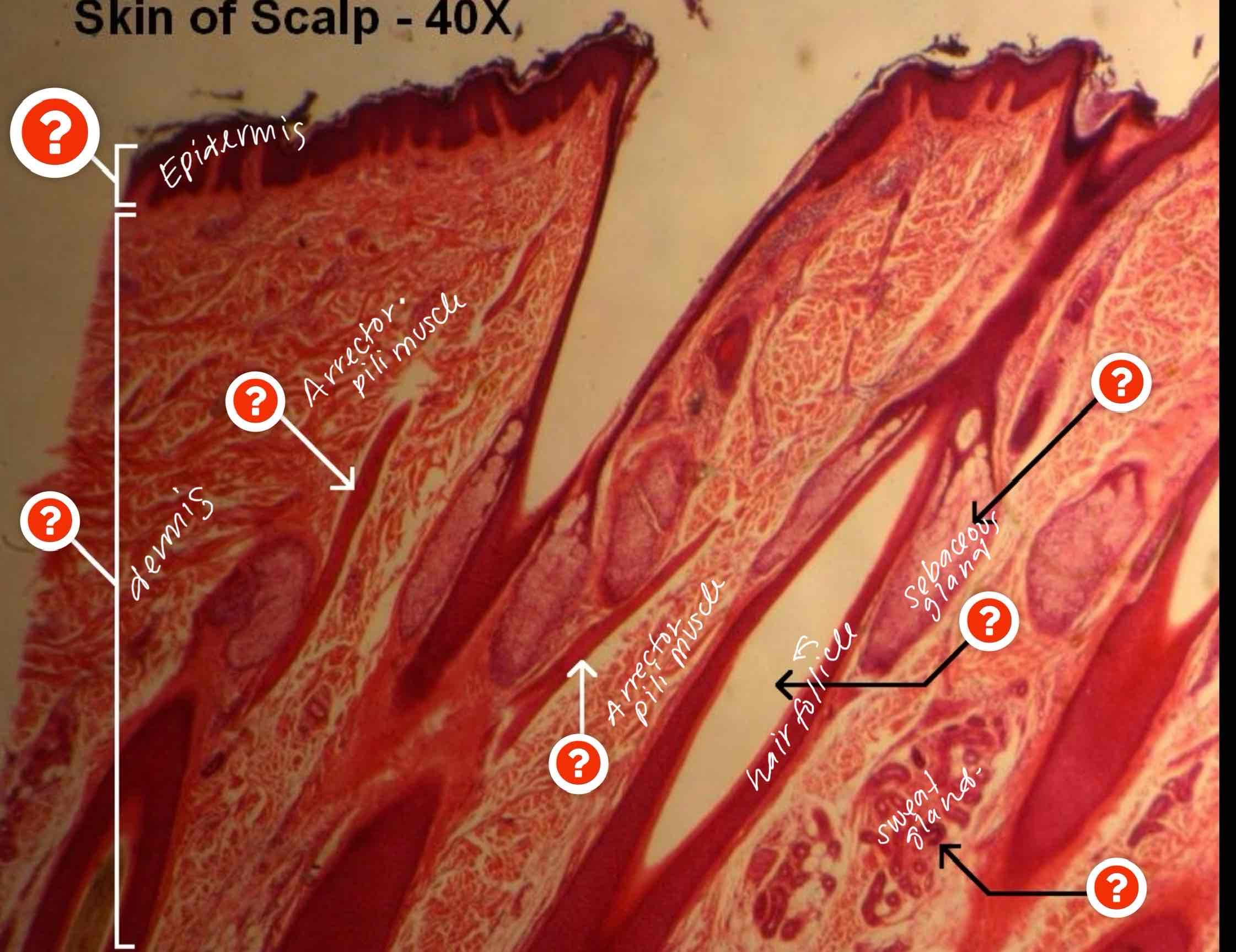
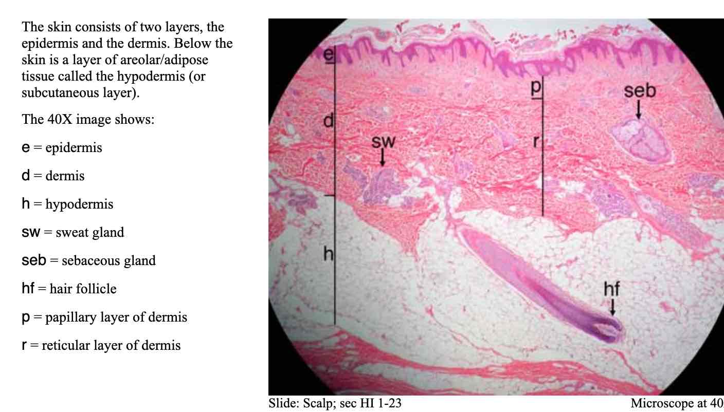
Can you identify the parts of the scalp
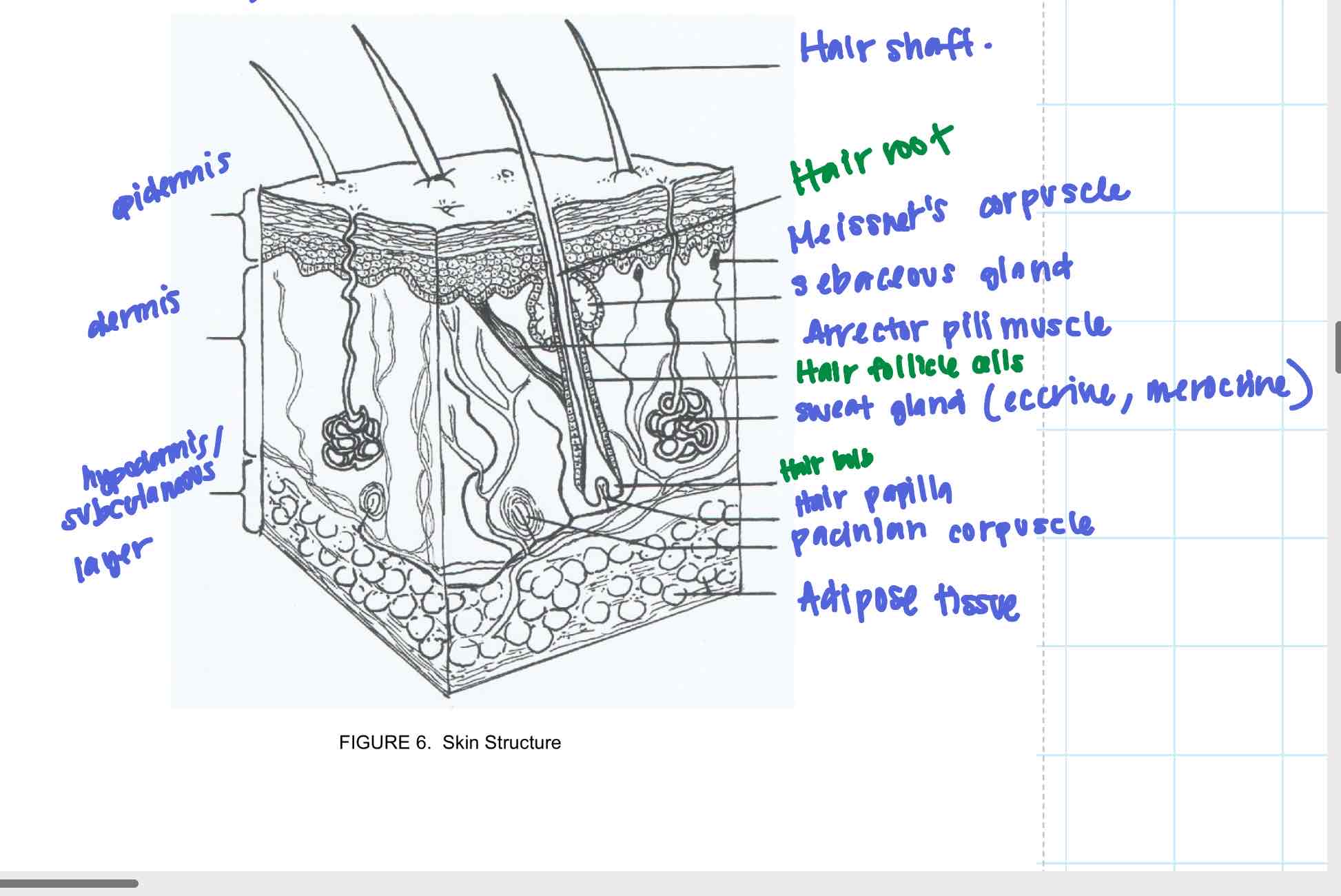
Can you explain the function and structure of this diagram
epidermis:
Structure: made of keratinized stratified squamous epithelium; areas of thick skin are composed of 5 sub layers; contains keratinocytes, melanocytes, and langerhans cells
Function: protection from physical, chemical and pathogens; prevents water loss; contains sensory receptors; melanin provides protection from UV
Dermis:
structure: made of connective tissue (collagen and elastin) provides elasticity and strength; divided into two layers (papillary layer; thin and vascular with dermal papillae) and reticular layer (thicker, contains collagen and elastin fibres); houses hair follicles, sweat glands, sebaceous glands, nerves, blood vessels and sensory receptors; reticular layer houses the pacinian corpuscle and the meissner’s corpuscle
function: provide structural support; houses sensory receptors (meissner’s and pacinian corpuscle); regulates blood flow and contains glands that secrete oil and sweat).
Sweat gland: coiled glands found in the dermis; with a duct that opens to the skin’s surface; acts to regulate body temperature through the secretion of sweat.
Hypodermis (subcutaneous) layer:
Structure: made of adipose tissue (fat) and areolar (loose connective tissue); houses blood vessels, lymphatic vessels, and nerves;
function: acts as shock absorber and insulator; and anchors the skin to underlying structures (muscles and bones).
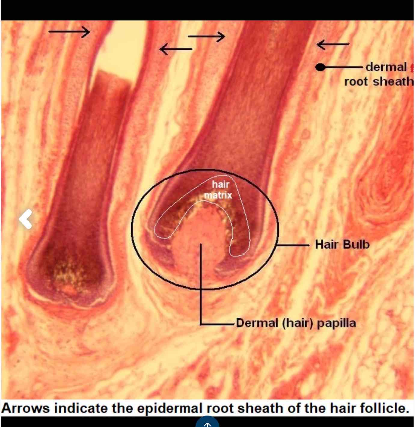
hair papilla:
Location: found in dermis
Structure: cap-shaped structure at the base of the hair follicle, made of connective tissue and blood vessels
Function: plays a role in hair growth; provide blood supply, oxygen and nutrients
hair bulb:
location: found in the dermis
structure: rounded base of the follicle where the hair is formed; consists of the hair matrix (responsible for making new hair cells).
Function: where hair growth begins; cells in the matrix divide and push upwards, forming hair shaft
Hair follicle:
location: in dermis
structure: a tube-like structure in the dermis that houses the hair root; includes the epithelial root sheath (inner — protection and outer layers — support to structure) — helps protect and guide the growing hair; contains stem cells for hair regeneration — and is surrounded by the dermal root sheath (which keeps the hair anchored to dermis).
Function: where hair grows. Contains all the necessary components for hair production, including the hair bulb and hair papilla
Arrector pili muscle:
location: in dermis, attached to the hair follicle.
Structure: a small smooth muscle
Function: causes goosebumps by contracting and making the hair stand upright (usually in response to cold or fear)
Sebaceous gland:
location: in dermis
structure: glands associated with hair follicles and skin; they secrete sebum (oil).
Function: lubricates the hair and skin; has anti bacterial properties that help protect the skin from harmful pathogens.
Hair root:
location: in the dermis
structure: the portion of the hair embedded in the hair follicle
function: anchors the hair to the skin and provides a base for the production of the hair shaft.
Can you identify this sensory receptor? where it’s found and what is it’s function?
Meissner’s corpuscle:
found in dermal papillae in thick skin (which is in the papillary layer; and that is in the dermis)
Function: touch receptors; nerve endings in skin responsible for sensitivity to light touch — found on eyelids, lips, fingertips
Structure: surrounded by connective tissue and nerve endings
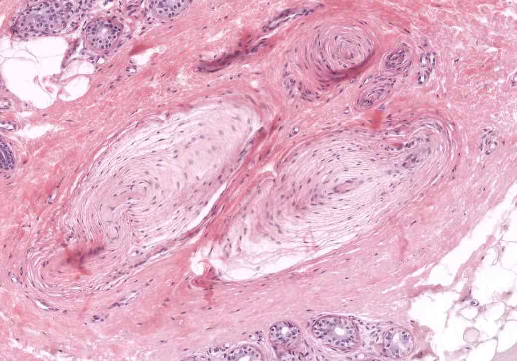
Can you identify this sensory receptor? provide function, location and structure
Pacinian Corpuscle:
function: detects deep pressures and vibrations
structure: large oval or spherical structures of 20 to 60 concentration lamallae; found in the dense irregular connective tissue (dermis) underneath the epithelium.
location: found in dermis and hypodermis — found in breast, genitals, deep dermis