Taste & Olfaction
1/30
There's no tags or description
Looks like no tags are added yet.
Name | Mastery | Learn | Test | Matching | Spaced |
|---|
No study sessions yet.
31 Terms
Deficit of smell/taste
Smell - hyposmia/anosmia
Taste - hypogeusia/ageusia
Which systems are involved in taste & olfaction?
Olfaction:
Olfactory (orthonasal) system
Taste:
Olfactory system - retronasal
Gustatory (taste) system
Trigeminal system - hot/cold texture
Basic pathway for both taste & olfaction
Physical/chemical environment --> Reception --> Transduction --> Encoding & Transmission --> Perception
5 taste receptors
Sweet, sour, salty, bitter, umami
Organs of the gustatory system
Tongue (main), pharynx, palate and epiglottis
Gustatory reception process
Food is broken down by enzymes in saliva during mastication (chewing) --> becomes solubilised --> diffuses down the crevices/sides of the papillae --> contacts taste receptors and cilia
Why is the tongue the main gustatory organs?
Has the highest receptor density
Papillae & Taste buds (definitions)
Papillae =taste-sensitive structures (bumps on the tongue).
Taste buds = bud of neurons containing several different taste receptor neurons with axons leaving the taste bud; found within papillae.
Types of papillae
Circumvallate papillae – largest, contain many thousands of taste buds, located at posterior.
Foliate papillae – elongated structure, contain hundreds of taste buds, lie along posterior lateral edge.
Fungiform papillae – smallest, contain one or two taste buds, widespread across anterior portion and tip of tongue.
Olfactory sensory (receptor) neurons: location, structure and surrounding structure
Location: Olfactory epithelium lining the nasal cavity, below the cribriform plate.
Structure: Bipolar - has a cell body and dendrite that extends into the mucus layer; have cilia on the end which is where olfactory receptors (ORs) are located - only one kind of OR is expressed.
Surrounding structures: Supporting cells surrounding the ORN, olfactory glands (secrete mucus), basal epithelial cells (carry out neurogenesis – continually divide to replenish ORNs; reduces with age).
Olfactory reception process
Odourants enter nasal cavity --> dissolves in secreted mucus coating the olfactory epithelium --> binds to receptors on olfactory sensory neurons cilia.
Retronasal olfaction
Mastication (chewing) pushes air into the nasal cavity engaging olfaction; allows comparisons between food and is why food lacks flavour when you have a cold.
Olfactory transduction
Odourants enter nasal cavity.
Dissolves in secreted mucus coating the olfactory epithelium.
Binds to olfactory receptors (G protein-coupled receptors) on olfactory sensory neurons cilia – ORs couple with G proteins upon binding of odourants.
Activates adenylate cyclase which converts ATP --> cAMP.
cAMP binds to and opens plasma membrane Na+ and Ca2+ channels – influx of Na+ and Ca2+
Ca2+ opens Cl- channels causing an efflux of Cl-.
Causes depolarisation and firing of APs.
APs pass along the olfactory nerve, formed from several olfactory receptor neuron axons, which passes through the cribriform plate and into the brain.
Types of olfactory receptor
Main groups are all G protein-coupled receptors (GPCRs):
Odorant receptors (ORs)
Vomeronasal receptors (V1Rs & V2Rs) - sense pheromones
Trace-amine associated receptors (TAARs)
Formyl peptide receptors (FPRs)
Guanylyl cyclase receptors (GC-D) - couple with guanylyl cyclase instead of adenylyl cyclase.
Gustatory transduction
Salt & sour channels:
Food in the mouth interacts with the ion channels present in taste receptor cells.
Causes depolarisation and firing of APs.
NT is released on to gustatory axons.
Gustatory axons fire APs which are transmitted to the rest of the brain.
Sweet, bitter & umami receptors:
Receptors couple to GQ protein type in response to the stimuli.
This activates phospholipase C and increase intracellular Ca2+.
Sour transduction (type III cells)
Otopetrin1 (OTOP1) is a Zn sensitive, H+ selective ion channel that allows H+ to pass through.
The acidification also inhibits Kir2.1 channel.
Causes depolarisation and firing of APs.
Leads to an influx of Ca2+ and synaptic vesicle fusion, releasing NT (possible ATP – unknown) onto the gustatory nerve.
Bitter, Sweet & Umami transduction (type II cells)
Taste GPCR receptor couples to the gustducin G protein.
This activates phospholipase C.
Causes IP3 receptor activation and releases intracellular Ca2+ stores.
Ca2+ opens the non-selective TRPM5 cation channel causing an influx of Na+.
This opens VG channels, causing depolarisation and firing of APs.
ATP is released through the pore of the V-G CALHM1/3 channels.
ATP binds to the P2X2/3 receptors located on gustatory afferents.
Type 2 cells lack the required proteins for traditional vesicular synapses e.g. SNAP-25, and so Ca2+ isn't required for NT release, only depolarisation.
Salty transduction (most likely type II cells)
Influx of Na+ occurs through the endothelial Na+ channels (ENaC).
Causes depolarisation and firing of APs
ATP is released through the pore of the V-G CALHM1/3 channels.
ATP binds to the P2X2/3 receptors located on gustatory afferents.
Bitter, sweet, umami receptors (GPCRs)
T2R – single GPCR that bitter tastants bind to, which activates G proteins (gustducin) and increases IP3 levels via the PLC cascade.
T1R family – sweet tastants bind to the GPCR heteromer T1R2 and T1R3 (bound together tightly), which couple with gustducin, etc.
Umami tastants are the same as sweet tastants except they bind to T1R1 and T1R3 GPCR complex.
Gustatory specificity
Afferents are sensitive to all tastants but have higher sensitivity to a ‘preferred’ tastant in the gustatory systems.
Olfactory specificity
Weak - poses a problem when identifying stimuli.
Olfactory encoding & transmission (olfactory bulb circuit)
Odourants activate olfactory sensory neurons, which pass through the cribriform plate.
ORNs with the same OR type target the same glomerulus in the olfactory bulb.
At the glomerulus, the ORN axons synapse on to other cell types – output neurons: mitral and tufted cells.
Output neurons are excited by ORNs and they only receive input from a single glomerulus - output neurons are activated by the odourant that activated the ORNs targeting their glomerulus.
Glomeruli
Neurons target 1-2 of these: a lateral and medial glomerulus.
Inhibitory interneurons
2 layers:
Periglomerular (PG) neurons – neurons around the glomerulus; receive input from olfactory nerve and modulate signals at the presynaptic terminals and postsynaptic dendrites.
Granule cells – deeper in the olfactory bulb; form reciprocal dendrodendritic synapses with mitral cell lateral dendrites.
Methods of imaging activity in olfactory bulb circuit
Axons - Calcium-sensitive fluorescent dye conjugated to a large sugar molecule gets transported along axons.
2-Photon Ca imaging - mice express a genetically encoded Ca indicator in neurons around glomeruli.
Olfaction - Concentration invariance
A single glomerulus will respond to several odourants.
Regardless of concentration of odourants, the response of the glomerulus will be the same, making it hard to distinguish between 2 stimulating odourants.
Glomeruli stimulation pattern and spatial correlation problem
•It is thought that the brain uses the stimulation pattern to determine the stimuli.
•As the concentration of the same odourant increases, more glomeruli are recruited.
•This means the glomeruli stimulation pattern changes as the concentration changes even though the same odour is being detected.
•Therefore, the spatial correlation map cannot confidently determine the odour.
How does the olfactory bulb compensate for the glomeruli stimulation pattern problem?
Output neurons - recording the stimulation of the output neurons shows much more correlation between different concentrations of the same odour.
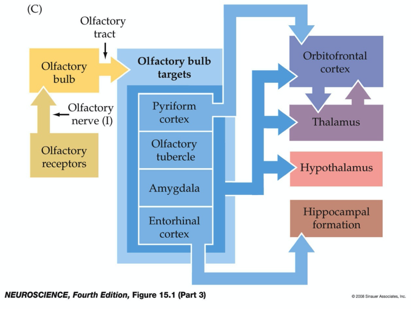
Olfactory signal targets
ORs in the nasal epithelium of the nose project along cranial nerve I —> through cribriform plate —> olfactory bulb —> mitral and tufted axons form the lateral olfactory tract —> directly target cortical and subcortical structures (blue) —> other cerebral structures.
Bypasses the thalamus.
Gustatory system afferents
Primary sensory axons
Cranial nerve VII (facial) - also innervates the tongue.
Cranial nerve IX (glosso-pharyngeal) - also innervates the tongue.
Cranial nerve X (superior laryngeal) - also innervates the epiglottis.
Central gustatory pathway
Information is carried from taste receptors --> brain stem --> nucleus of solitary tract (NST) in the medulla --> ventral posterior medial nucleus of the thalamus --> gustatory cortex (insula).
It is thought to be the gustatory cortex where information from the taste receptors is integrated with retronasal olfaction.