3.2 TRANSPORT IN ANIMALS
1/111
There's no tags or description
Looks like no tags are added yet.
Name | Mastery | Learn | Test | Matching | Spaced | Call with Kai |
|---|
No analytics yet
Send a link to your students to track their progress
112 Terms
3.2.1
TRANSPORT IN ANIMALS
Double circulatory system
One in which the blood flows through the heart twice for each circuit of the body
Single circulatory system
One in which the blood flows through the heart once for each circuit of the body
Transport
The movement of substances such as oxygen, nutrients, hormones, waste and heat around the body
There are three main factors that influence the need for a transport system:
- size
- surface area
- level of metabolic activity
Size
The cells inside a large organism are further from its surface - the diffusion pathway is increased. The diffusion rate is reduced, and diffusion is too slow to supply all the requirements. Also, the outer layers of cells use up the supplies, so that less will reach the cells deep inside the body.
Surface area to volume ratio
Small animals have a small surface area to volume ratio which means that in their body they have a sufficient area of body surface through which exchange can occur. However, larger animals have a smaller surface area to volume ratio. This means that each gram of tissue has a smaller area of body surface for exchange.
Level of metabolic activity
this factor influences the need for a transport system because animals get energy from food and getting this energy requires oxygen. If any animals is very active - cells need good supply of oxygen and nutrients to supply the energy needed
An effective transport system will include:
- A fluid or medium to carry nutrients, oxygen and wastes around the body- this is the blood.
- A pump to create pressure that will push the fluid around the body- this is the heart.
- Exchange surfaces that enable substances to enter the blood and leave it again where they are needed - these are the capillaries.
An efficient transport system:
- Tubes or vessels to carry the blood by mass flow
- Two circuits- one to pick up oxygen and another to deliver oxygen to the tissues.
Which animal has a single circulatory system?
Fish
In a single circulatory system, the blood takes the following route:
heart --> gills --> body --> heart
Which species has a double circulatory system?
Mammals
In the double circulatory system, the blood takes the following route:
heart --> body --> heart --> lungs --> heart
Why is the single circulatory system beneficial for fish?
- The blood pressure drops as blood passes through the tiny capillaries of the gills.
- Blood has a low pressure as it flows towards the body, and will not flow very quickly.
- The rate at which oxygen and nutrients are delivered to respiring tissues, and carbon dioxide and urea are removed, is limited.
Fish are not as metabolically active as mammals, as they do not maintain their body temperature. Therefore, they need less energy. Their single circulatory system delivers sufficient oxygen and nutrients for their needs.
Why is the double circulatory system beneficial for mammals?
- the blood pressure must not be too high in the pulmonary circulation, otherwise it may damage the delicate capillaries in the lungs.
- The heart can increase the pressure of the blood after it has passed through the lungs, so the blood is under higher pressure as it flows to the body and flows more quickly.
- The systematic circulation can carry blood at a higher pressure than the pulmonary circulation.
Mammals are active animals and maintain their body temperature. Supplying the energy for activity and the heat needed to keep the body warm requires energy from food. The energy is released from food in process of respiration. To release a lot of energy, the cells need a good supply of both nutrients and oxygen, as well as the removal of waste products.
3.2.2
BLOOD VESSELS
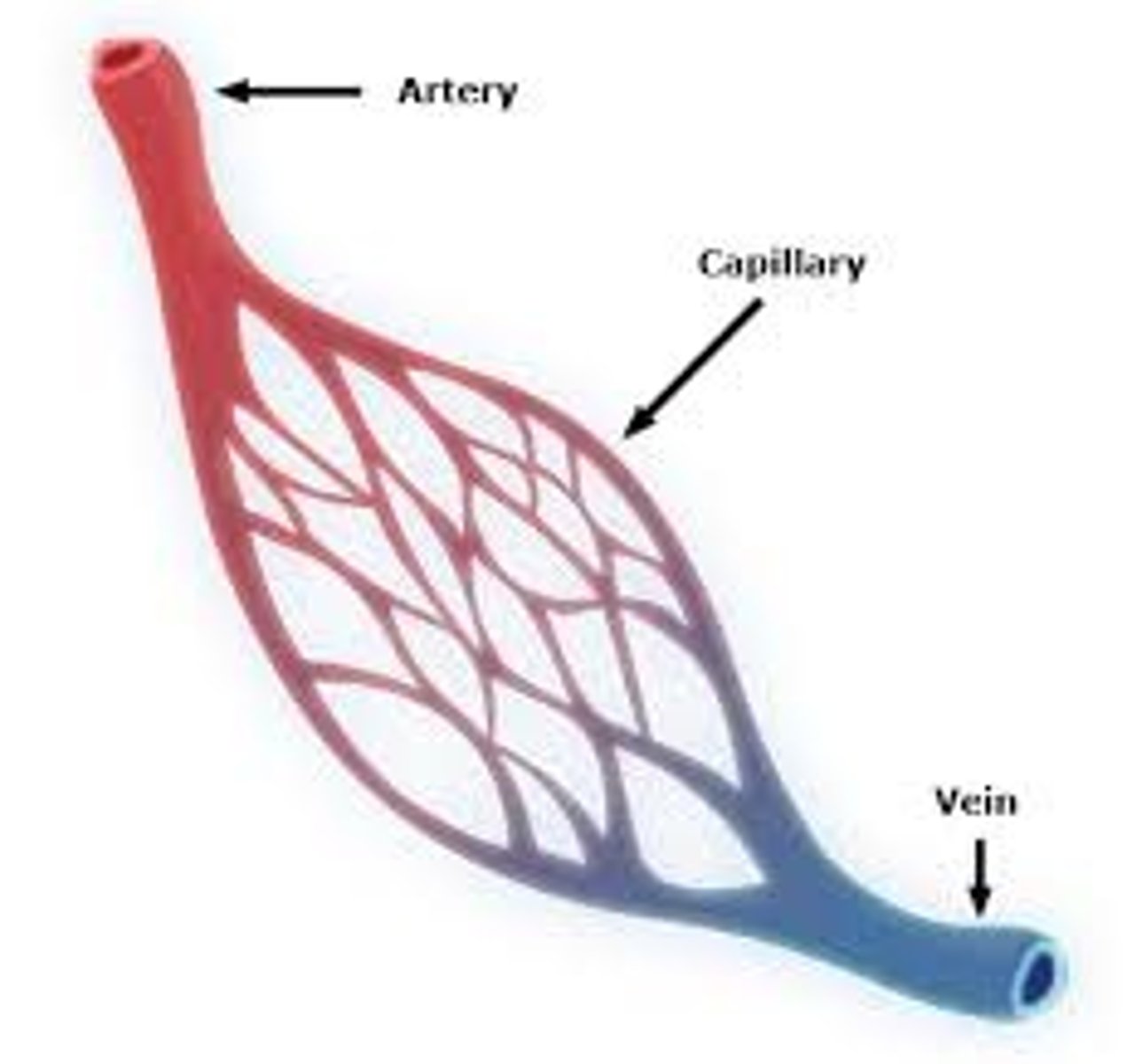
Arteries
Blood vessels that carry blood away from the heart
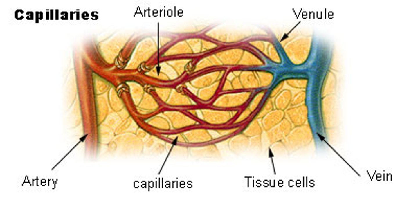
Arterioles
small blood vessels that distribute the blood from an artery to the capillaries
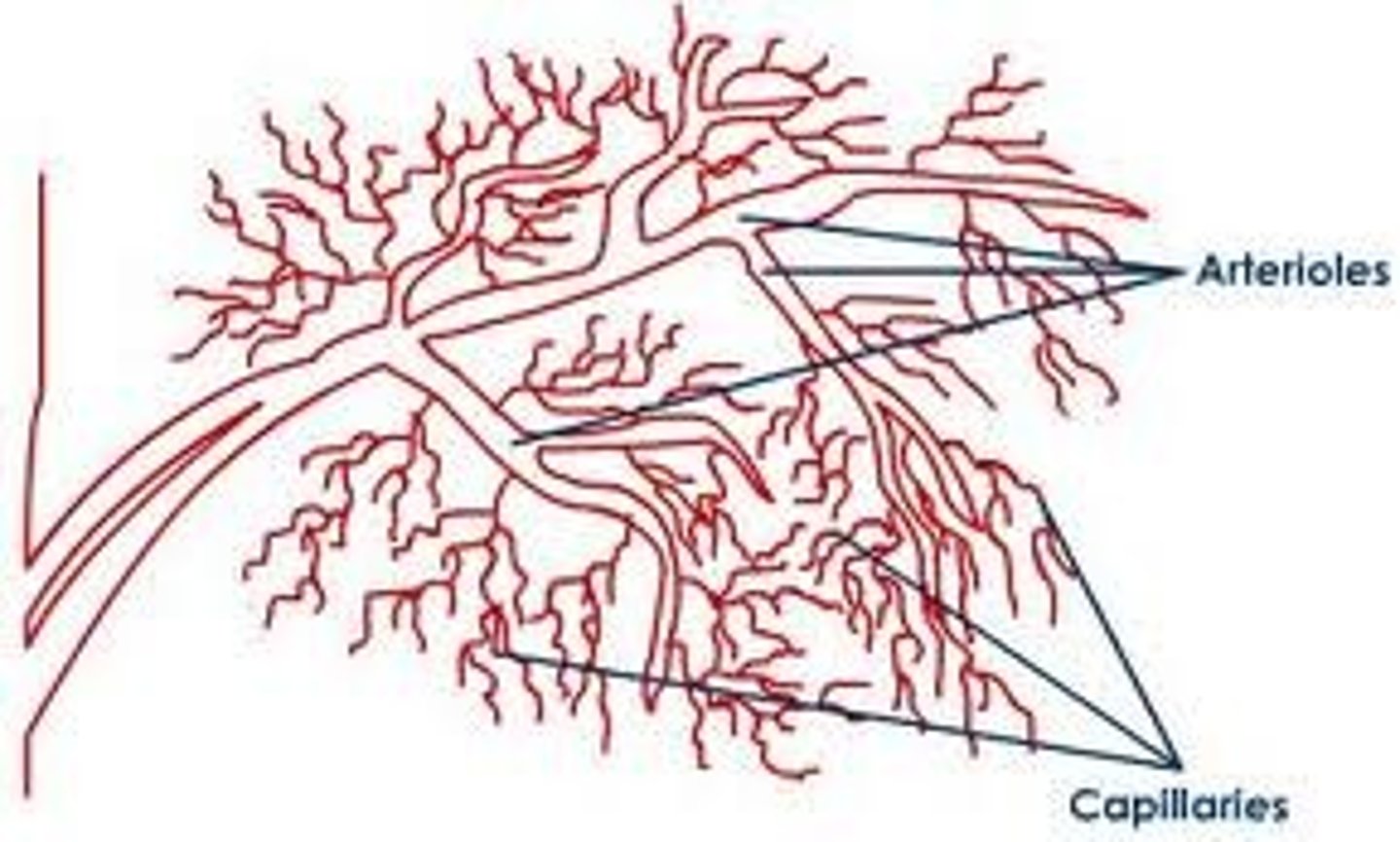
Capillaries
very small vessels with very thin walls
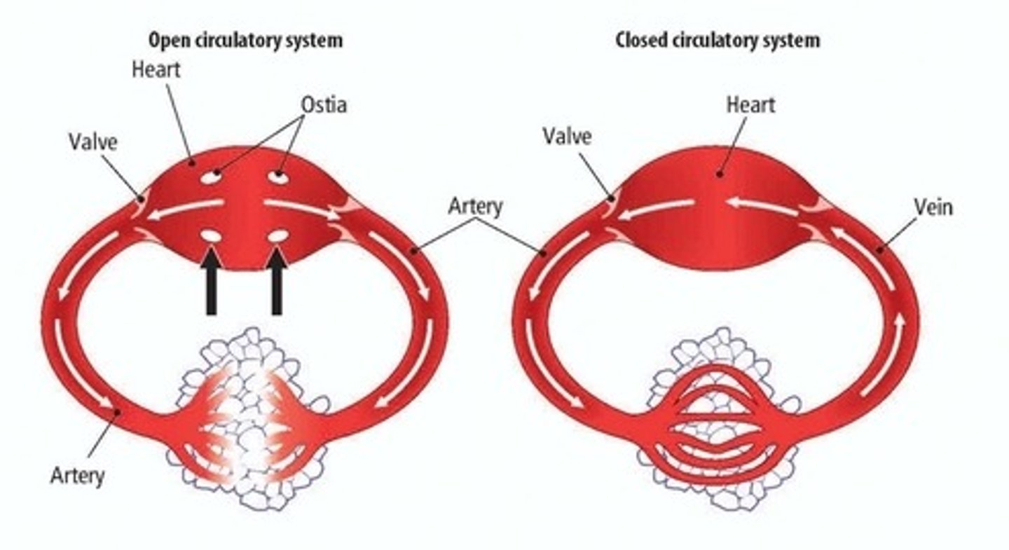
Closed circulatory system
One in which the blood is held in vessels

Open circulatory system
One in which the blood is not held in vessels
Veins
vessels that carry blood back to the heart
Venules
small blood vessels that collect blood from the capillaries and lead into the veins
Circulation in insects
There is a muscular pumping organ, much like the heart. This is a long, muscular tube that lies just under the dorsal (upper) surface of the body. Blood from the body enters the heart through pores called ostia. The heart then pumps blood towards the head by peristalsis. At the forward end of the heart (nearest the head), the blood simply pours out into the body cavity. This circulation can continue when the insect is at rest, but body movements may still affect circulation.
Some larger and more active insects, such as locusts, have open ended tubes attatched to the heart. These direct the blood towards active parts of the body, such as the leg and wing muscles.
Open circulatory systems have certain disadvantages:
- Blood pressure is low and blood flow is slow
- Circulation of blood may be affected by body movements or lack of body movements.
What fluid bathes the tissues and cells in a closed circulatory system?
Tissue fluid
Advantages of a closed circulatory system to an open circulatory system
- Higher pressure, so that blood flows more quickly.
- More rapid delivery of oxygen and nutrients
- More rapid removal of CO2 and other wastes
- Transport is independant of body movements
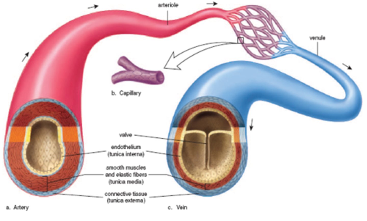
What is the endothelium?
An inner lining in all blood vessels, made up of a single layer of cells. This is a thin layer that is particularly smooth in order to reduce friction with the flowing blood.
How do the arteries withstand the high pressure of the blood?
- Thick walls
- Small lumen
- The inner wall of the artery is folded to allow the lumen to expand as blood flow increases
The wall consists of three layers:
- Inner layer (tunica intima) consists of a thin layer of elastic tissue which allows the wall to stretch and then recoil to help maintain blood pressure.
- Middle layer (tunica media) consists of a thick layer of smooth muscle.
- Outer layer (tunica adventitia) is a relatively thick layer of collagen and elastic tissue. This provides strength to withstand the high pressure, and recoil to maintain the pressure.
How do arterioles increase resistance to flow and reduce the rate of flow of blood?
Contraction of this muscle will constrict the diameter of the arteriole. Constriction of the arteriole walls can be used to divert the flow of blood to regions of the body that are demanding more oxygen.
How do capillaries allow for the exchange of materials between the blood and tissue fluid?
- The lumen is very narrow - its diameter is about the same as that of a red blood cell. The red blood cells may be squeezed against the walls of the capillaries; this helps with the transfer of oxygen, as it reduces the diffusion path to the tissues. It also increases resistance and reduces the rate of flow
- The walls consist of a single layer of flattened endothelial cells. This reduces the diffusion distance for the materials being exchanged.
- The walls are leaky. They allow blood plasma and dissolved substances to leave the blood.
How do veins ease the flow of blood?
Large lumen
Structure of veins
- The walls are thinner then arteries. The walls have thinner layers of collagen, smooth muscle and elastic tissue as they do not need to stretch and recoil, and are not actively constricted in order to reduce blood flow.
- They have valves to prevent the backflow of blood
3.2.3
EXCHANGE AT THE CAPILLARIES
Blood
The fluid used to transport materials around the body
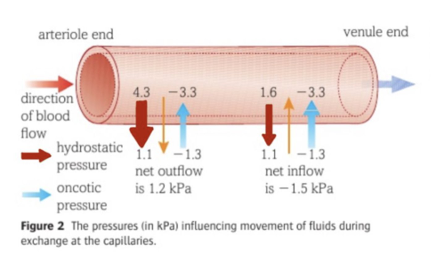
Hydrostatic pressure
The pressure that a fluid exerts when pushing against the sides of a vessel or container
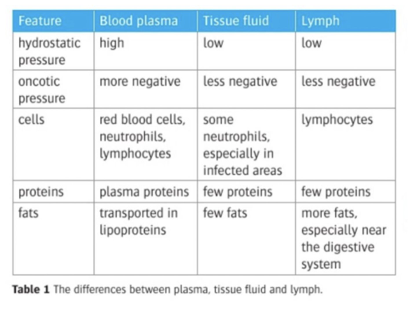
Lymph
The fluid held in the lymphatic system, which is a system of tubes that returns excess tissue fluid to the blood system
Oncotic pressure
The pressure created by the osmotic effects of the solutes
Plasma
The fluid portion of the blood
Tissue fluid
The fluid surrounding the cells and tissues
Formation of tissue fluid
Pressure Filtration :
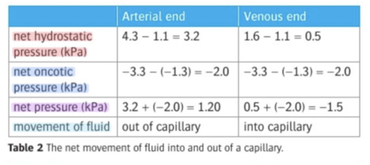
1. At the start of the capillary bed (ARTERIOLE END) there is a high hydrostatic pressure inside the capillaries and therefore fluid is forced out.
2. As the fluid is forced out the pressure decreases and therefore at the VENULE END fissure fluid is no longer formed.
3. Due to the loss of water from the capillary the water potential decreases and it means the water re-enters the capillary by osmosis.
What happens to the tissue fluid that hasn't re-entered the blood?
- Some tissue fluid is directed into another tubular system called the lymph system or lymphatic system.
- This drains excess tissue fluid out of the tissues and returns it to the blood system in the subclavian vein in the chest.
- The fluid in the lymphatic system is called lymph and is similar in composition to the tissue fluid. It will contain more lymphocytes, as these are produced in the lymph nodes.
What are lymph nodes?
Swellings found at intervals along the lymphatic system, which have an important part to play in the immune response.
The differences between plasma, tissue fluid and lymph
Pressures (kpa) influencing movement of fluids during exchange at capillaries
The net movement of fluid into and out of the capillary
3.2.4
THE STRUCTURE OF THE HEART
Atrio-ventricular valves
valves between the atria and the ventricles, which ensure that the blood flows in the correct direction
Cardiac muscle
Specialised muscle found in the walls of the heart chambers
Semilunar valves
valves that prevent blood re-entering that heart from the arteries
Structure of the heart
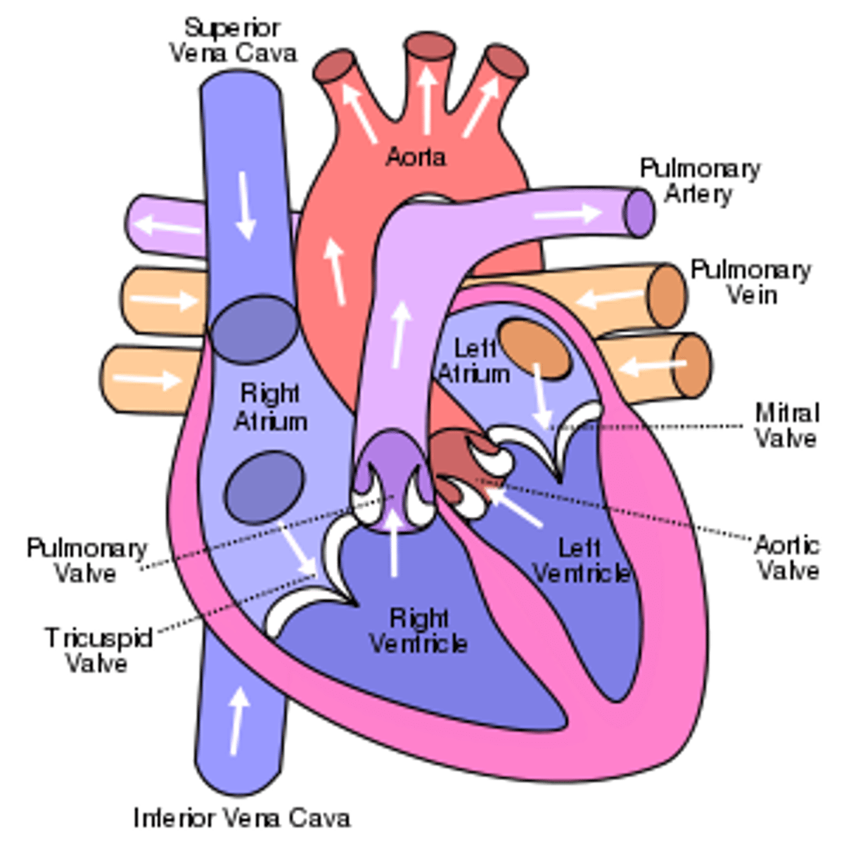
Blood flow through the heart
Deogygenated blood flows through the vena cava into the right atrium. Oxygenated blood from the lungs flows through the pulmonary vein into the left atrium. From the atria , blood flows down through the atrio-ventricular valves into the ventricles. Attatched to the valves are tendinous chords, which prevent the valves from turning inside out when the ventricles contract. A wall of muscle called the septum separates the ventricles from each other. This ensures that the oxygenated blood in the left side of the heart and the deoxygenated blood on the right side of the heart are kept separate. Deoxygentated blood leaving the left ventricle flows into the aorta. This carries blood to a number of arteries that supply all parts of the body. At the base of major arteries, where they exit the heart, are the semi-lunar valves. These prevent blood returning to the heart as the ventricles relax.
Blood pressure in the atria
- The muscles of the atrial wall is very thin, this is because these chambers do not need to create much pressure.
- Their function is to receive blood from the veins and push it into the ventricles
Blood pressure in the right ventricle
-The walls of the right ventricle are thicker than the walls of the atria
-This enables the right ventricle to pump blood out of the heart
-The right ventricle pumps deoxygenated blood to the lungs - the lungs are in the chest cavity beside the heart, so the blood does not need to travel very far. Also, the alveoli in the lungs are very delicate and could be damaged by very high blood pressure.
Blood pressure in the left ventricle
the walls of the left ventricle can be two or three times thicker than the right one
-the blood is pumped out the aorta and needs sufficient pressure to overcome the resistance of the systhemic circulation.
3.2.5
THE CARDIAC CYCLE
Cardiac cycle
The sequence of events in one full beat of the heart
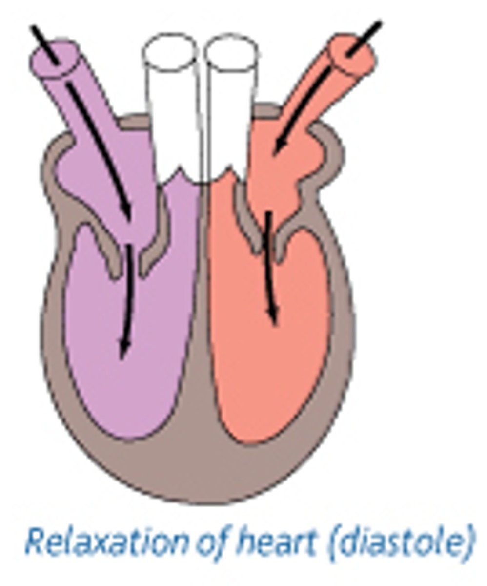
Diastole
The muscular walls of all four chambers relax. Elastic recoil causes the chambers to increase in volume, allowing blood to flow in from the veins
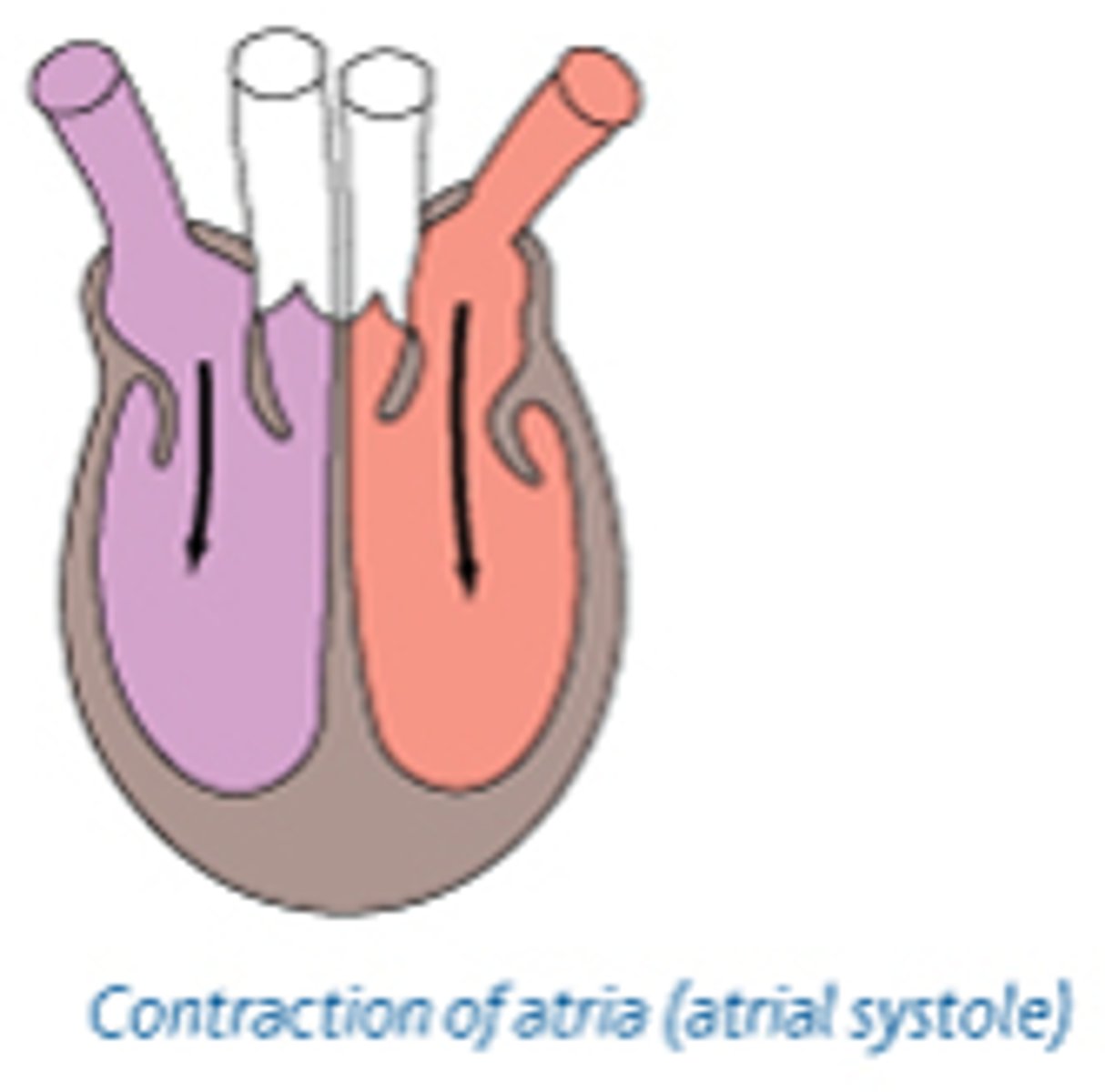
Atrial systole
Both left and right atria contract together. The muscle in the walls is thin, so this contraction creates only a small increase in pressure. This helps push blood into the ventricles, stretching their walls and ensuring they are full of blood.
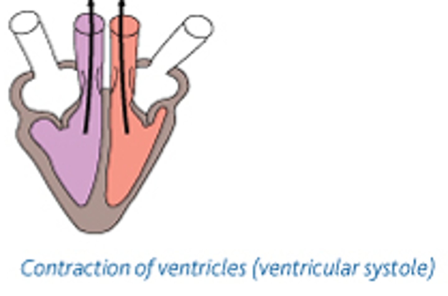
Ventricular systole
Both right and left ventricles pump together. Contraction starts at the apex (base) of the heart so that blood is pushed upwards towards the arteries.
The action of the atrio-ventricular valves diagram
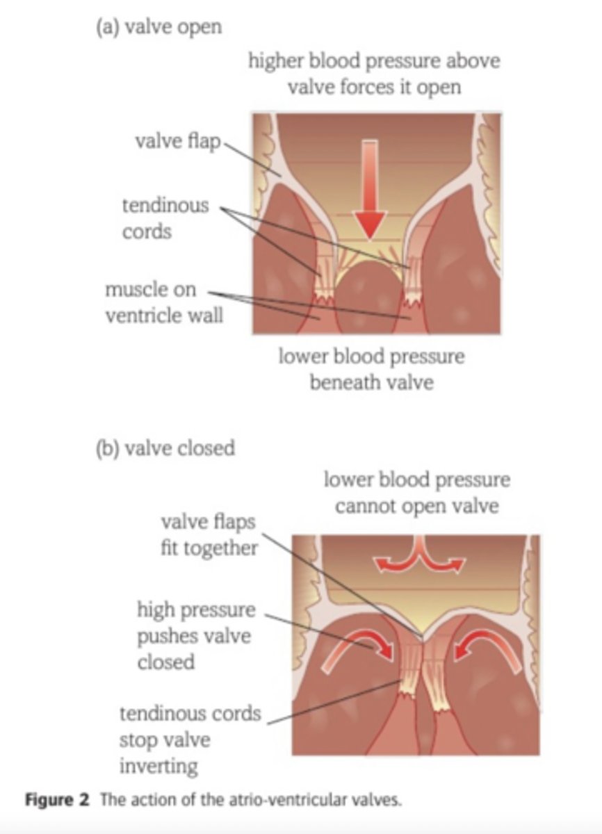
When does the atrio-ventricular valves open?
When the atria's contract and push blood to the ventricles.
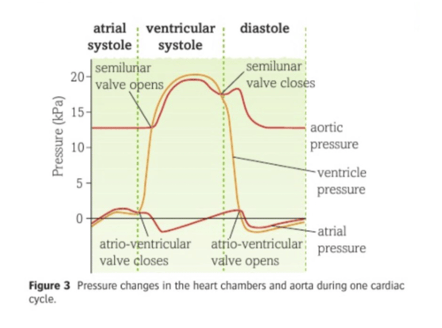
When do the semilunar valves open?
when the pressure in the ventricles exceeds the pressure in the arteries
When do the semilunar valves close?
When the pressure in the arteries is greater than the pressure in the ventricles
Pressure changes in the heart chambers and the aorta during one cardiac cycle diagram
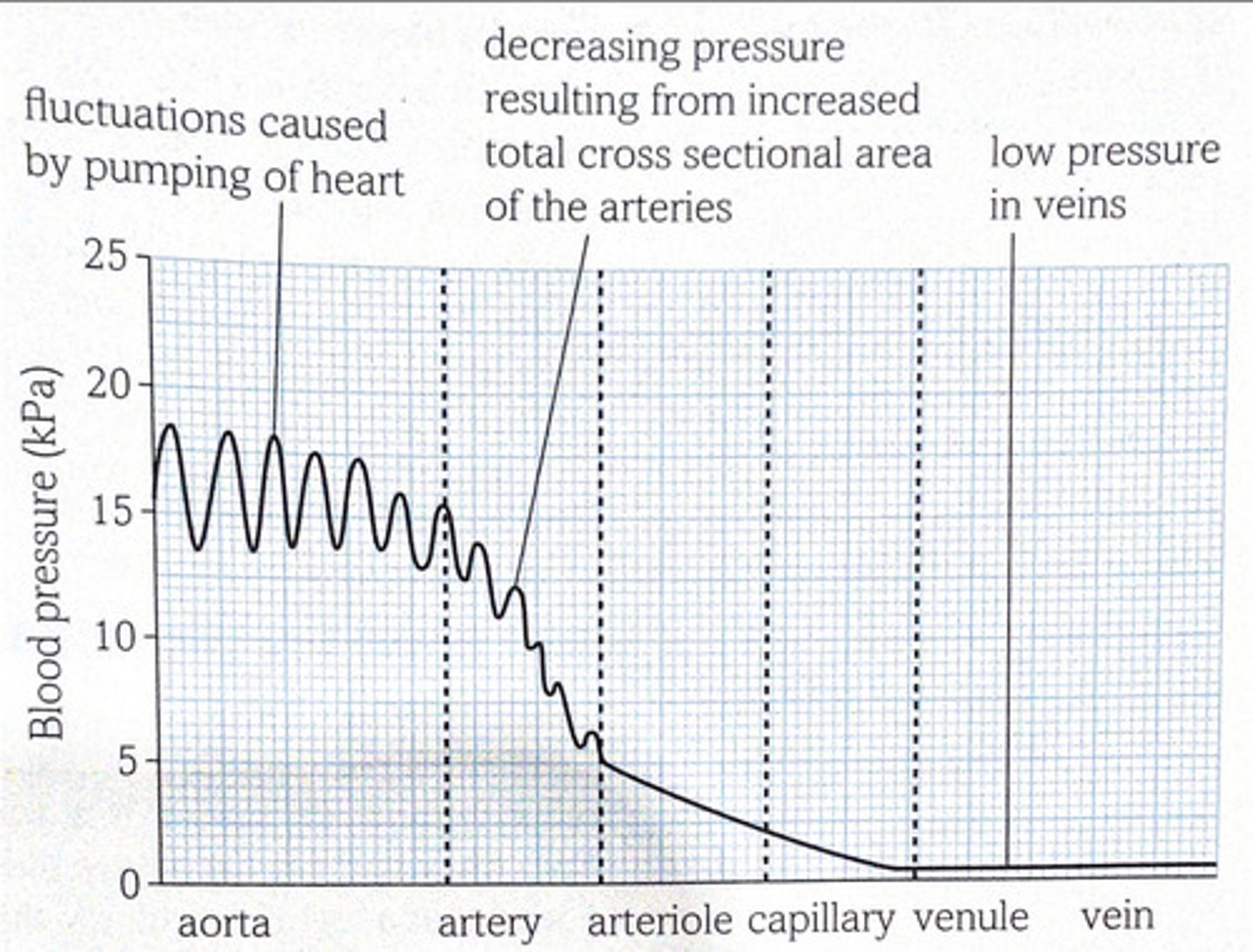
The structure of the artery walls plays a large part in creating a more even flow:
- The artery walls close to the heart have a lot of elastic tissue.
- When blood leaves the heart, these walls stretch.
- As blood moves on and out of the aorta, the pressure in the aorta starts to drop.
- The elastic recoil of the walls helps to maintain the blood pressure in the aorta.
- The further the blood flows along the arteries, the more the pressure drops and the flunctuations become less obvious.
- It is important to maintain the pressure gradient between the aorta and the arterioles, as this is what keeps the blood flowing towards the tissues.
The blood pressure at different parts of the blood system
3.2.6
COORDINATION OF THE CARDIAC CYCLE
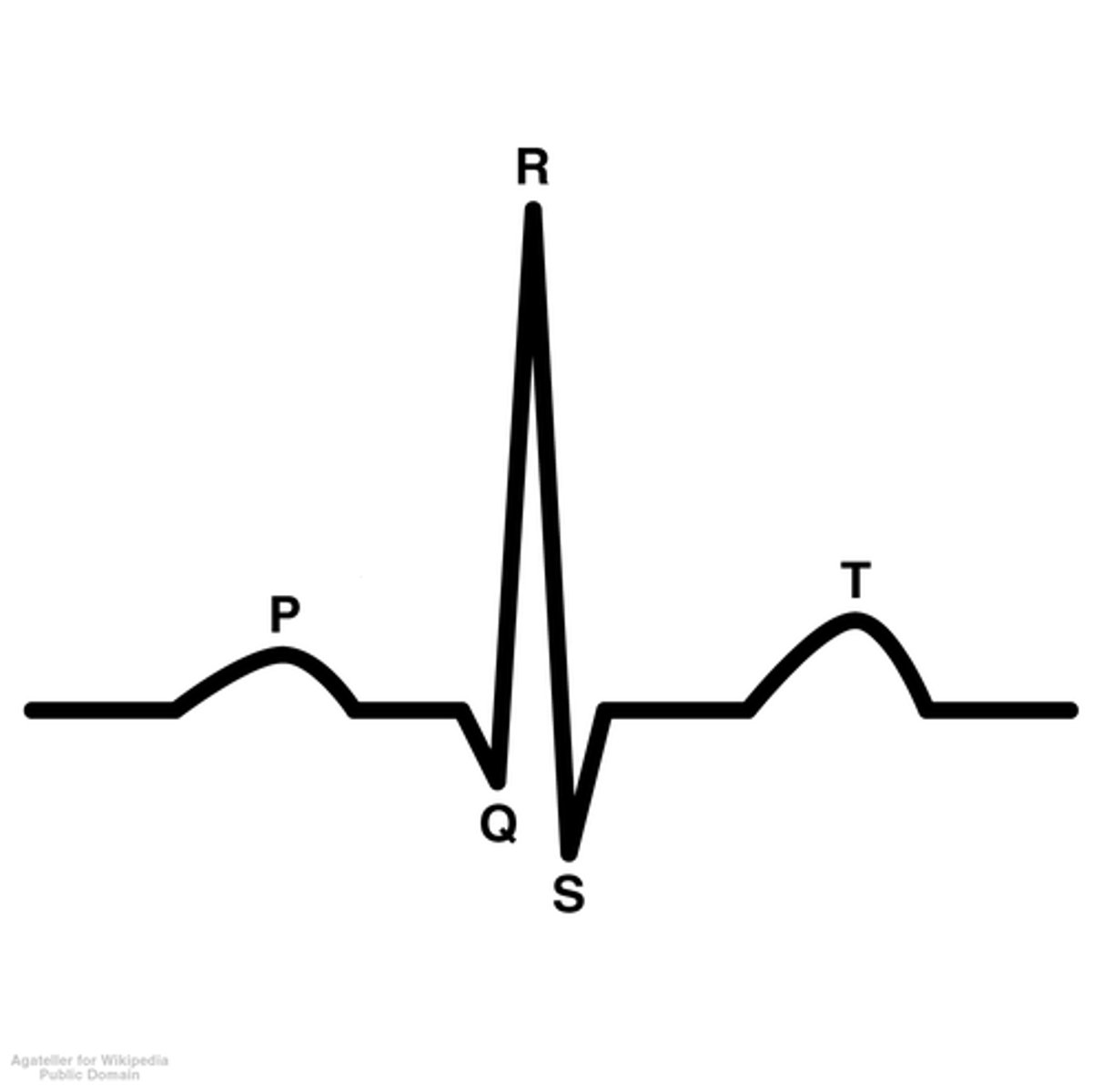
Electrocardiagram
A trace that records the electrical activity of the heart
Fibrillation
Uncoordinated contraction of the atria and ventricles
Myogenic muscle
Muscle that can initiate its own contraction
Purkyne tissue
Consists of specially adapted muscle fibres that conduct the wave of excitation from the AVN down the septum to the ventricles.
Sino-atrial node (SAN)
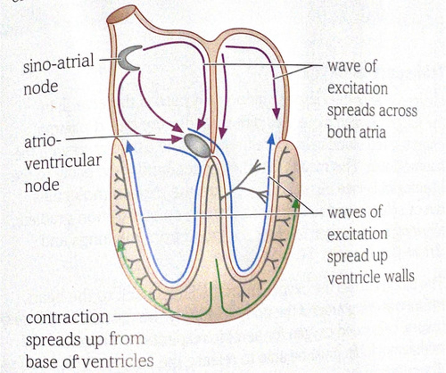
The hearts pacemaker. It is a small patch of tissue that sends out waves of electrical excitation at regular intervals in order to initiate contractions
Contraction of the atria
- the wave of excitation quickly spreads over the walls of both atria
-it travels along the membranes of the muscle tissue
-as the wave of excitation passes it causes the cardiac muscle cells to contract
-this is an atrial systole
Why can the wave of excitation not pass down to the ventricles?
The tissue at the base of the atria is unable to conduct the wave of excitation
Atrio-ventricular node (AVN)
This is the only route that can conduct the wave of excitation through to the ventricles. The wave of excitation is delayed in the node. This allows time for the atria to finish contracting and for the blood to flow down into the ventricles before they begin to contract.
Contraction of the ventricles
- After a short delay, the wave of excitation is carried away from the AVN and down the Purkyne tissue.
- This runs down the septum.
- At the base of the septum, the wave of excitation spreads from the base, causing the muscles to contract.
- As the excitation spreads upwards from the base of the ventricles, it causes the muscles to contract.
- This means that the ventricles contract from the base upwards.
- This pushes the blood up towards the major arteries at the top of the heart.
Coordination of the cardiac cycle diagram
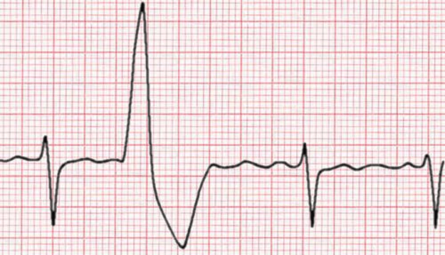
Wave P in electrocardiagram
Shows excitation of the atria
QRS wave in electrocardiagram
indicates the excitation of the ventricles
Wave T in electrocardiagram
Shows diastole
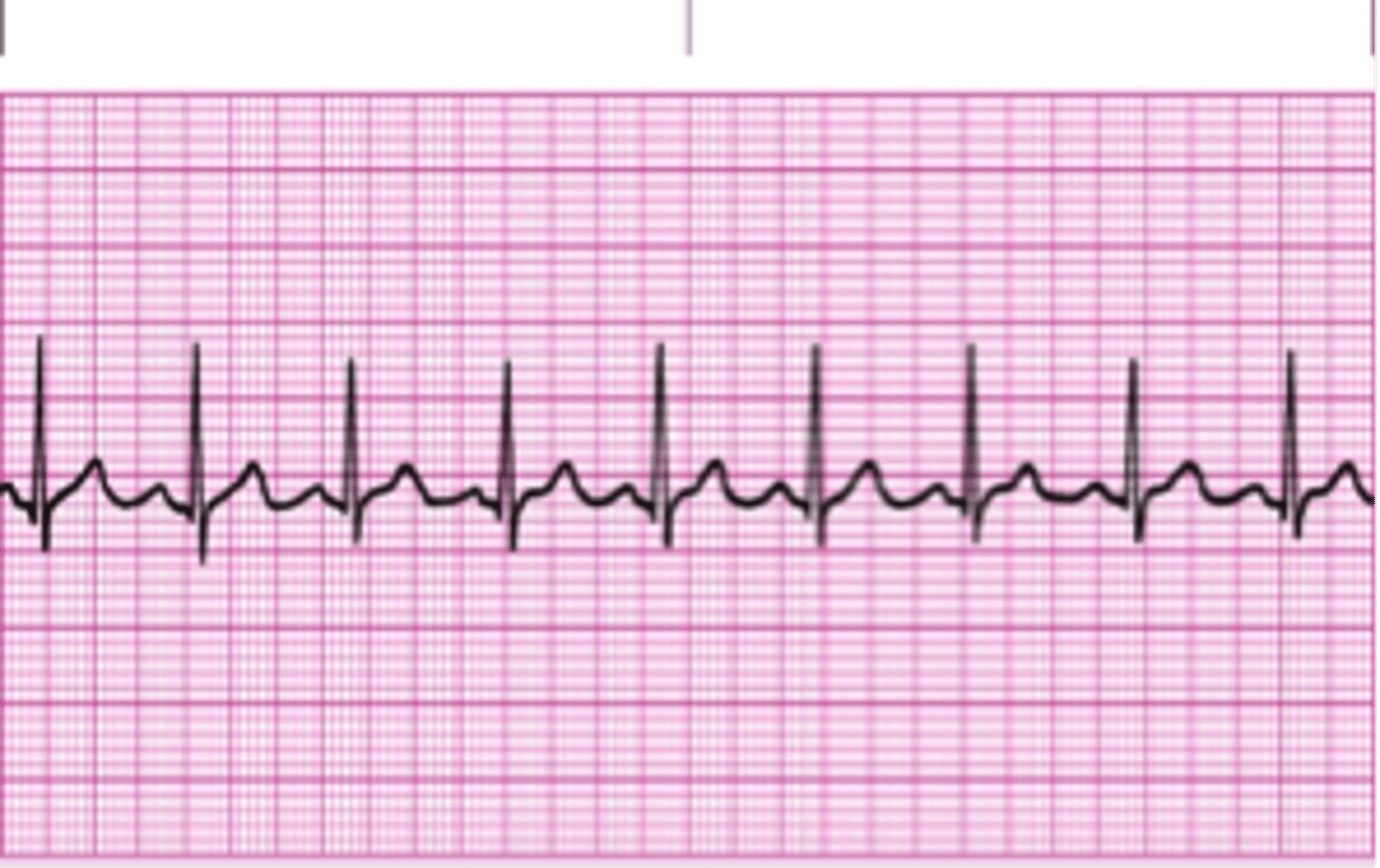
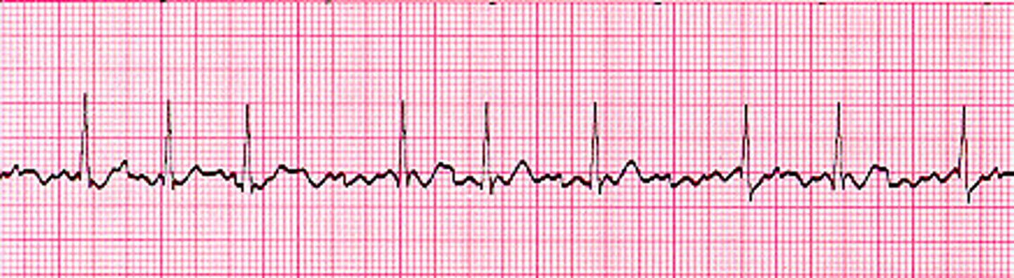
An ECG
Sinus rhythm
normal heart rhythm
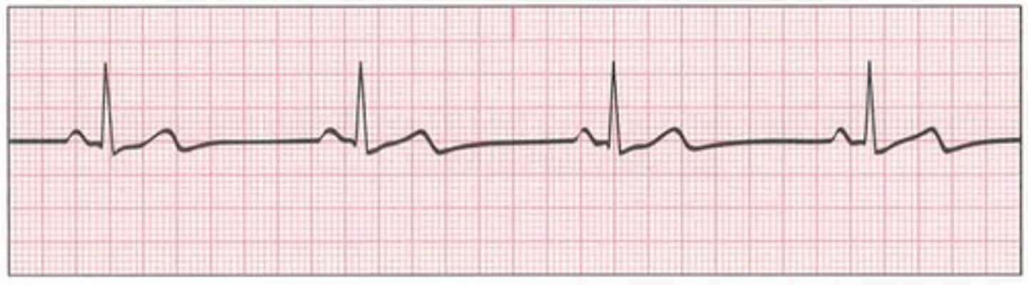
Bradychardia
slow heart rate
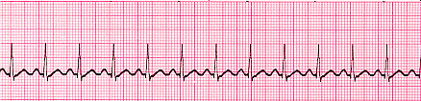
Tachycardia
fast heart rate
Atrial fibrillation
Atria beating more frequently then ventricles - no clear P waves seen
Ectopic heartbeat
An extra beat or an early beat of the ventricles.
3.2.7
TRANSPORT OF OXYGEN
Affinity
A strong attraction
Dissociation
Means releasing the oxygen from the oxyhaemoglobin
Fetal haemoglobin
The tyoe of haemoglobin usually found only in the fetus
Haemoglobin
The red pigment used to transport oxygen in the blood
When haemoglobin takes up oxygen, it becomes...
Oxyhaemoglobin
Reversible binding of oxygen by haemoglobin diagram
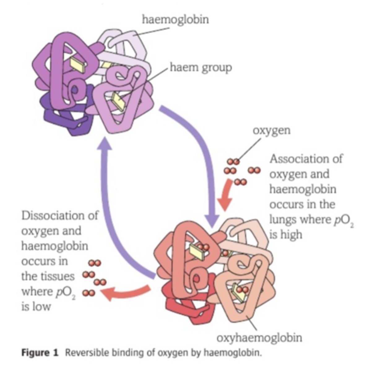
Transport of oxygen
Oxygen is absorbed into the blood as it passes the alveoli in the lungs. Oxygen molecules diffusing into the blood plasma enter the red blood cells. Here they become associated with the haemoglobin. The blood carries the oxygen from the lungs back to the heart, before travelling around the body to supply the tissues. In the body tissues, cells need oxygen for aerobic respiration. Therefore the oxyhaemoglbin must be able to release the oxygen. This is called dissociation.
What happens to the haemoglobin dissociation curve when oxygen tension rises?
The diffusion gradient into the haemoglobin molecule increases. Eventually, one oxygen molecule enters the haemoglobin molecule and associates with one of the haem groups. This causes a slight change in the shape of the haemoglobin, known as conformational change . It allows more oxygen to enter the haemoglobin molecule and associate with the other haem groups relatively easily. This accounts for the steepness of the curve as oxygen tension rises.
The haemoglobin dissociation curve
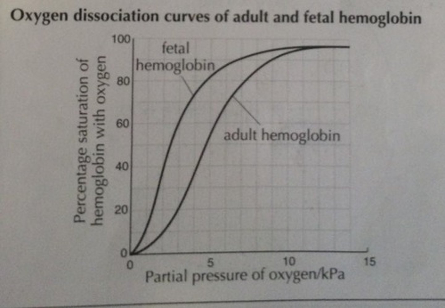
Fetal haemoglobin Vs Adult haemoglobin
Higher affinity for oxygen than adult haemoglobin because fetal haemoglobin must be able to associate with oxygen in an environment where the oxygen tension is low enough to make adult haemoglobin release oxygen.