Exercise 2 - 9 TABLES AND POINTERS
1/148
Earn XP
Description and Tags
Bacteriology
structures and composition of microorganisms
examination of living bacteria in fresh preparation
staining of microorganisms
bacterial smear
differential stains
isolation of pure culture of organisms
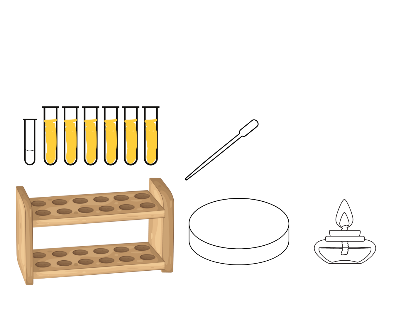
inhibition and distruction of microorganisms by physical agents
action of disinfection on bacteria
antimicrobial suscentibility testing
staphylococcus
University/Undergrad
Name | Mastery | Learn | Test | Matching | Spaced | Call with Kai |
|---|
No analytics yet
Send a link to your students to track their progress
149 Terms
Four-quadrant streak plate method
identifier: colonies in the 4th quadrant
Identify method of isolation pure culture
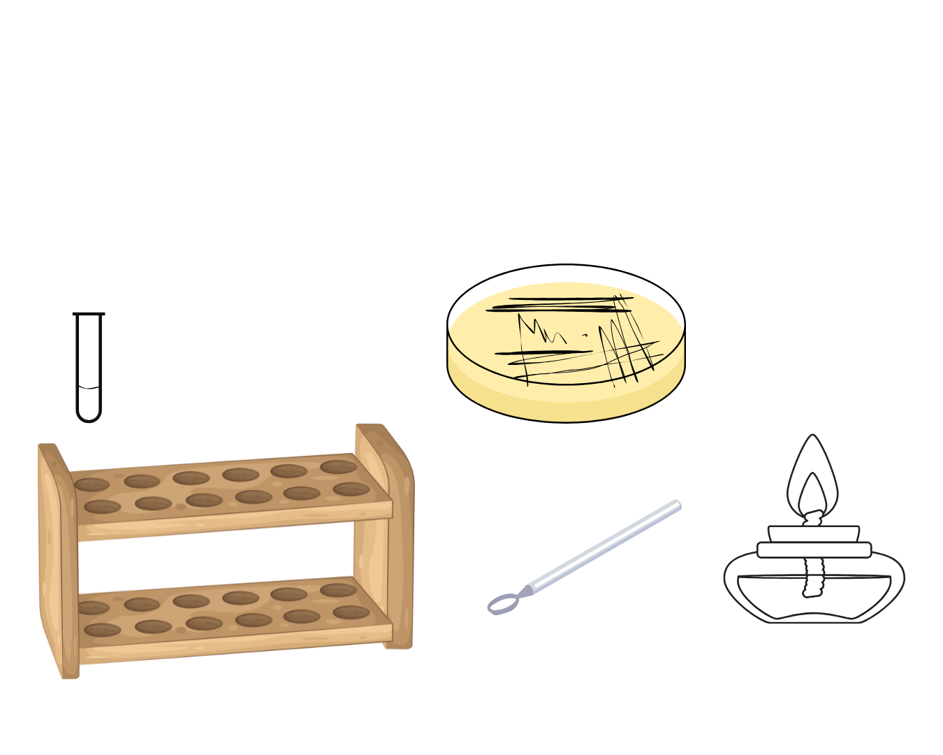
Aseptic technique
Purpose of alcohol lamp

Nutrient agar
Culture medium used

Fourth Quadrant
which quadrant are colonies isolated

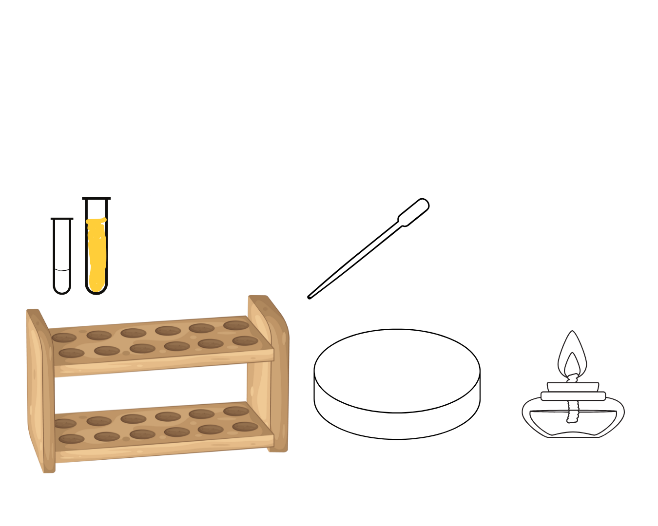
Serial Dilution Pour Plate Method
Identifier: there is label for serial dilution in the longer test tube
Identify method of isolation pure culture

Melted nutrient agar
Culture medium used

Ten-folds dilution
Two-folds dilution
Type of dilution used in the test
1:1000
1:60

Quantitative: Serial Dilution Pour Plate Method
Qualitative: Four-quadrant streak plate method
which is the quantitative and qualitative method for isolation of pure culture
37°Celsius for 24 hours
Incubation requirement for isolation of pure culture
Quebec colony counter
Principle: pressure sensing device
What equipment is used to quantitate the colonies in this test (include its principle)
Colony Forming Units/mL = colony count x reciprocal of dilution
Too few to count formulae (you are counting all the colony in the petri dish)
Average number of colonies in five squares x 62.5 x reciprocal of dilution=Colony Forming Units/mL
Too numerous to count formulae
Serial Dilution Pour Plate Method
Identify method of isolation pure culture

Too many to count
First three dilutions interpretation
1:10
1:100
1:1 000
Too few to count
last three dilutions interpretation
1:10 000
1:100 000
1:1 000 000

As the dilution increases the colony count decreases
As the dilution _ the colony count _
62.5
diameter of petri dish
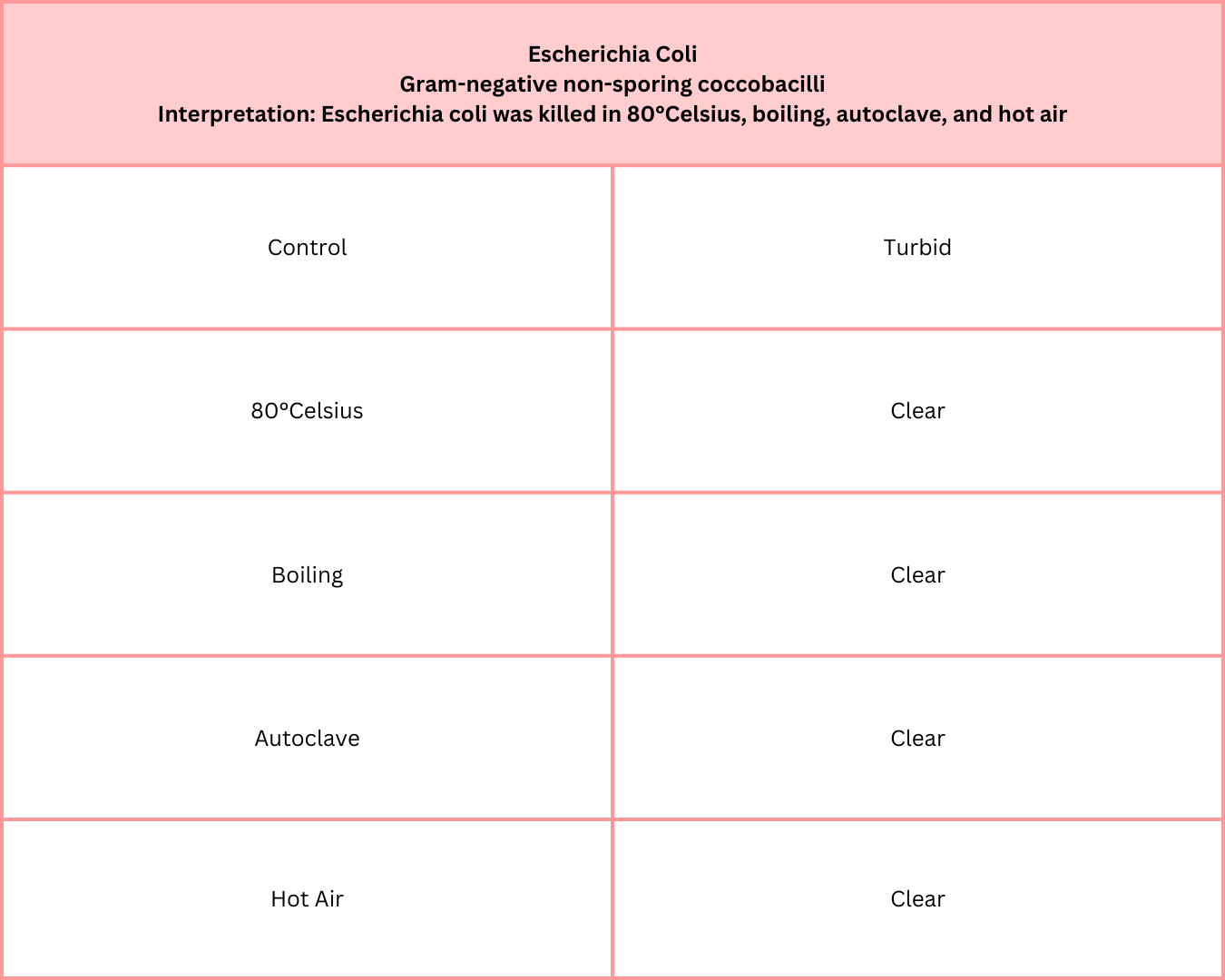
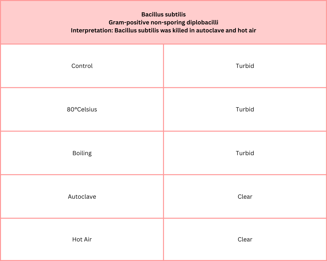
Disinfection
bacteriostatic; inhibition
Sterilization
bacteriocidal; killing
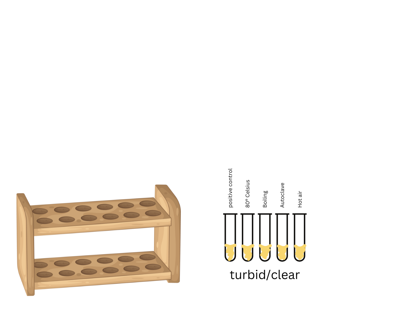
80°Celsius
Boiling: 100°Celsius
Autoclave: 121°Celsius
Hot air: 180°Celsius
temperature of physical agents
_
boiling
autoclave
hot air
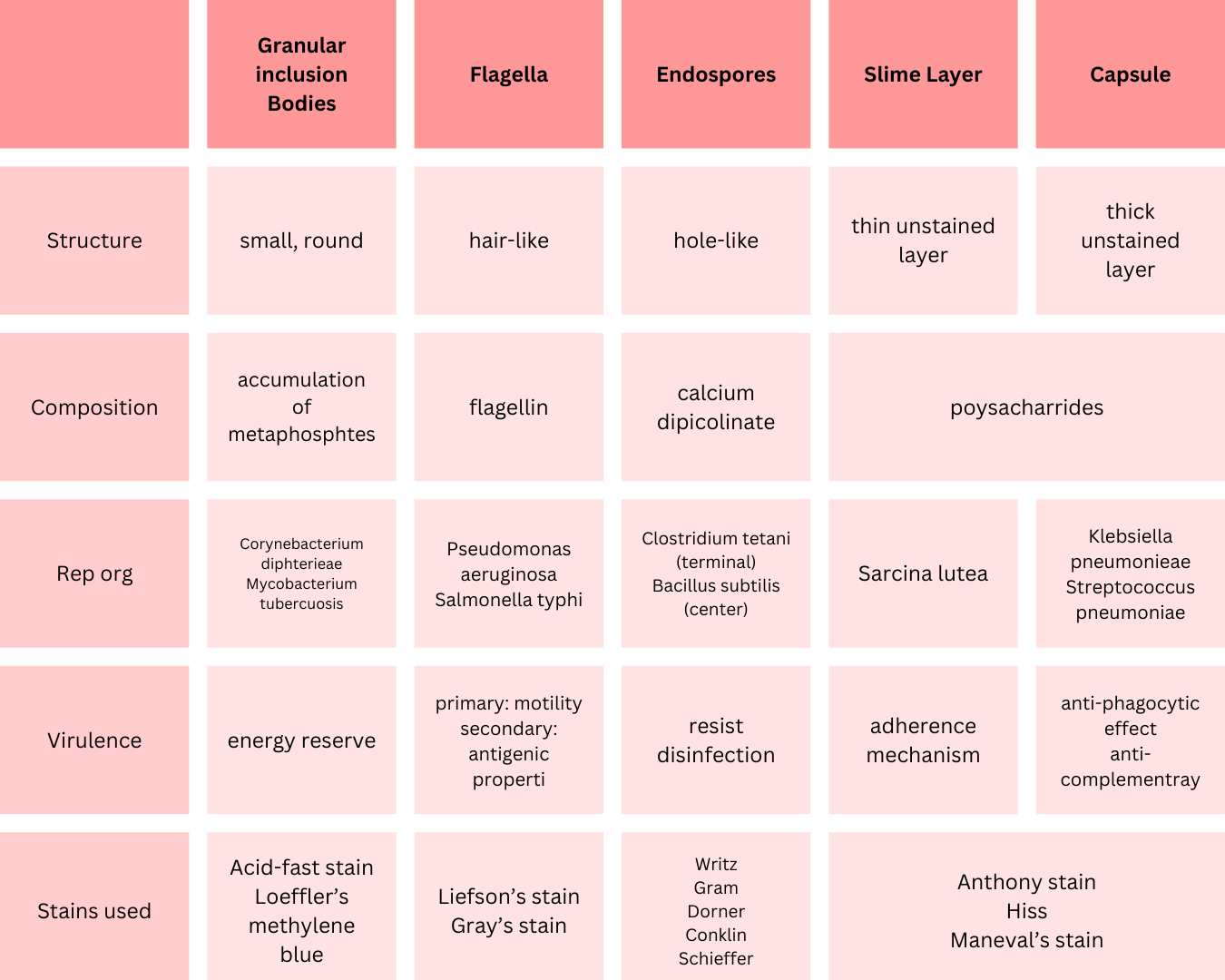
Endospores; Resists disinfection
What is responsible for the characteristic result of the organism of letter A to this test and its action


Nutrient broth
medium used

80°Celsius
Boiling: 100°Celsius
What are the physical agents of disinfection

Autoclave: 121°Celsius
Hot air: 180°Celsius
What are the physical agents of sterilization
Physical agents of disinfection
80°Celsius
Boiling: 100°Celsius
Physical agents of sterilization
Autoclave: 121°Celsius
Hot air: 180°Celsius
Turbidity
Only evidence of bacterial growth of this test

Turbidity
change of pH in the medium
presence of Hydrogen gas in the Durham’s fermentation tube
(if any of these are indicators are positive there is growth of bacteria)
3 evidence of bacterial growth

(disinfectant) killed the bacteria in (time)
How do you interpret the result
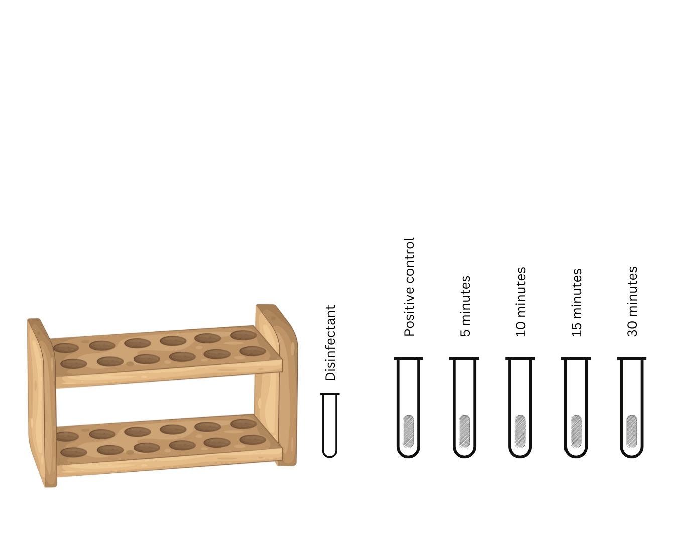
answer the time only (ex. 30 mins)
Contact time of the disinfectant to kill the organism
To trap Hydrogen gas
Purpose of Durham’s fermentation tube in the culture medium

Andrade’s indicator
peach
pink
pH indicator in the culture medium or the responsible component of the change in color of the medium
name
original color
change in color if there is change in pH

Lactose broth with Durham’s fermentation tube
culture medium

Coliform
Gram-negative non-sporogenous lactose fermenter organisms
non-sporogenous: there is no endospores
type organisms that produce HYDROGEN gas
name
description
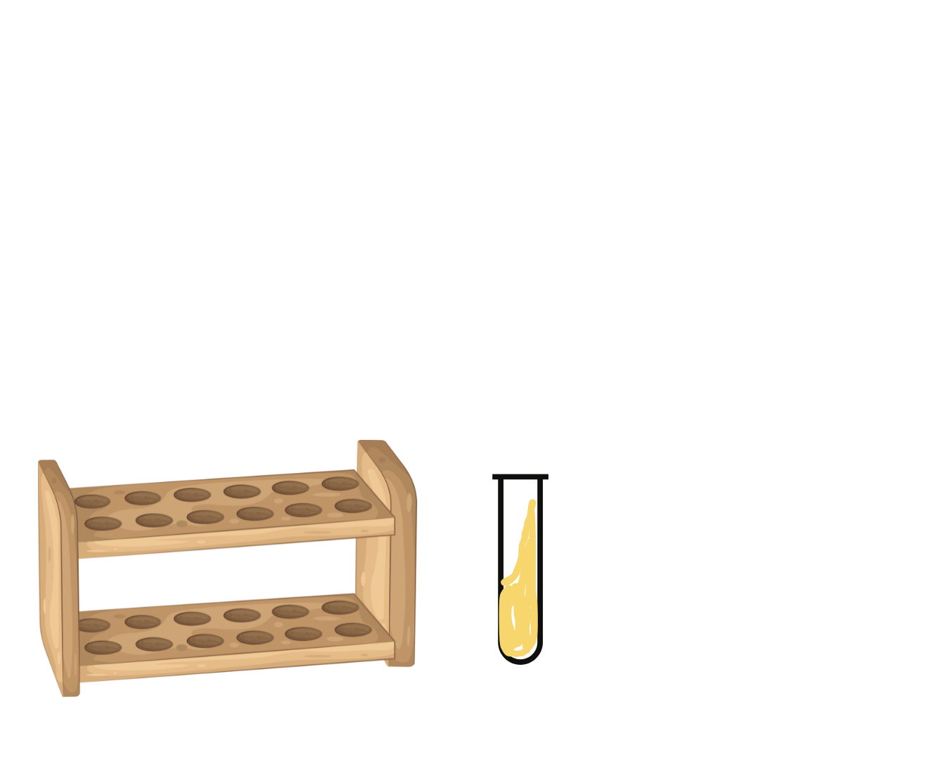
Antimicrobial susceptibility test
identify test

Kirby-Bauer method
identify method used in the test

0.5 McFarland standard
Composition
Volume: 99.5 ml of 1% sulfuric acid; 0.5 ml of 1.175% barium chloride
Concentration:1.5 x 10⁸ CFU/ml
bacterial suspension used in this test is compared to the turbidity of what material and its composition

Mueller-Hinton agar
culture medium used in the test
Beef Extract – 2.0 g/L
Acid Hydrolysate of Casein (Peptone) – 17.5 g/L
Starch – 1.5 g/L
Agar – 17.0 g/L
Distilled Water – 1,000 mL
Magnesium Ions (Mg²⁺) – 20–25 mg/L
Calcium Ions (Ca²⁺) – 50 mg/L
Final pH – 7.2-7.4 at 25°C
trace amounts of thymidine, usually less than 1 mg/L
Supplements in Mueller Hinton agar
Gram’s Staining/ Hucker’s method
identify method
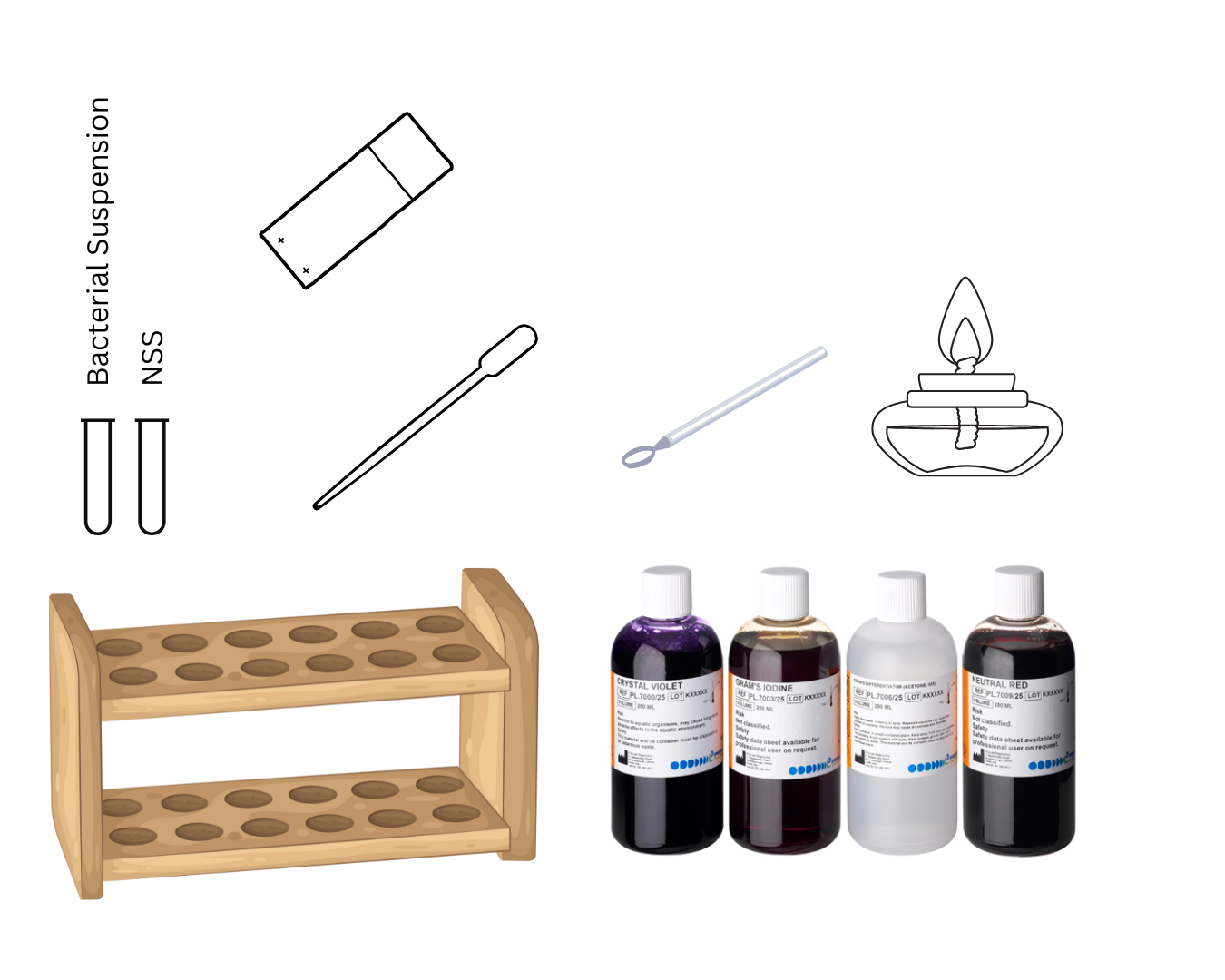
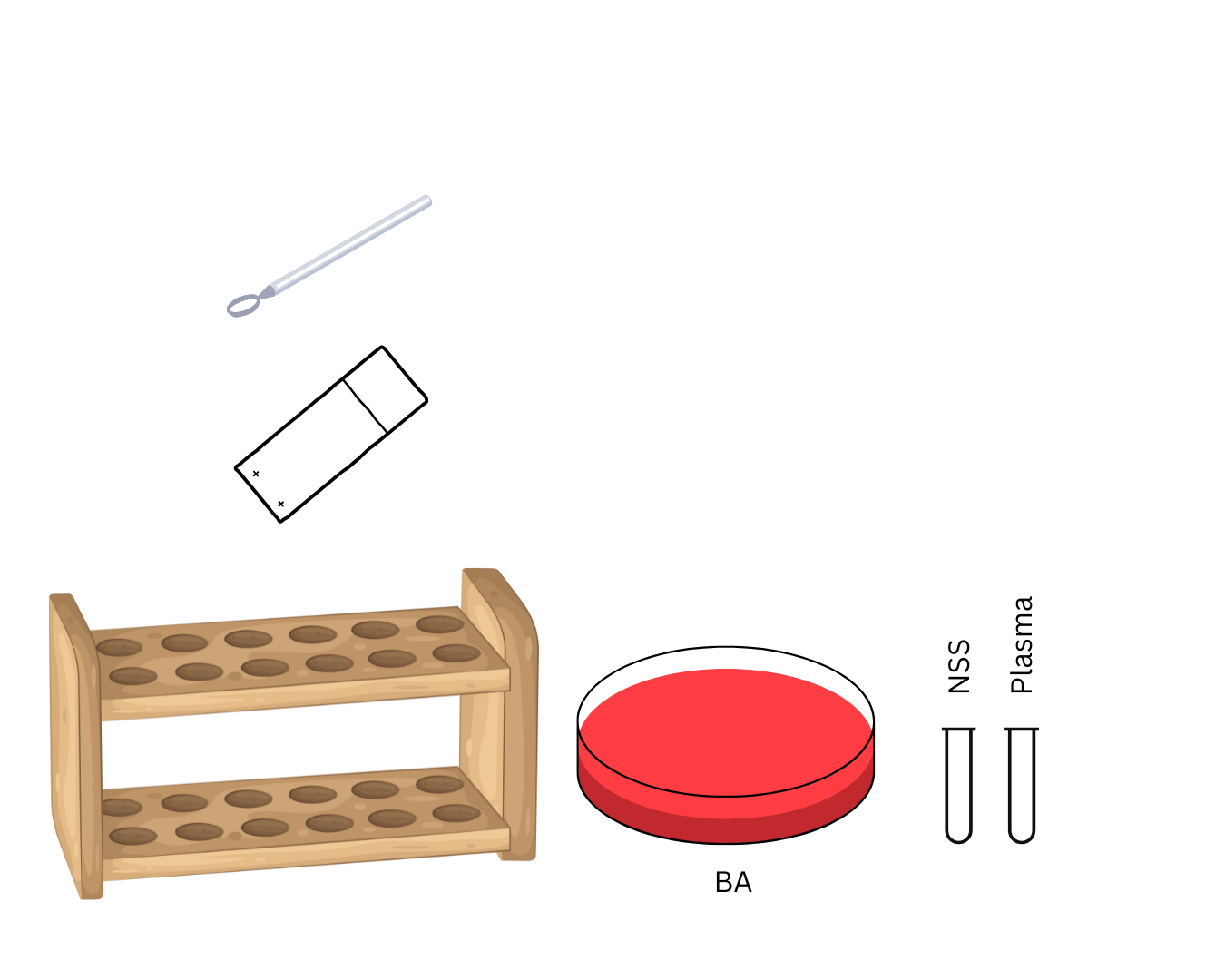
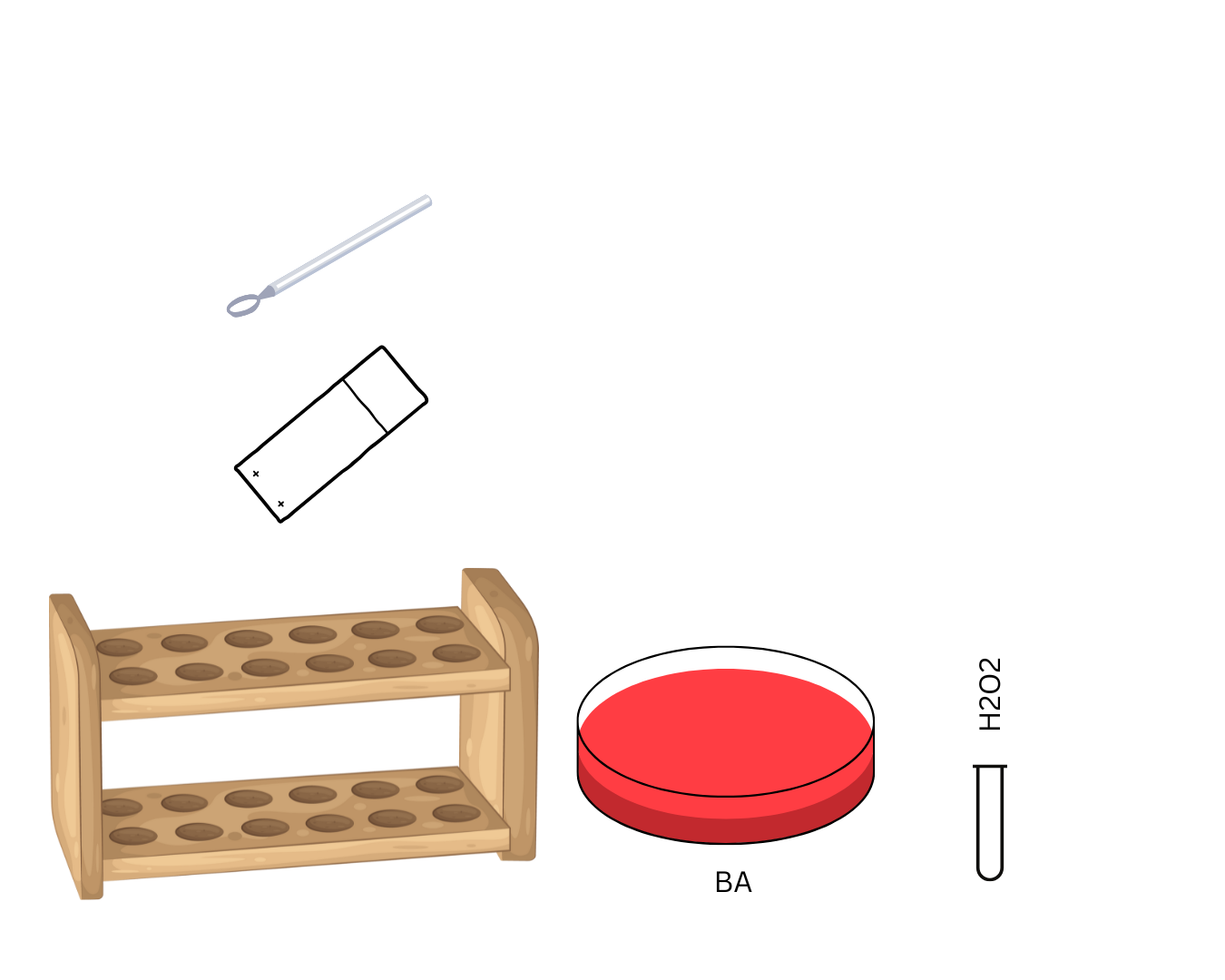
Staphylococci
identify organism investigated
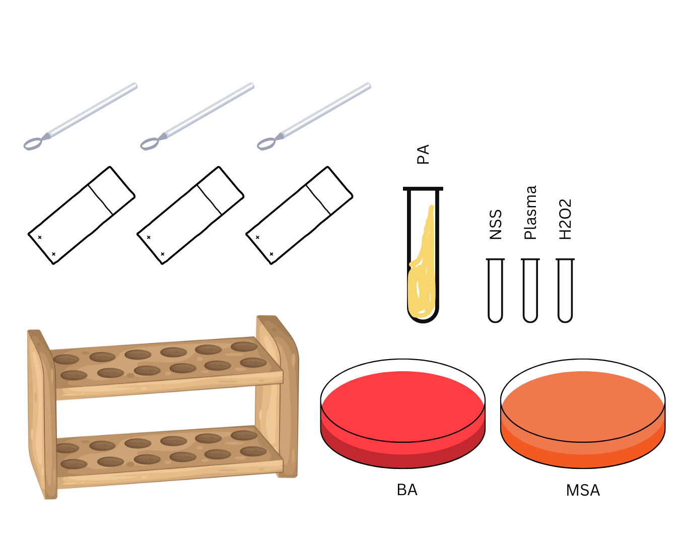
pigment production
Presumptive test
identify test and protocol

Catalase test
Presumptive test
identify test and protocol

Coagulase test
Confirmatory test
identify test and protocol
Slant portion; aerobic condition
Butt portion; anaerobic condition
parts in the tube and its conditions
simple streaking
streaking used

Oxygen
element responsible for the positive result
complete hemolysis (BA turns clear color of TSA is seen)
beta hemolysis (and its description)
Partial hemolysis (greenish brown)
alpha hemolysis (and its description)
no hemolysis (no change in culture media)
gamma hemolysis (and its description)
VIRULENCE
Ability of the microorganism to cause infection or disease.
STRUCTURES & COMPOSITION
Things or substances present within the organism which they utilize for them to cause infection to human host
irregular shaped circles
GRANULAR
inside the body
INCLUSION
ACCUMULATION OF METAPHOSPHATES
composition of the bacterial structure, granular inclusion body
NEUTROPHILS, EOSINOPHILS, & MONOCYTES
3 types of phagocytes
Metachromasia
A special property of granular inclusion bodies/granules where it appears purple when stained with methylene BLUE
metachromatic granules
GIB are also called
diphtheria and leprae
causative agent: Corynebacterium diphtheriae
Babes-Ernst granules
Chinese letter appearance
specific name of the metachromatic granules of Corynebacterium diphtheriae and its morphology
fibrous protein
structure of composition
flagellin
composition

Tannic acid
specific reagent necessary for the demonstration of the flagella
MORDANT
what reagent is necessary for the demo of flagella
NO OF FLAGELLA PRESENT IN ORGANISM
LOCATION OF THE FLAGELLA IN THE BODY OF THE ORGANISM
criteria of MESSEA’s classification
Monotrichous - one flagella
Lophotrichous - several flagella in a tuft(bundle)
Amphitrichous - one flagella each side
Peritrichous - flagella all over the body
Atrichous - no flagella
Monotrichous
Lophotrichous
Amphitrichous
Peritrichous
Atrichous
Pseudomonas aeruginosa - monotrichous
Salmonella typhi - peritrichous
Representative Organisms and their flagella characteristics
Drumstick appearance
Terminal spores description
scanty amount
slime layer amount
copious amount
capsule amount
NEGROSIN - detects capsule that produces semi-opaque background
QUELLUNG REACTION - ab-ag (antiserum) swells the capsule
TESTS DONE for capsules and description
COMPLEMENT SYSTEM
second line and highest form of defense against any invading antigen
CELL-LYSIS OR CELL DEATH
end action of the complement system when the immune system is activated
SIMPLE STAINING
1 dye only
morphology of the bacteria in films
direct staining
DIFFERENTIAL STAINING
2 or more dyes
principle of dfs: bacteria differ chemically and physically and may react differently to a given staining procedure
principle of dfs: bacteria differ _ and _ and may react _ to a given staining procedure
1 uL
volume a wire loop can hold
Primary Staining
Crystal violet(HEXAMETHYL-PARA-ROSANILINE CHLORIDE)
alternative: Gentian violet
1 min
Mordant
Gram’s iodine (brown-colored stain)
1 min
Decolorization (crucial step)
Acetone alcohol
3-5 seconds
Counter Staining
Safranin (red-colored stain)
45 secs
DFS steps, stain used, time
THICK PEPTIDOGLYCAN LAYER WITH TEICHOIC ACID (amino acids and disaccharides)
Gram-positive organisms’ cell wall
THIN PEPTIDOGLYCAN LAYER WITH LIPOPOLYSACCHARIDES (lipids and polysaccharides)
Gram-negative organisms’ cell wall
simultaneously kills the microorganism
allows the smear to adhere
heat fixing
preserves morphology
air drying
MORDANT
intensifies the color of the primary stain to the cell wall
CRYSTAL VIOLET-IODINE COMPLEX
iodine molecule forms an attachment with the crystal violet molecule making…
may appear gram negative
gram positive organism and the crystal violet was rinsed vigorously
PURE CULTURE
only one species of bacteria
NUTRIENT AGAR
simple culture medium used for the isolation of non fastidious organisms
Pepton Digest of animal tissue: 5 grams per liter
Beef extract 1.5 grams per liter
NaCl 5 grams per liter
Agar 15 grams per liter
Sterile water
NUTRIENT AGAR composition
Used to quantitate colonies
Count the surface and subsurface colonies
pressure-sensing device
beeping sound
Quebec Counter
use
counts the what colonies
principle
indicator
psychrophiles
incubation is unsuitable for


principle of Four quadrant streak plate method: by streaking, dilution gradient is established on the surface of the plate as cells are deposited on the agar surface
principle of Four quadrant streak plate method: by _ , dilution _ is established on the _ of the plate as _ are deposited on the agar _
appearance
color
edge
elevation
shape
size
Criteria used to characterize bacterial growth
pasteur pipette
joseph lister
used for dispensing to the petri dish in serial dilution pour plate method invented by
0.85 g; 100mL
isotonic
preserves morphology
normal saline solution
composition
type of solution
action
STERILIZATION
All forms of microbial life including bacterial endospores are killed.
DISINFECTION
inhibits pathogenic organisms but not necessarily all microorganisms or endospores are destroyed.