Cog Neuro Exam 1
5.0(1)
5.0(1)
Card Sorting
1/231
Earn XP
Description and Tags
Study Analytics
Name | Mastery | Learn | Test | Matching | Spaced |
|---|
No study sessions yet.
232 Terms
1
New cards
gray matter
cell bodies, dendrites
2
New cards
white matter
myelinated axons
- white matter tracts - 'highways of the brain"
- white matter tracts - 'highways of the brain"
3
New cards
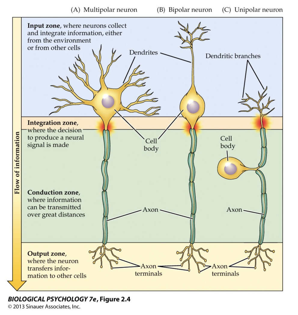
types of neurons
- multipolar
- unipolar
- bipolar
- unipolar
- bipolar
4
New cards
central nervous system
includes brain and spinal cord
5
New cards
peripheral nervous system
includes everything except the brain and spinal cord
6
New cards
glial cells
glial (glue) cells support neuronal activity
7
New cards
astrocytes
star-shaped glial cells with many processes that receive neuronal input and monitor activity
8
New cards
microglial cells (microglia)
small cells that remove debris from injured cells
9
New cards
oligodendrocyte
myelinates axons in the CNS
- cytoplasm of oligodendrocyte wraps around the axon)
(Schwann cells in PNS)
- cytoplasm of oligodendrocyte wraps around the axon)
(Schwann cells in PNS)
10
New cards
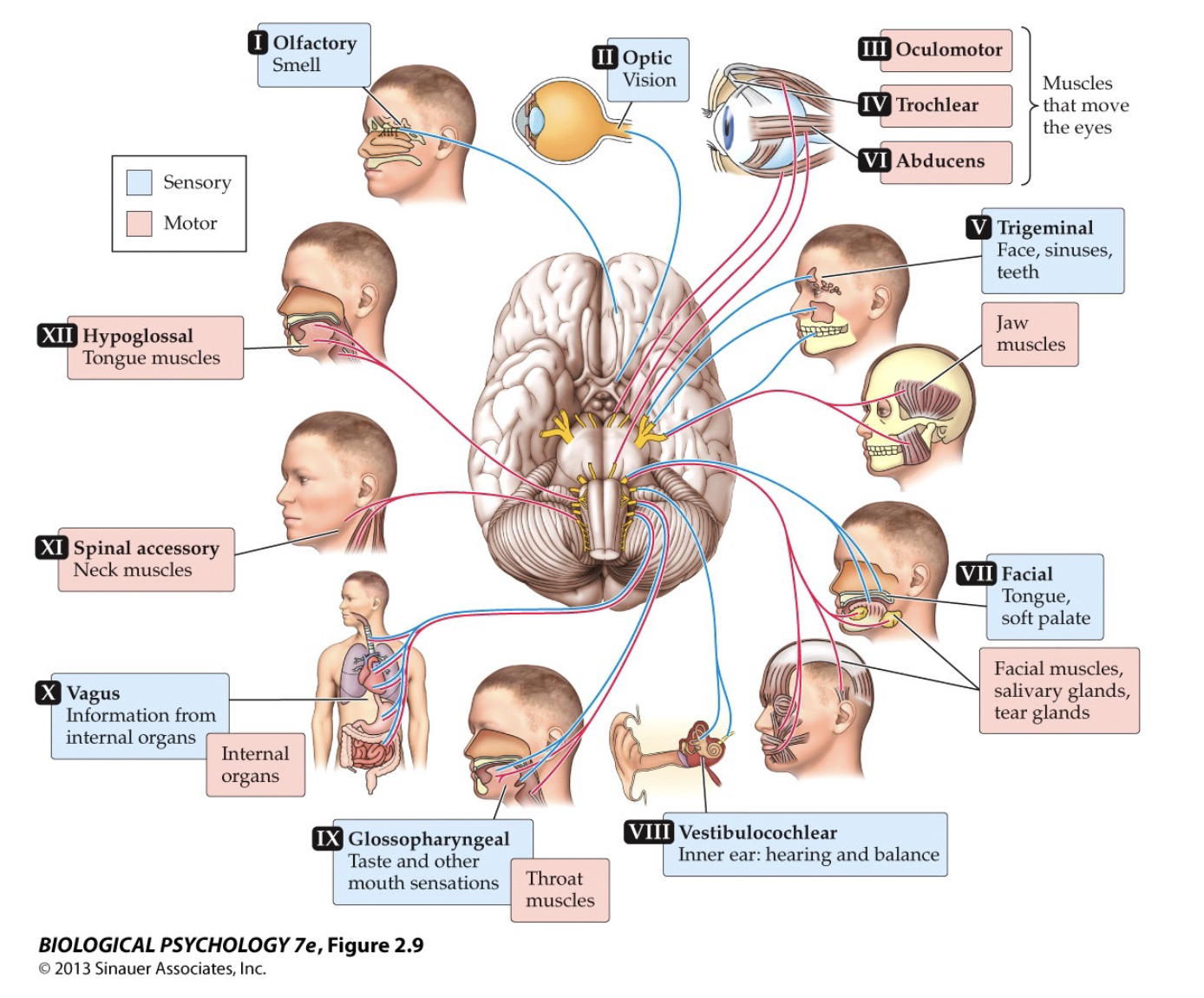
cranial nerves
connected directly to the brain
- 12 total, with sensory and motor functions
- 12 total, with sensory and motor functions
11
New cards
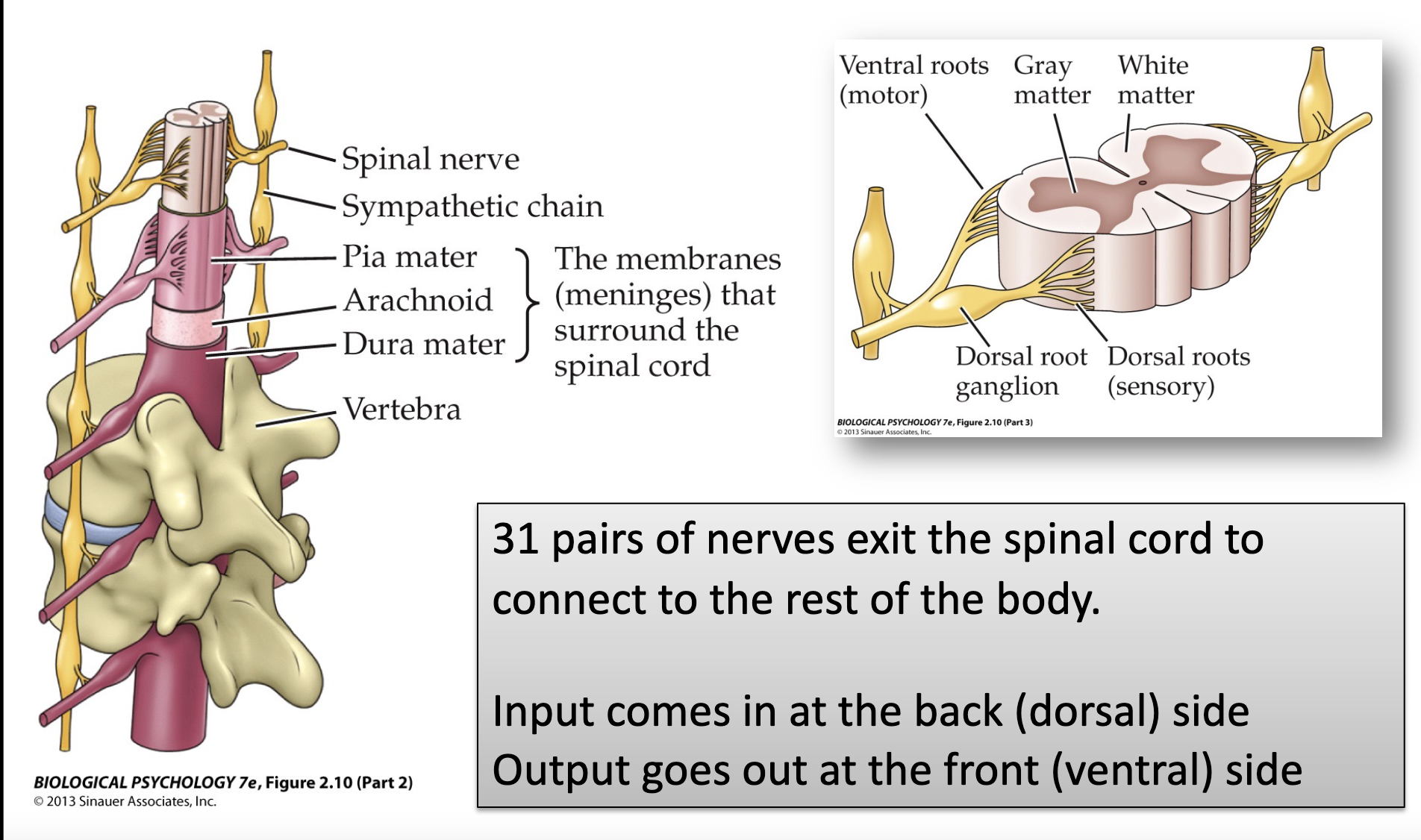
spinal (somatic) nerves
connected to the spinal cord
- 31 pairs
- input comes in the dorsal side
- output goes out ventral side
- 31 pairs
- input comes in the dorsal side
- output goes out ventral side
12
New cards
superior/inferior
superior up, towards the top of the skull
inferior = down, towards the spine
*can also use dorsal/ventral
inferior = down, towards the spine
*can also use dorsal/ventral
13
New cards
dorsal/ventral
dorsal = up, towards top of skull
ventral = down, towards spine
*makes more sense if you imagine humans walking on all 4's
- means the same as superior/inferior
ventral = down, towards spine
*makes more sense if you imagine humans walking on all 4's
- means the same as superior/inferior
14
New cards
rostral/caudal
rostral = font, towards the face
caudal = back, away from the face
*means the same as anterior/posterior
caudal = back, away from the face
*means the same as anterior/posterior
15
New cards
anterior/posterior
anterior = font, towards the face
posterior = back, away from the face
- can also use rostral/caudal
posterior = back, away from the face
- can also use rostral/caudal
16
New cards
medial/lateral
medial = inwards, towards the midline
lateral = outwards, toward the ears
lateral = outwards, toward the ears
17
New cards
autonomic nervous system
division of the peripheral nervous system into sympathetic and parasympathetic
- primarily controls glands and internal organs
- involuntary actions of smooth muscles and heart and glands
- primarily controls glands and internal organs
- involuntary actions of smooth muscles and heart and glands
18
New cards
sympathetic and parasympathetic nervous system
sympathetic: flight or flight
- norepinephrine (adrenaline)
parasympathetic: rest and digest
- acetylcholine
- norepinephrine (adrenaline)
parasympathetic: rest and digest
- acetylcholine
19
New cards
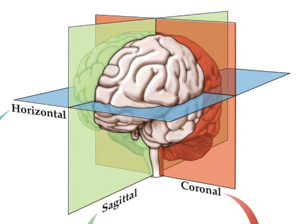
views of the brain
- horizontal
- coronal - vertical, front/back view of brain
- sagittal - vertical, side view of brain
- coronal - vertical, front/back view of brain
- sagittal - vertical, side view of brain
20
New cards
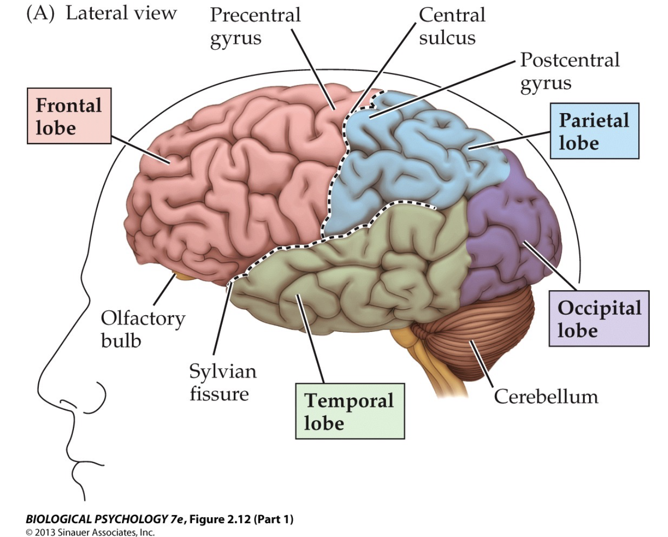
frontal lobe
part of the cerebral cortex on the anterior part of the brain
- borders temporal lobe in the lateral/ventral/posterior side, and the parietal lobe to the posterior sides
- lies right behind the forehead
- borders temporal lobe in the lateral/ventral/posterior side, and the parietal lobe to the posterior sides
- lies right behind the forehead

21
New cards
parietal lobe
part of the cerebral cortex on the posterior, dorsal part of the brain
-borders frontal lobe in the anterior side, temporal lobe in the lateral/ventral side, and the occipital lobe in the posterior/ventral side
- lies below the crown of the head
-borders frontal lobe in the anterior side, temporal lobe in the lateral/ventral side, and the occipital lobe in the posterior/ventral side
- lies below the crown of the head

22
New cards
occipital lobe
part of the cerebral cortex on the posterior part of the head
- borders parietal lobe in the dorsal/anterior side, and the temporal lobe in the lateral/anterior side
- lies in the back of the head
- borders parietal lobe in the dorsal/anterior side, and the temporal lobe in the lateral/anterior side
- lies in the back of the head
23
New cards
temporal lobe
part of the cerebral cortex on the lateral sides of the brain
- borders the frontal lobe in the dorsal/anterior/medial side, the parietal lobe in the dorsal/posterior/medial side, and the occipital lobe in the posterior side
- lies inside the temples of the head
- borders the frontal lobe in the dorsal/anterior/medial side, the parietal lobe in the dorsal/posterior/medial side, and the occipital lobe in the posterior side
- lies inside the temples of the head

24
New cards
olfactory bulb
one of two enlargements at the terminus of the olfactory nerve at the base of the brain just above the nasal cavities
- on the ventral side of the frontal lobes
- on the ventral side of the frontal lobes

25
New cards
precentral gyrus
on the posterior edge of the frontal lobe (border w/ parietal lobe), bounded in the back by the central sulcus
- contains the motor area
- contains the motor area

26
New cards
central sulcus
sulcus dividing the frontal and parietal lobes
- anterior side: precentral gyrus (frontal lobe)
- posterior side: postcentral gyrus (parietal lobe)
- anterior side: precentral gyrus (frontal lobe)
- posterior side: postcentral gyrus (parietal lobe)

27
New cards
postcentral gyrus
on the anterior edge of the parietal lobe (border w/ frontal lobe), bounded in the front by the central sulcus

28
New cards
sylvian fissure
separates the temporal lobe from the frontal and parietal lobes
- dorsal to the temporal lobe
- dorsal to the temporal lobe

29
New cards
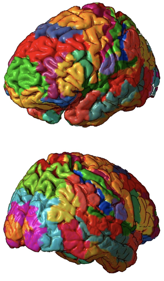
gyrus and sulcus
gyri (singular - gyrus): the folds or bumps in the brain
sulci (singular - sulcus): the indentations or grooves in the brain
- folding of the cortex increases surface are
sulci (singular - sulcus): the indentations or grooves in the brain
- folding of the cortex increases surface are
30
New cards
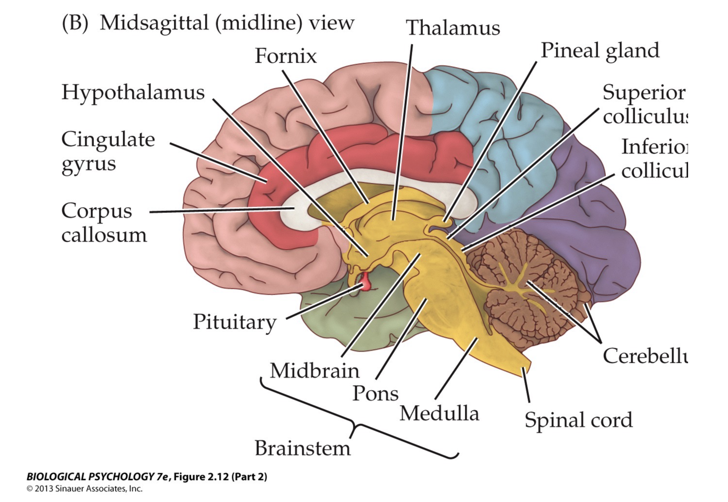
brain structures
31
New cards
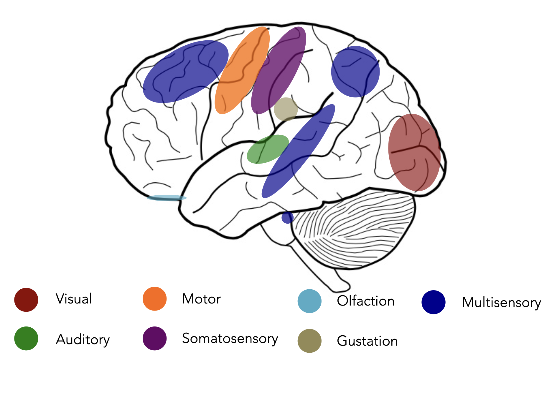
locations of motor, visual, auditory, and somatosensory corteces (and more)
- motor cortex: movement
- somatosensory cortex: somatic sensation (sense of touch)
- visual/striate cortex: vision
- auditory cortex: hearing
- somatosensory cortex: somatic sensation (sense of touch)
- visual/striate cortex: vision
- auditory cortex: hearing
32
New cards
Brodmann areas
division of the brain based on (cyto)architecture
- (cytoarchitecture -cellular composition of the central nervous system's tissues under the microscope)
- (cytoarchitecture -cellular composition of the central nervous system's tissues under the microscope)
33
New cards
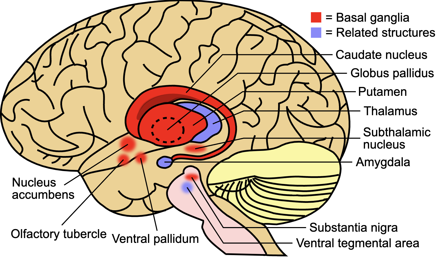
basal ganglia structures
several motor-related structures
- thalamus - sesory processing
- tail of caudate, head of caudate (caudate nucleus)
- globulus pallidus
- nucleus accumbens
- putamen
-subthalamic nucleus
etc.
*amygdala is definitely anatomically connected, but not really part of it
- thalamus - sesory processing
- tail of caudate, head of caudate (caudate nucleus)
- globulus pallidus
- nucleus accumbens
- putamen
-subthalamic nucleus
etc.
*amygdala is definitely anatomically connected, but not really part of it
34
New cards
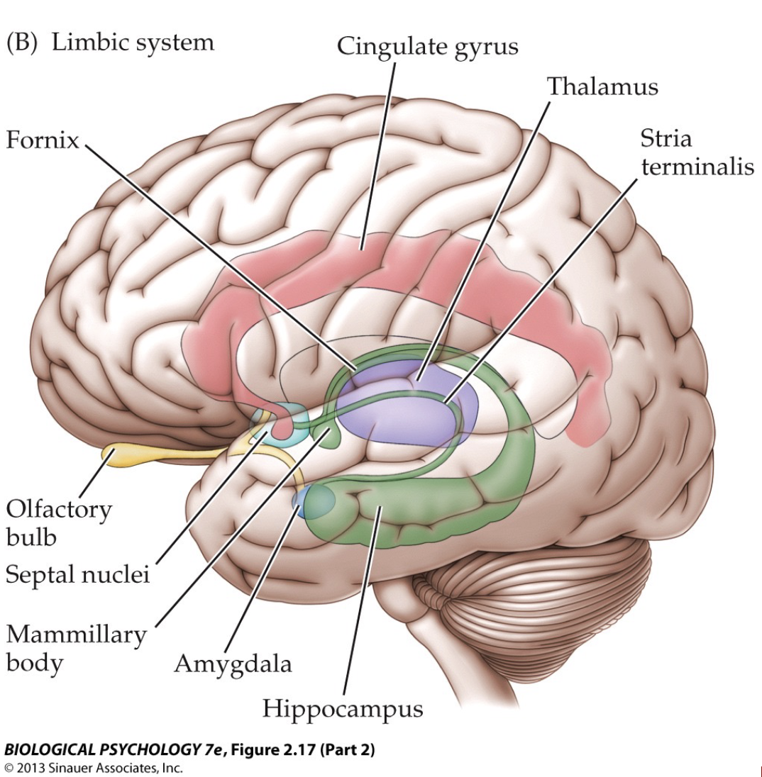
limbic system
several structures related to emotional processing
- hippocampus - very important for memory
- amygdala
-olfactory bulbs
- cingulate gyrus - reward processing
- thalamus
etc.
- hippocampus - very important for memory
- amygdala
-olfactory bulbs
- cingulate gyrus - reward processing
- thalamus
etc.
35
New cards
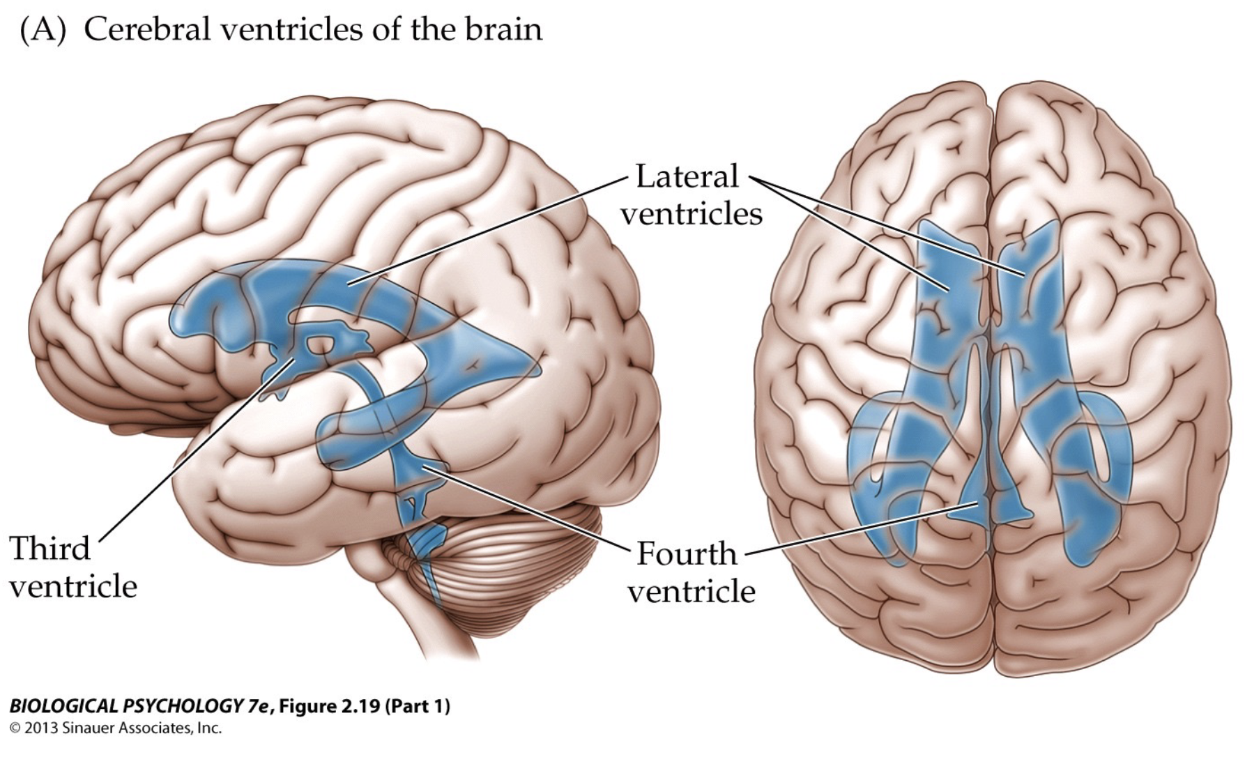
ventricles
protection and supplies
- fluid filled
- shock absorbers
- exchange of nutreints etc. between blood vessels and brain tissue
- choroid plexus crucial for producing cerebrospinal fluid
- network of blood vessels and cells in the ventricles that are covered by a thin layer of cells that make cerebrospinal fluid
-CSF drains out of the bottom of the ventricles and surrounds the brain and spinal cord and it is also gradually recycled into the blood
36
New cards
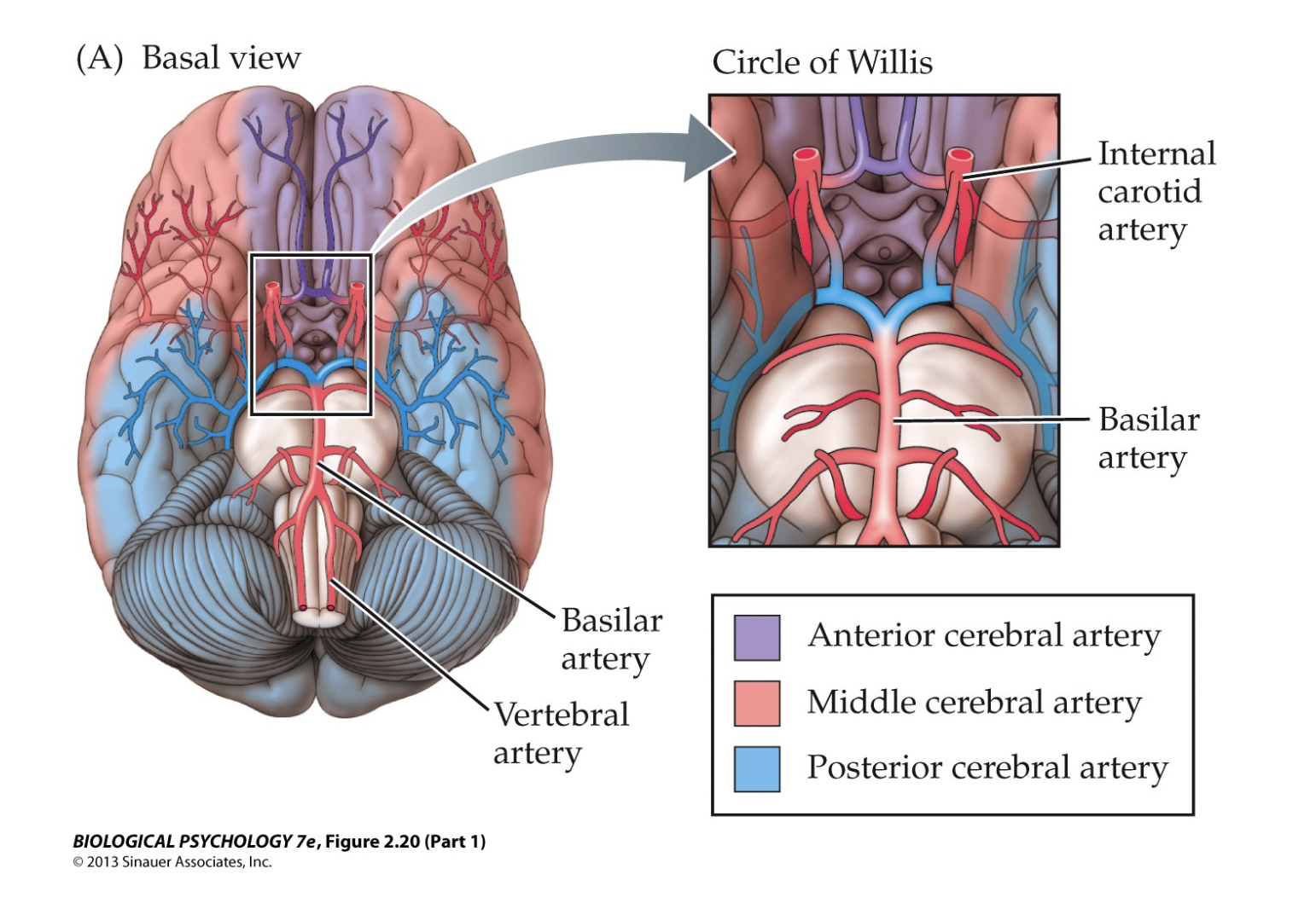
blood vessels
oxygen supply
- internal carotid arteries are connected to many blood vessels in the brain
- anterior cerebral artery - feeds the medial, anterior, and dorsal parts of brain
- middle cerebral artery - feeds the lateral parts of the brain
- posterior cerebral artery - feeds dorsal and posterior parts of the brain
- circle of Willis: the joining area of several arteries at the bottom (inferior) side of the brain (forms a 'circle')
37
New cards
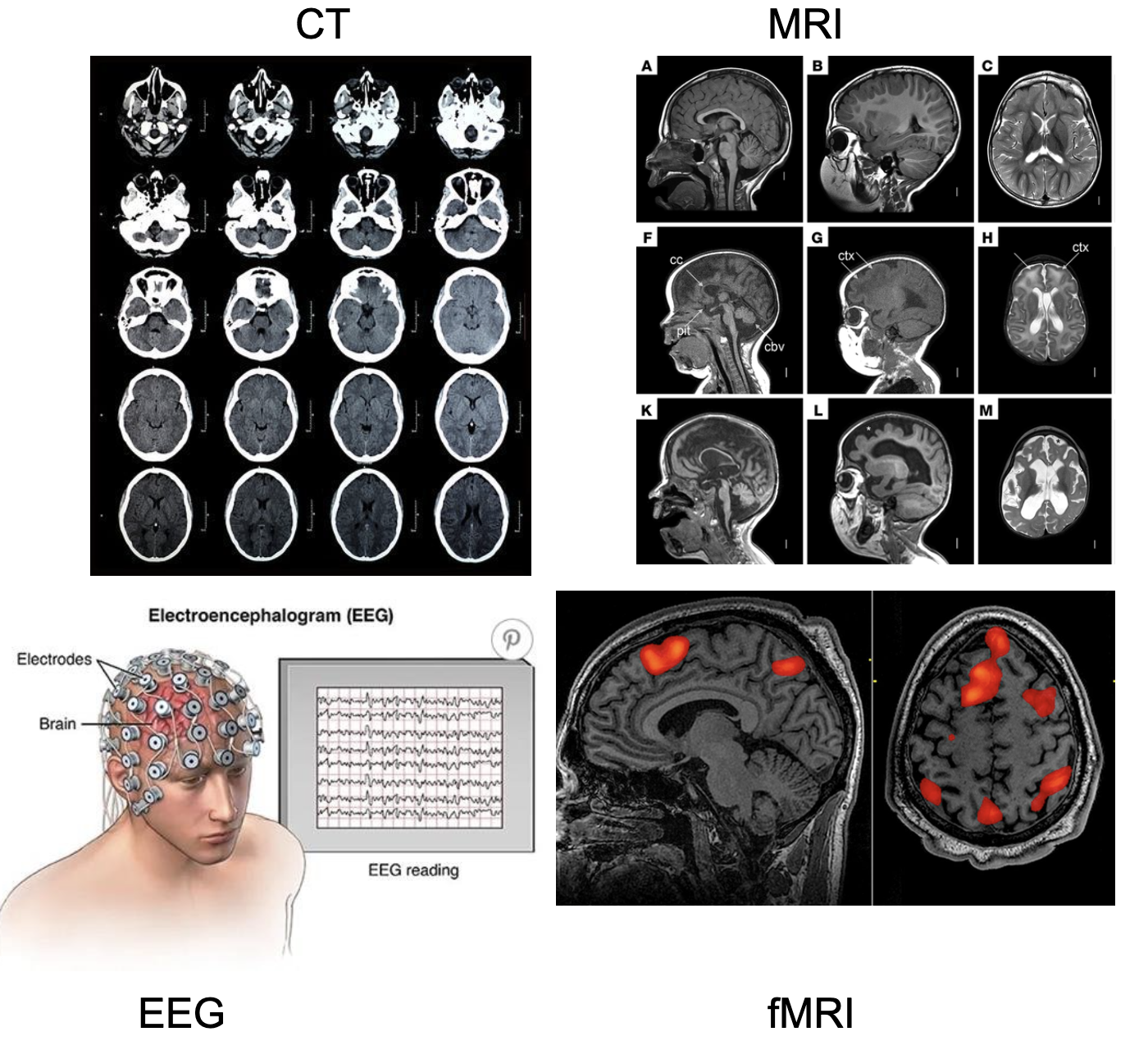
brain imaging techniques
- MRI (magnetic resonance imaging) - uses strong magnetic fields, magnetic field gradients, and radio waves to generate images of the organs in the body
-fMRI (functional MRI) - measures brain activity by detecting changes associated with blood flow
- CT (computed tomography) - combines a series of X-ray images taken from different angles around the body and uses computer processing to create cross-sectional images (slices) of the bones, blood vessels and soft tissues inside the body
- EEG (electroencephalogram) - records/measures electrical activities through electrodes attached to the scalp
38
New cards
neurophysiology
the study of electrical and chemical processes in neurons
- information flow:
- within neurons - electrical signals
- between neurons - chemical signals
- information flow:
- within neurons - electrical signals
- between neurons - chemical signals
39
New cards
electrical signaling
- fast over long distances
- neurons contain mostly anions --> inside of a neuron more negatively charged than the outside
- neurons contain mostly anions --> inside of a neuron more negatively charged than the outside
40
New cards
ion channels
membrane-spanning transport protein for ions
- cell membrane itself is impermeable for water soluble molecules such as ions that are present intra- and extracellularly
- selective permeability of a neuron
- ex: non-gated potassium (K+) channels selectively allow K+ into the cell
- passive transport: does not cost energy
- cell membrane itself is impermeable for water soluble molecules such as ions that are present intra- and extracellularly
- selective permeability of a neuron
- ex: non-gated potassium (K+) channels selectively allow K+ into the cell
- passive transport: does not cost energy
41
New cards
membrane resting potential
difference in electrical potential across the membrane of a cell when it is inactive - about -65mV in neurons
- reflects 'balancing act' between opposing forces that drive K+ in and out of the cell
- diffusion - movement of molecules from areas of high concentration to low concentration
- electrostatic forces - tendency of charged molecules or ions to move towards areas with the opposite charge
- ions constantly moving back and forth across membrane through ion channels and pumps (ex. K+ channels, Na+/K+ pumps)
- but, at some point, the opposing forces are at equilibrium and there is no *net* flow
42
New cards
important ions for neural signaling
- sodium: Na+
- potassium: K+
- chloride: Cl-
- calcium: CA++ or CA2+
- magnesium: Mg++ or Mg2+
at rest: more K+ inside the cell
more Na+ and Cl- outside the cell
- potassium: K+
- chloride: Cl-
- calcium: CA++ or CA2+
- magnesium: Mg++ or Mg2+
at rest: more K+ inside the cell
more Na+ and Cl- outside the cell
43
New cards
sodium/potassium pump
(Na+/K+)
(Na+/K+)
exchanges 3 Na+ for 2 K+ ions
- K+ is pumped in
- Na+ is pumped out
*active transport: costs energy
- can move ions across membrane, creating a large concentration difference (gradient)
- K+ is pumped in
- Na+ is pumped out
*active transport: costs energy
- can move ions across membrane, creating a large concentration difference (gradient)
44
New cards
equilibrium potential
voltage difference across a permeable membrane needed to counterbalance diffusion forces
- The electrical potential at which a given concentration gradient across the membrane is stable, when the membrane is permeable for X.
- voltage difference needed to counteract diffusion forces
- The electrical potential at which a given concentration gradient across the membrane is stable, when the membrane is permeable for X.
- voltage difference needed to counteract diffusion forces
45
New cards
membrane resting potential of a neuron (value)
about ~65 mV
- K+, Cl-, and Na+ are all fighting to reach their equilibrium potential, but there are more passive K+ channels, than Cl- and Na+ channels, so K+ is "winning"
- K+ has an EP of -85 mV, but the movement of Cl- and Na+ brings the MRP to about -65mV
- there is constant movement of ions passively through channels, since non of the ions are at their EP
46
New cards
hyperpolarization
increasing negativity of membrane potential
47
New cards
depolarization
decreasing negativity of membrane potentail
48
New cards
generating a potential
-potentials/neuronal electrical activity - deviations from the resting potential
- caused by temporarily changing, very locally, the permeability for an ion by opening gated ion channels
- other ion channels briefly open (ex. Na+), so the membrane potential changes
-gets closer to EP of K+, +67 mV ---> depolarization ---> potential
- caused by temporarily changing, very locally, the permeability for an ion by opening gated ion channels
- other ion channels briefly open (ex. Na+), so the membrane potential changes
-gets closer to EP of K+, +67 mV ---> depolarization ---> potential
49
New cards
electrotonic conduction
passive propagation of a potential
- signals spread along the membrane in all directions extremely fast
- but, it also leaks away because of non-gated ion channels that are trying to restore membrane resting potential (it is 'lossy')
- passive process, and signals decrease over space
- signals spread along the membrane in all directions extremely fast
- but, it also leaks away because of non-gated ion channels that are trying to restore membrane resting potential (it is 'lossy')
- passive process, and signals decrease over space
50
New cards
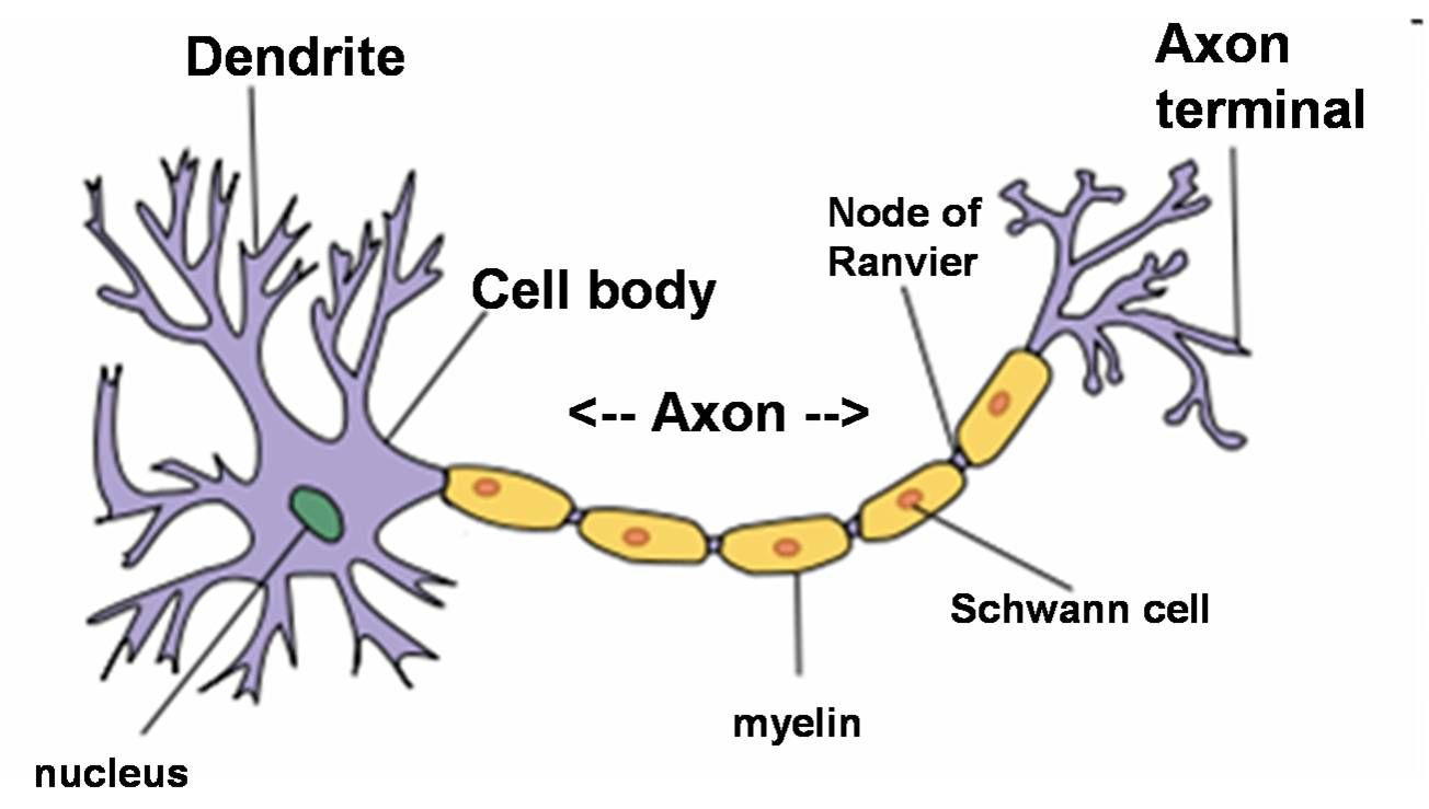
structure of a (multipolar) neuron
- dendrites
- cell body + nucleus
- axon hillock - base of the axon where action potential starts
- axon
- axon terminal
- cell body + nucleus
- axon hillock - base of the axon where action potential starts
- axon
- axon terminal
51
New cards
graded local potentials
spread passively from synapses (dendrites) to axon hillock, and from the tend of the axon to axon terminals
- electrotonic conduction
- flexible in shape and magnitude
- electrotonic conduction
- flexible in shape and magnitude
52
New cards
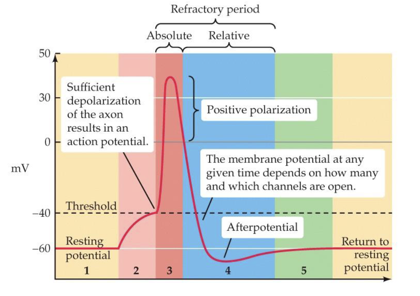
action potential
brief but radical changes in polarization that send an electrical charge down the neuron
- threshold: about -40mV
- fundamental unit for electrical communication
- uniform in shape and magnitude
- larger depolarizations produce more action potentials, not bigger or longer ones
- information is encoded in the (change in) frequency of action potentials
- action potentials propagate actively over the axon
53
New cards
generating an action potential
1. neurotransmission causes depolarization at the synapse
2. electrotonic conduction of graded potential (charge flows through the inside of the neuron)
3. axon hillock: voltage-gated Na+ channels open ---> action potential starts here
2. electrotonic conduction of graded potential (charge flows through the inside of the neuron)
3. axon hillock: voltage-gated Na+ channels open ---> action potential starts here
54
New cards
absolute vs. relative refractory periods
after an action potential passes through, it is impossible (or difficult) for that section of membrane to fire again
- absolute refractory period: Na+ channels already open, or they are INactivated for 1ms
- a new action potential absolutely can not be produced
- relative refractory period: when Na+ channels DEactivate, the membrane potential is hyperpolarized at -80 mV, so a larger depolarization is needed to trigger a new action potential
55
New cards
Hodgkin-Huxley cycle
- Na+ channels open at threshold membrane potential (about -40 mV) --> Na+ rushes into the cell
- after about 1 millisecond (at +30 mV) the Na+ channel is inactivated (not closed) --> absolute refractory period
- now K+ channels open --> K+ leaves the cell (concentration and electrical gradient)
- membrane potential decreases, then becomes negative until about -80 mV, and K+ channels close
- Na+ channels are now deactivated (closed) ---> relative refractory period
- normal resting potential is restored at -65 mV
56
New cards
action potential propagation
electrotonic conduction not sufficient
- the depolarization of an action potential is strong enough to cause threshold depolarization in the next adjacent segment --> the action potential is regenerated along the axon
- the action potential cannot flow 'backwards' because the previous segment is in the refractory period
- the depolarization of an action potential is strong enough to cause threshold depolarization in the next adjacent segment --> the action potential is regenerated along the axon
- the action potential cannot flow 'backwards' because the previous segment is in the refractory period
57
New cards
myelin
oligodendrocytes produce myelin that insulate the axon (cytoplasm of oligodendrocyte wraps around the axon)
- node of Ranvier - small gaps between myelinated sections
- action potential 'jumps' from node to node
- action potential can travel farther in a myelinated axon
-prevents 'leakage' of ions during electrotonic conduction
- very important for communication between neurons
- node of Ranvier - small gaps between myelinated sections
- action potential 'jumps' from node to node
- action potential can travel farther in a myelinated axon
-prevents 'leakage' of ions during electrotonic conduction
- very important for communication between neurons
58
New cards
saltatory conduction
action potential 'jumps' from node to node (nodes of ranvier)
- electrotonic conduction of AP along myelinated sections, then the AP is regenerated at the node
- faster way to travel down an axon than traveling in an axon without myelin.
- electrotonic conduction of AP along myelinated sections, then the AP is regenerated at the node
- faster way to travel down an axon than traveling in an axon without myelin.
59
New cards
toxins
some toxins block ion channels and prevent neuronal signaling
- ex: tetrodoxin (found in puffer fish) - voltage-gated Na+ channel blocker
- ex: tetrodoxin (found in puffer fish) - voltage-gated Na+ channel blocker
60
New cards
multiple sclerosis
autoimmune disease in which the immune system attacks the myelin sheath or the cells that produce and maintain it
- particularly the optic nerve, the deep cerebral white matter, the cerebellar peduncles, and particular parts of the brainstem and spinal cord.
- particularly the optic nerve, the deep cerebral white matter, the cerebellar peduncles, and particular parts of the brainstem and spinal cord.
61
New cards
how many neurons are there in the brain?
100 billion neurons
-10,000 connections each
- 1 quadrillion (10^15, or 1,000,000,000,000) total synapses in the brain!
-10,000 connections each
- 1 quadrillion (10^15, or 1,000,000,000,000) total synapses in the brain!
62
New cards
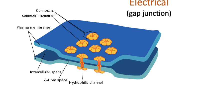
2 types of synapses
- electrical (gap junction)
- chemical
- chemical
63
New cards
electrical synapse (gap junction)
current just flows between cells - it is 'passive'
- bi-directional
- fast (no delay)
- used for synchronization (ex. heart cells, release of neural hormomes - a bunch of cells need to do the same thing)
- about ~2 nanometers of space between neurons
- minority in the brain
- disadvantage: no computations take place. not doing anything complex, just creating more of the same signal across more cells.
64
New cards
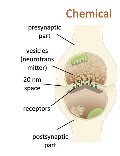
chemical synapse (definition)
new potential created in postsynaptic cell - it is 'active'
- one-directional
- slow - about ~0.5-1.0 ms of delay between arrival of the action potential at axon terminal and the creation of a postsynaptic action potential.
- involves neurotransmitters
- used for integration/computation in the postsynaptic neuron
- one-directional
- slow - about ~0.5-1.0 ms of delay between arrival of the action potential at axon terminal and the creation of a postsynaptic action potential.
- involves neurotransmitters
- used for integration/computation in the postsynaptic neuron
65
New cards
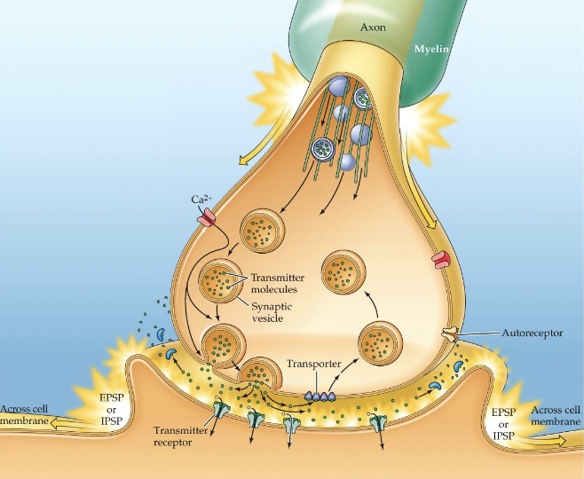
chemical synapse (overview)
presynaptic part:
- depolarization from action potential triggers voltage-gated Ca2+ channels
-Ca2+ influx leads to vesicles releasing neurotransmitter into the cleft
synaptic cleft:
- released neurotransmitter binds to receptor on postsynaptic membrane
- neurotransmitter is then either degraded by enzymes or taken up again in the presynaptic part
postsynaptic part:
- neurotransmitter activates the receptor to do something, for example to open an ion channel, leading to a postsynaptic potential, either excitatory (EPSP) or inhibitory (IPSP).
66
New cards
neurotransmitter degradation and reuptake
degradation: neurotransmitters are rapidly broken down / deactivated by a special enzyme
-NTs may be recycled to make more NT in the axon terminal
reuptake: neurotransmitters are rapidly cleared from the synaptic cleft by being taken up into the presynaptic cell
- special receptors (transporters) bring the NT back inside the cell
- may be repacked into newly formed synaptic vessicles
-NTs may be recycled to make more NT in the axon terminal
reuptake: neurotransmitters are rapidly cleared from the synaptic cleft by being taken up into the presynaptic cell
- special receptors (transporters) bring the NT back inside the cell
- may be repacked into newly formed synaptic vessicles
67
New cards
excitatory post synaptic potential (EPSP)
local postsynaptic membrane DEpolarization in the postsynaptic neuron
- caused by excitatory synapses
- pushes the postsynaptic cell a bit closer to threshold membrane potential (Na channels open, Na+ into cell)
-EPSPs caused by many neurons that converge on the postsynaptic cell --> action potential
- caused by excitatory synapses
- pushes the postsynaptic cell a bit closer to threshold membrane potential (Na channels open, Na+ into cell)
-EPSPs caused by many neurons that converge on the postsynaptic cell --> action potential
68
New cards
inhibitory postsynaptic potential
local postsynaptic membrane HYPERpolarization in the postsynaptic neuron
- caused by inhibitory synapses
- pulls the postsynaptic cell further away from threshold membrane potential (Cl channels open, Cl- into cell)
- caused by inhibitory synapses
- pulls the postsynaptic cell further away from threshold membrane potential (Cl channels open, Cl- into cell)
69
New cards
postsynaptic potential (PSP)
neurotransmitters released into the synapse briefly alter the membrane potential of the postsynaptic cell
- *graded* potential: bigger stimulus --> more hyper/depolarization. longer stimulus --> longer lasting hyper/depolarization (no increase in size)
- PSPs last much longer than action potentials (more than 10 ms)
- (a neuron can receive 100s of synapses from other cells --> 100s or 1000s of PSPs
- a balance of excitatory and inhibitory ESPS is vital in neural processing of information (over-excitation --> seizure. under-excitation --> coma/death)
- excitatory/inhibitory effects are sometimes caused by which neurotransmitter is present.
- action potential generation in the postsynaptic cell is determined by the (im)balance of the number of excitatory and inhibitory signals received.
70
New cards
botox
prevents fusion of vesicles to the presynaptic membrane by splitting SNARE proteins, hence no transmitter release (exocytosis)
- botox is a neuromodulator
- botox is a neuromodulator
71
New cards
neurotransmitter
a chemical released from the presynaptic axon terminal that serves as the basis of communication between neurons
- generally easy to synthesize from amino acids in diet
- amino acids are most common NT in the brain
- amines - based on modifications of a single amino acid by enzymes
- generally easy to synthesize from amino acids in diet
- amino acids are most common NT in the brain
- amines - based on modifications of a single amino acid by enzymes
72
New cards
amino acid neurotransmitters
glutamate: fast excitatory, memory
- main excitatory NT in the brain
GABA: fast inhibitory, memory
- main inhibitory NT in the brain
- subtypes of GABA receptors exhibit quite different properties
- GABA A, GABA B, GABA C receptors
(gamma-aminobutyric acid)
- main excitatory NT in the brain
GABA: fast inhibitory, memory
- main inhibitory NT in the brain
- subtypes of GABA receptors exhibit quite different properties
- GABA A, GABA B, GABA C receptors
(gamma-aminobutyric acid)
73
New cards
amine neurotransmitters
dopamine (DA): reward,
- involved in schizophrenia and Parkinson's disease
norepinephrine (NE)
epinephrine (EP)
serotonin (5-HT): mood, sleep depression
- involved in schizophrenia and Parkinson's disease
norepinephrine (NE)
epinephrine (EP)
serotonin (5-HT): mood, sleep depression
74
New cards
acetyl choline (ACh)
neurotransmitter: neuromuscular
- first NT to be identified
- receptors:
nicotinic (nACh): ionotropic (muscles)
and muscarinic (mACh) metabotropic
- Alzheimer's disease: widespread loss of cholinergic neurons
- first NT to be identified
- receptors:
nicotinic (nACh): ionotropic (muscles)
and muscarinic (mACh) metabotropic
- Alzheimer's disease: widespread loss of cholinergic neurons
75
New cards
ligand
a molecule that can bind to a receptor protein
- can activate or block it
endogenous ligands: neurotransmitters and hormones made inside of the body
- agonist
exogenous ligands: drugs and toxins from outside the body
- receptor agonist
- competitive antagonist
- non-competitive agonist/antagonist (neuromodulator): does not bind to the same receptor site
76
New cards
antagonist (ligand)
an exogenous ligand that all together stops the receptor from producing a response
- ex: poisons can block acetylcholine (ACh) receptors in the brain
- ex: poisons can block acetylcholine (ACh) receptors in the brain
77
New cards
agonist (ligand)
molecules (can be drugs) that bind to receptors and mimic the action of a neurotransmitter.
- ex: nicotine
- ex: nicotine
78
New cards
types of ion channels
-non-gated
-voltage-gated
-ligand-gated (also called chemically-gated ion channels or ionotropic receptors)
-stretch gated (mechanosensitive)
-voltage-gated
-ligand-gated (also called chemically-gated ion channels or ionotropic receptors)
-stretch gated (mechanosensitive)
79
New cards
non-gated ion channels
- resting membrane potential
- always open
- always open
80
New cards
voltage-gated ion channels
- triggered by a voltage change
- can open and close
- can open and close
81
New cards
mechanosensitive (stretch-gated) ion channels
sensitive to mechanical stress
82
New cards
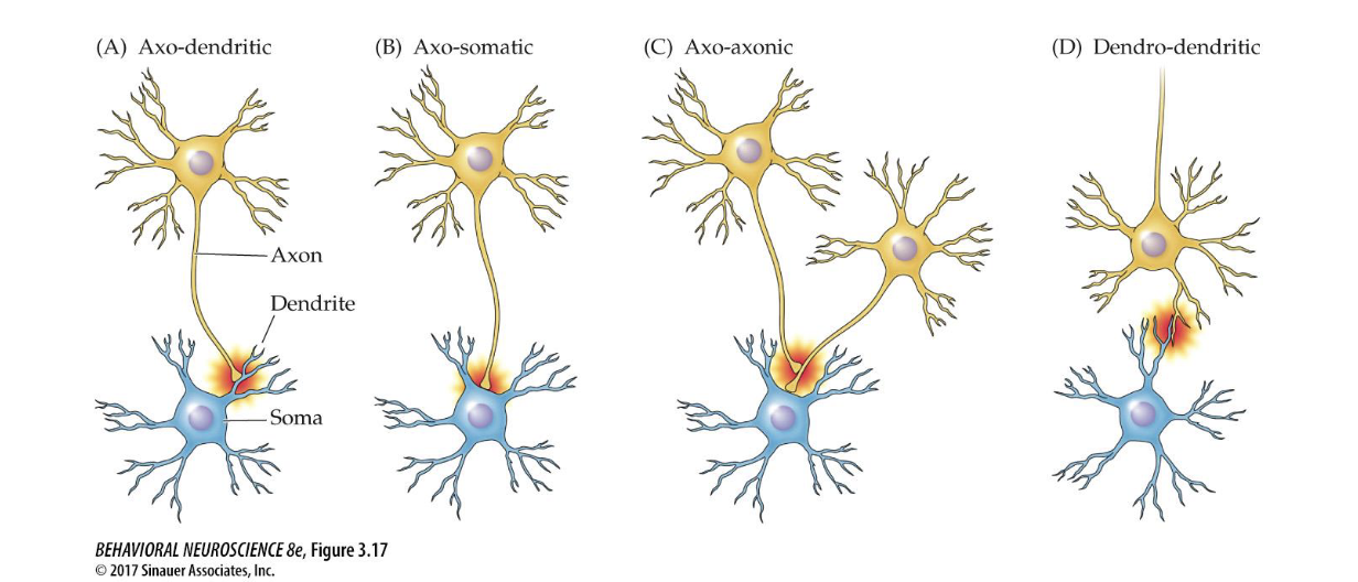
different chemical synapses
- axo-dendritic
- axo-somatic
- axo-axonic: allows the presynaptic neuron to regulate neurotransmitter release of the postsynaptic neuron
- dendro-dendritic - allows coordination of cells' activities
- axo-somatic
- axo-axonic: allows the presynaptic neuron to regulate neurotransmitter release of the postsynaptic neuron
- dendro-dendritic - allows coordination of cells' activities
83
New cards
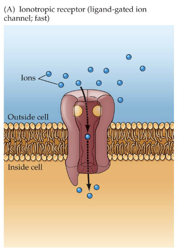
ionotropic receptors
(also called chemically-gated or ligand-gated ion channels)
postsynaptic receptor proteins that include an ion channel, which is opened when an agonist binds to it .
- fast communication
- open when some chemical binds to them (could be a neurotransmitter or some 2nd messenger)
postsynaptic receptor proteins that include an ion channel, which is opened when an agonist binds to it .
- fast communication
- open when some chemical binds to them (could be a neurotransmitter or some 2nd messenger)
84
New cards
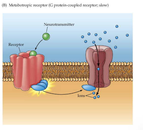
metabotropic receptors
postsynaptic receptor proteins that do not contain an ion channel, but may (when activated) activate a G-protein
- G-protein: acts as a '2nd messenger' inside the cell. it amplifies the effect of the 1st messenger (the neurotransmitter) and can initiate processes that affect postsynaptic membrane potential
- slower communication
- amplify and prolong synaptic signals
- G-protein: acts as a '2nd messenger' inside the cell. it amplifies the effect of the 1st messenger (the neurotransmitter) and can initiate processes that affect postsynaptic membrane potential
- slower communication
- amplify and prolong synaptic signals
85
New cards
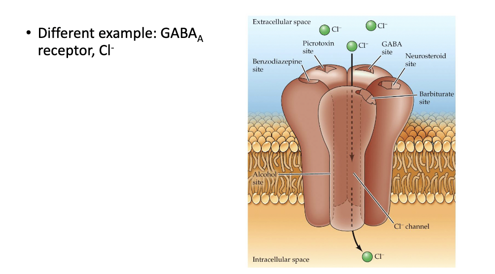
different example of postsynaptic receptor: GABA A receptor, Cl- channel (and example of neuromodulation)
- alcohol and bind to it (noncompetitive ligand) and modulate the effect of neurotransmitters binding to the receptors
- alcohol is a neuromodulator
- alcohol is a neuromodulator
86
New cards
up-regulation
compensatory increase in receptor availability at the synapse of a neuron
87
New cards
down-regulation
compensatory decrease in receptor availability at the synapse of a neuron
88
New cards
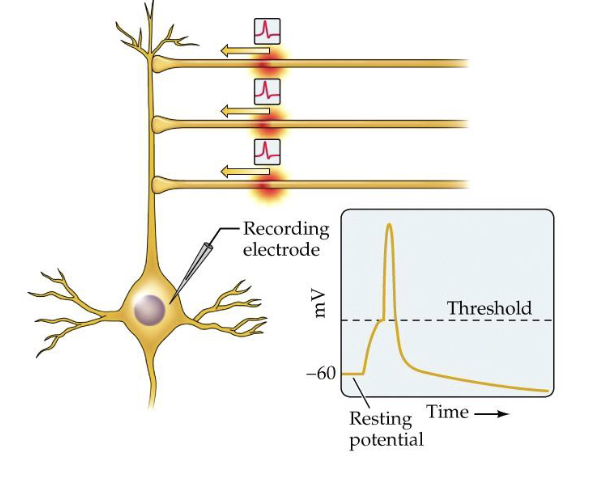
spatial summation
summation of postsynaptic potentials from different synapses (different physical locations across the cell body) that overlap in time
- physically closer together --> increased summation (and vice versa)
- physically closer together --> increased summation (and vice versa)
89
New cards
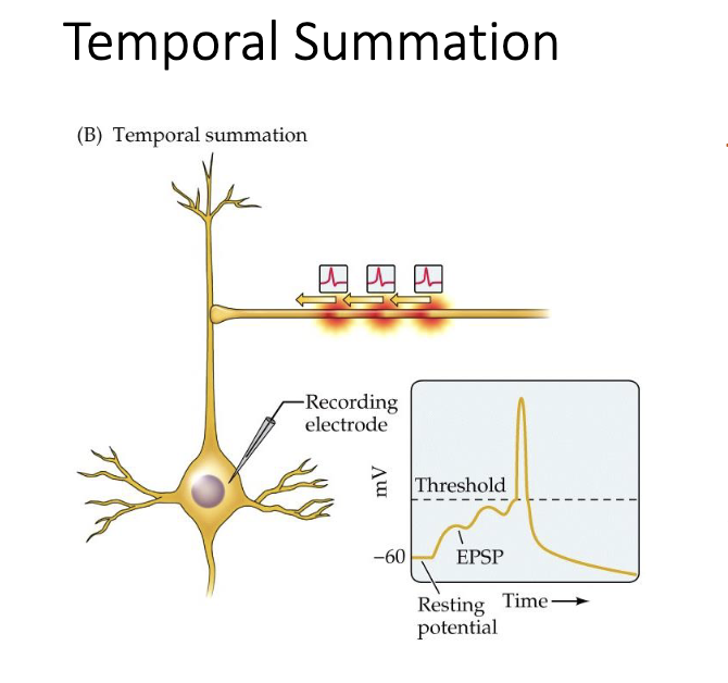
temporal summation
summation of potentials from one synapse that overlap in time
- closer together in time --> increased summation (and vice versa)
- closer together in time --> increased summation (and vice versa)
90
New cards
information processing
- graded postsynaptic potentials spread passively from the dendrites ober the cell body towards the axon hillock (in multi-and bi-polar cells)
- if a depolarization is strong enough (exceeds the threshold) reaches the axon hillock --> action potential produced
- spatial and temporal summation determine whether an action potential is triggered
- if a depolarization is strong enough (exceeds the threshold) reaches the axon hillock --> action potential produced
- spatial and temporal summation determine whether an action potential is triggered
91
New cards
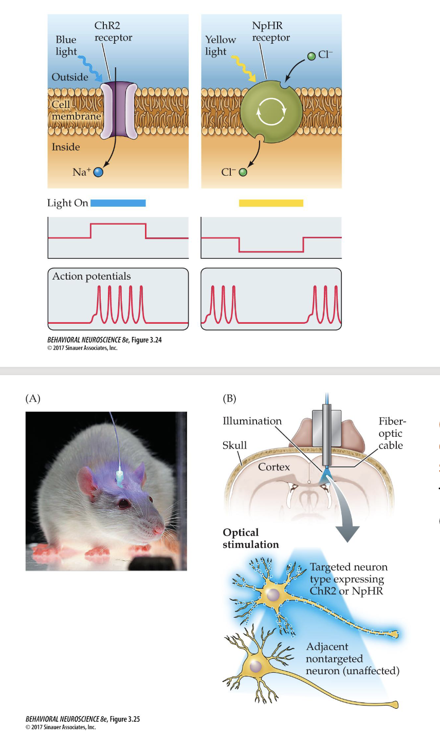
optogenetics
inducing EPSPs and IPSPs experimentally
- advantage over electrical stimulation: targets specific cells, controlled PSPs
- advantage over electrical stimulation: targets specific cells, controlled PSPs
92
New cards
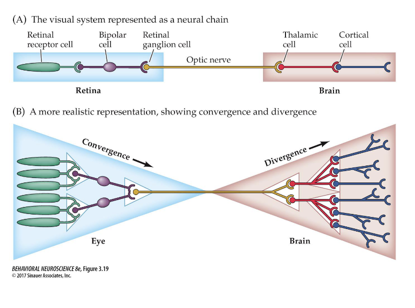
convergence and divergence
convergence: neuronal connections in which many cells send signals to a single cell
- range fractionation: information from receptors of different sensitivities is sent to one cell and integrated to code for the intensity of a stimulus
divergence: one cell sends signals to many other cells
- allows for an impulse to be amplified in order to produce a response over a widespread area
93
New cards
neural chain
a simple kind of neural circuit in which neurons are attached linearly, end to end
- ex: knee jerk reflex: sensory neuron synapses directly onto motor neuron (synapse is in the spinal cord). involves myelinated axons of large diameter.
- ex: knee jerk reflex: sensory neuron synapses directly onto motor neuron (synapse is in the spinal cord). involves myelinated axons of large diameter.
94
New cards
analog- vs. digital-like signals
- analog: vary in strength (ex. graded potential)
- digital: all-or-none, vary in frequency (ex. action potential)
- digital: all-or-none, vary in frequency (ex. action potential)
95
New cards
event related potentials (ERPs)
(also called evoked potential)
gross potential changes evoked by a discrete sensory stimulus, such as light flashes
- ex: auditory evoked potentials can be recorded with an EEG, and can diagnose deafness or hearing impairments in infants.
gross potential changes evoked by a discrete sensory stimulus, such as light flashes
- ex: auditory evoked potentials can be recorded with an EEG, and can diagnose deafness or hearing impairments in infants.
96
New cards
neurochemistry
branch of neuroscience concerned with the fundamental composition and processes OF the nervous system
- endogenous processes
- endogenous processes
97
New cards
neuropharmacology /(psychopharmacology)
the scientific field concerned with the discovery and study of compounds that selectively AFFECT the function of the nervous system
98
New cards
neuropeptide neurotransmitters
endorphins, orexin, oxytocin
99
New cards
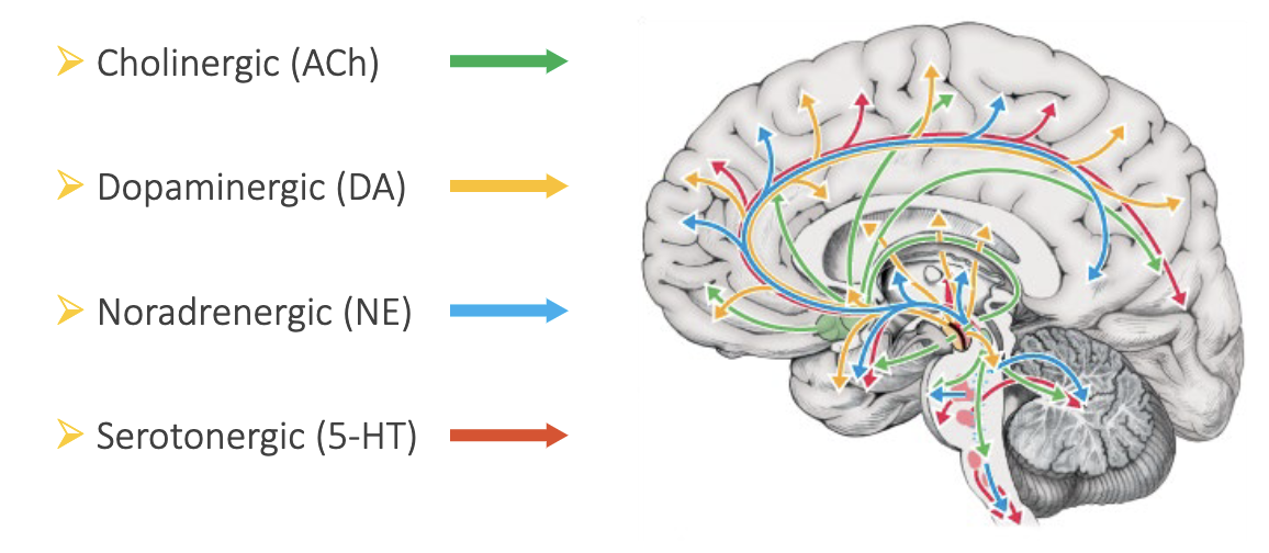
four major pathways
- Cholinergic (ACh)
- Dopaminergic (DA)
- Noradrenergic (NE)
- Serotonergic (5-HT)
- Dopaminergic (DA)
- Noradrenergic (NE)
- Serotonergic (5-HT)
100
New cards
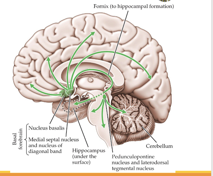
Cholinergic pathways
acetylcholine (ACh)
From: basal forebrain, (PPT/LDT - pedunculopontine nucleus and laterodorsal tegmental nucleus)
To: hippocampus, amygdala, cortex
Involved in muscle control and memory
- Alzheimer's disease: ACh deficiency
nicotinic receptors: ionotropic
- important in muscular system
- curare (antagonist) --> paralysis
muscarinic receptors: metabotropic
- atropine (antagonist) --> confusion, memory problems