Exam 2 (Part 2, 3/5, 3/7 Gastrulation+Neural Induction
1/28
There's no tags or description
Looks like no tags are added yet.
Name | Mastery | Learn | Test | Matching | Spaced | Call with Kai |
|---|
No analytics yet
Send a link to your students to track their progress
29 Terms
When does gastrulation occur in humans?
2nd-3rd week post-fertilization, after cleavage stages
What are the 3 major steps in gastrulation and the end result?
1) Massive cell movement leading to rearrangement or formation of germ layers
2) Induction of nervous system
3) Formation(verts)/elaboration(inverts) of body axes
End result: organism has basic body plan, A-P, D-V, L-R axes
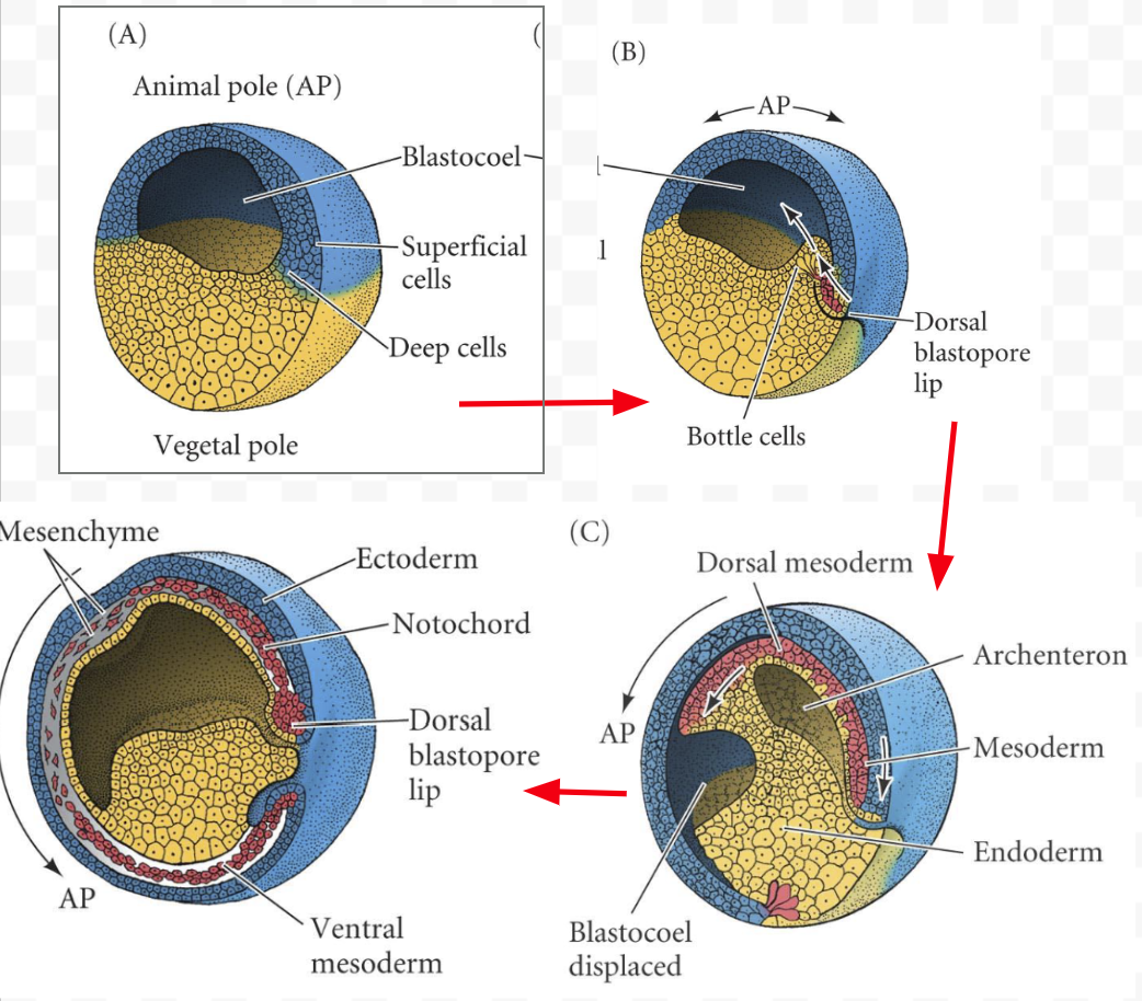
Gastrulation in amphibians (draw start, middle, end)
Dorsal mesoderm invaginates between the endoderm, then on the ventral side also.
By the end, should be ectoderm on outside only, mesoderm a concentric sphere around the endoderm which is inside.
Forms archenteron, the body cavity that becomes mouth-anus path kept throughout life
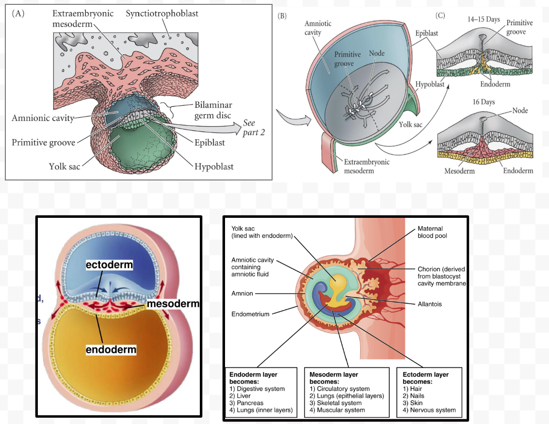
Gastrulation in mammals
dual layers → 3 layers. Mesoderm goes through node, is induced as it goes (later than amphibians, but using same proteins)
Embryo secreting VegF to attract vascular supply, local suppression of immune system
Pre-gastrulation embryo in mammals
-2 layers
-not spherical like amphibians, more flat out
-mesoderm invaginating btwn endoderm and ectoderm still
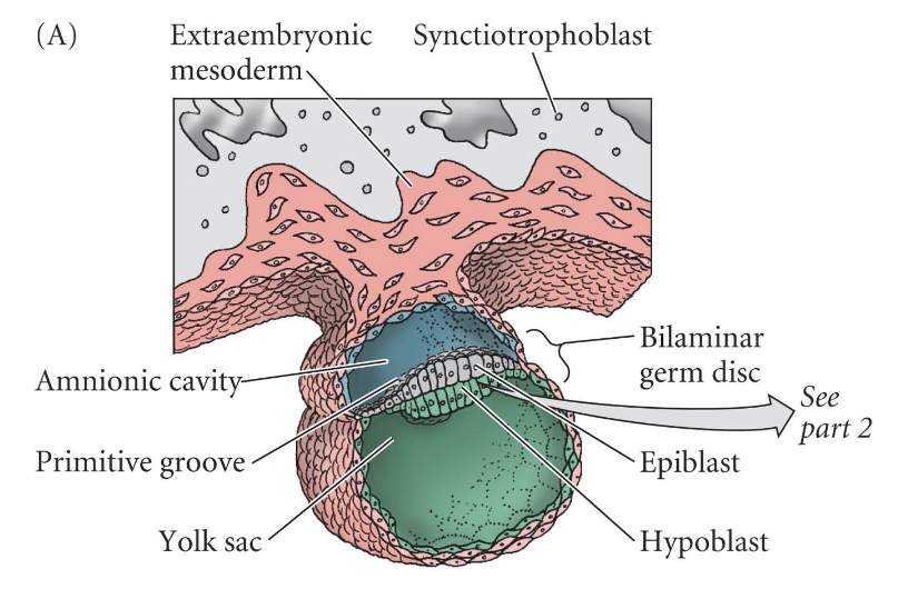
Mammal gastrulation drawings
Early (pre-organizer) experiments for neural induction
-demonstrated the key role that the dorsal mes-endoderm plays in the induction of the nervous system and the elaboration of the body axis
-early in development, many extracellular matrix molecules aren’t yet present, but junctions are. Nodal signaling breaks cell junctions, changes cytoplasm/cytoplasmic molecules to allow cell movement
Bottom line: dorsal region of embryo plays some important role in neural induction
Experiments:
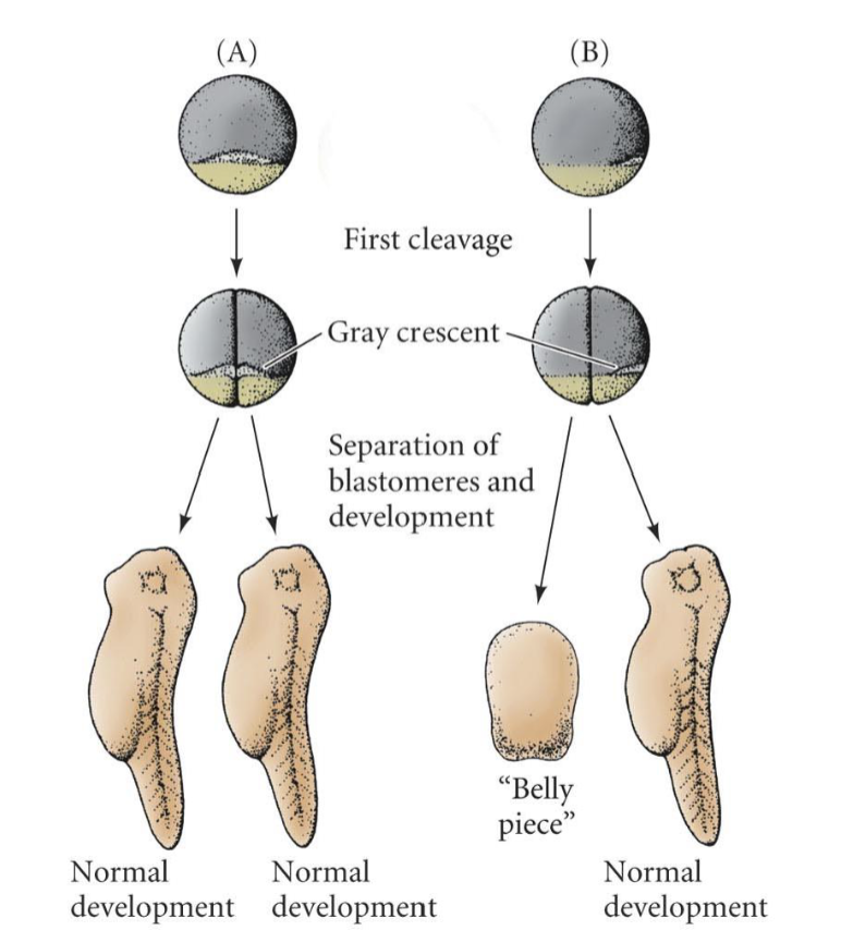
1) Division of grey crescent in amphibian embryos
2) Nervous system region transplantation
Division of grey crescent in amphibian embryos
-a pre-organizer neural induction experiment showing that the grey crescent is necessary for development of neural and D-V axis
-The gray crescent marks the future dorsal side of the embryo
-The dorsal lip will form from some of this area
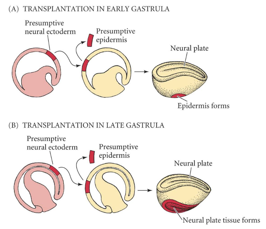
Nervous system region transplantation experiment
-a pre-organizer neural induction experiment showing that at some point during gastrulation, the nervous system is induced
-Transplantation of the region that would become the nervous system into the epidermis results in all epidermis when done early in gastrulation, but neural tissue if done late in gastrulation.
-Therefore, specification occurs mid-gastrulation
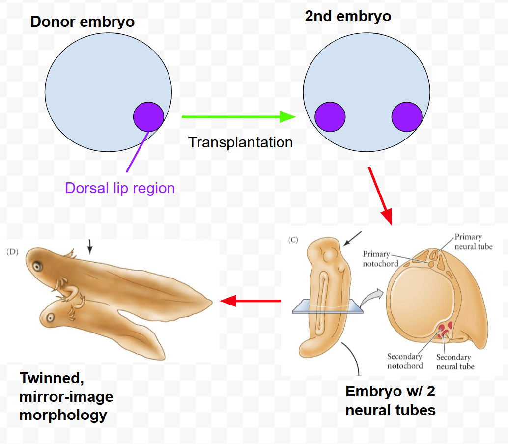
The organizer experiment
-Spemann and Mangold: Nobel in medicine
-Dorsal lip is termed organizer
-Transplanted dorsal lip region of pigmented embryo to ventral region of unpigmented embryo, resulted in
(1) induction of neural tissue
(2) organization of a new axis
-Only a tiny bit of dorsal lip region can result in a complete nervous system and basically a complete secondary embryo!!
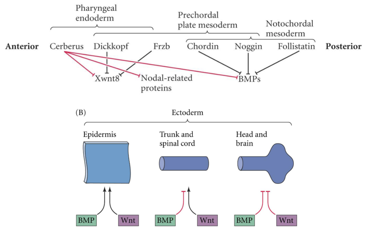
Tissue interactions mediating neural induction
Vertical signaling by mesoderm: organizer induces neural
—signals from the mesoderm to the ectoderm.
—Chordin, Noggin, Follistatin
Planar signaling by mesoderm: organizer patterns neural identity
—through the plane of the ectoderm. (evidence: removal of a continuous sheet of mesoderm-ectoderm and culture flat in vitro, assay for presence of neural markers shows they are there)
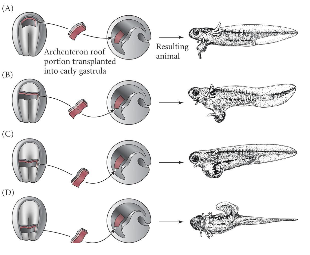
Regional neural induction
-mesoderm from different AP levels of the embryo were transplanted into the blastocoel of gastrula stage embryos.
-Result: different AP levels of the central nervous system were induced.
-NOTE: this was totally wrong. posterior mesoderm doesn’t need to form posterior regions, anterior mesoderm doesn’t need to form anterior regions. At early stages, any piece of mesoderm is able to induce a wide range of AP neural structures. The results of the experiment were mainly due to timing.
Experimentation to identify neural organizer molecule
1)Assayed substances for their ability to form neural tissue
-Pieces of embryo/live tissue, tissue from other organisms, dead embryo tissue, chemical agents (steroids, formaldehydes, ammonia, turpentine) → all induced neural tissue
2)Sublethal cytolysis
-Lysed cells w/ high pH, low salt, mechanical damage.
-All could induce neural induction in embryos
-(mechanical damage shows why previous experiments all worked!)
-Dissociation/damage of the animal cap induces neural because it releases the molecule inducer from the ectoderm
Determination of the Organizer Process
Dorsal has enriched B-catenin.
Nieuwkoop center (mes-endoderm that induces the Spemann Organizer region) determined by the overlap of TGFB (Vg1)/VegT signaling and B-catenin
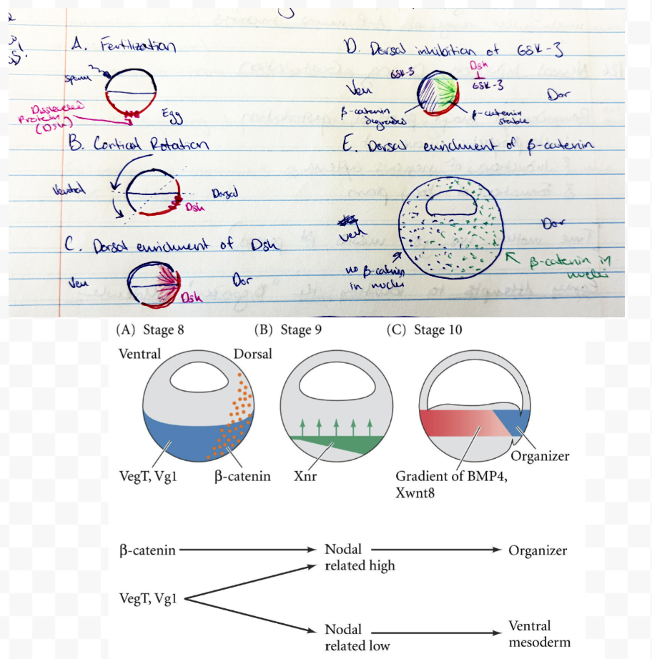
How does B-catenin turn on organizer genes?
-B-catenin complexes with Tcf-3 and binds to promoters of organizer genes like Siamois, which binds to goosecoid promoter. Goosecoid activates other organizer genes
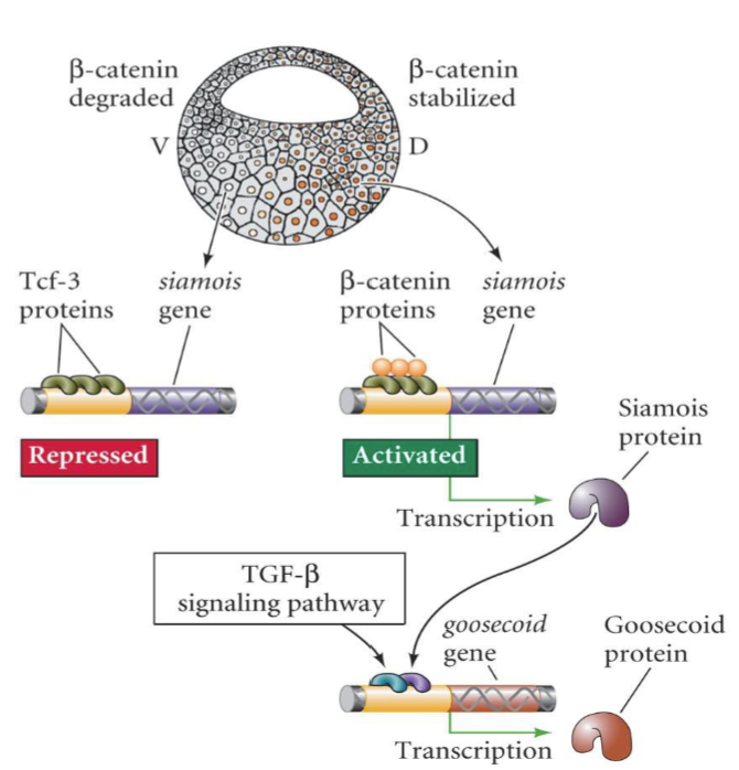
Experiments demonstrating the role of goosecoid
-in situ to find location of expression
-over express (results in duplicated axes, secondary neural tube to embryo)
-knock out (significant developmental defects, esp. head)
What genes are activated to lead to neuralization of tissue?
-BMP, Wnt inhibitors
-Noggin
-Chordin
-Follistatin
Noggin
Expression screening experiment:
-created a cDNA library (Rev. trans. mRNAs) from lithium-treated dorsalized embryos
-RNA synthesized from this library, subdivided, and injected into ventralized embryos
-if embryo dorsalized, the inducer mRNA was in that group. Further subdivided group. Repeat until 1 mRNA identified
-in situ to confirm it is expressed in dorsal lip
-also in dorsal mesoderm as it ingresses into embryo
-Noggin can rescue UV-irradiated (ventralized) embryos when injected
-Noggin inhibits BMP by binding to it and preventing it from binding to receptors
No noggin → can result in nervous system anyway due to redundancy
Right amount of noggin → normal development
Too much noggin → all nervous system, missing posterior structures
What is the default: neural or no neural?
Neural is default.
Noggin blocks BMP pathway from operating on dorsal side. BMP induces epidermis. When blocked, default occurs and default is neural.
Noggin is not a growth factor, BMP is.
Chordin
-isolated in differential screen for dorsal-specific genes
-is a secreted factor
-can rescue ventralized UV embryos
-Can bind BMPs like Noggin, Follistatin does (redundancy)
Follistatin
-Inhibits BMP signaling like Chordin, Noggin
-Back when the activin receptor knockout experiment (recall from mesoderm induction) was performed, 2 results occured:
(a) No mesoderm formed
(b) Much of embryo turned into neural tissue!
(“No muscle, but what a brain!”)
-Follistatin binds to activin/TGF-B/BMP to inhibit it.
BMP
-induce epidermal fate, inhibit neuralization
-A TGF-B family protein (like activin/Nodal)
-Must be inhibited by neural proteins (Noggin, Follistatin, Chordin) to form neural
Evolutionary conservation of neuralization pathway
Drosophila Sog homologous to vertebrate Chordin
Drosophila Dpp homologous to vertebrate BMP4
Wnt Inhibitors
-Anterior structures require inhibition of Wnt and BMP. Wnt should only be expressed in posterior.
-Cerebus, Dickkopf
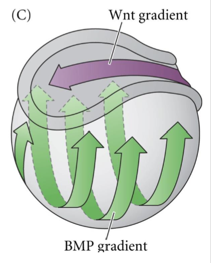
Cerebus
-Wnt, BMP inhibitor. Promotes anterior structures, can induce multiple heads when overexpressed
FRZB, Dickkopf
-Wnt inhibitors
-Excessive FRZB leads to excessive head structures. Depletion of Dickkopf leads to small heads
Summary diagram of Wnt, BMP and AP body axes organization
FGF Pathway
-Some organisms also require FGF pathway for neural induction, e.g. birds
-Form of multiple assurance to get importan structures
How do neuralization pathways lead to action potentials and neurons?
-Secondary messengers activate TFs
-TFs change gene expression