26. Ruminant Post-Diaphragmatic Digestive System
1/80
There's no tags or description
Looks like no tags are added yet.
Name | Mastery | Learn | Test | Matching | Spaced | Call with Kai |
|---|
No analytics yet
Send a link to your students to track their progress
81 Terms
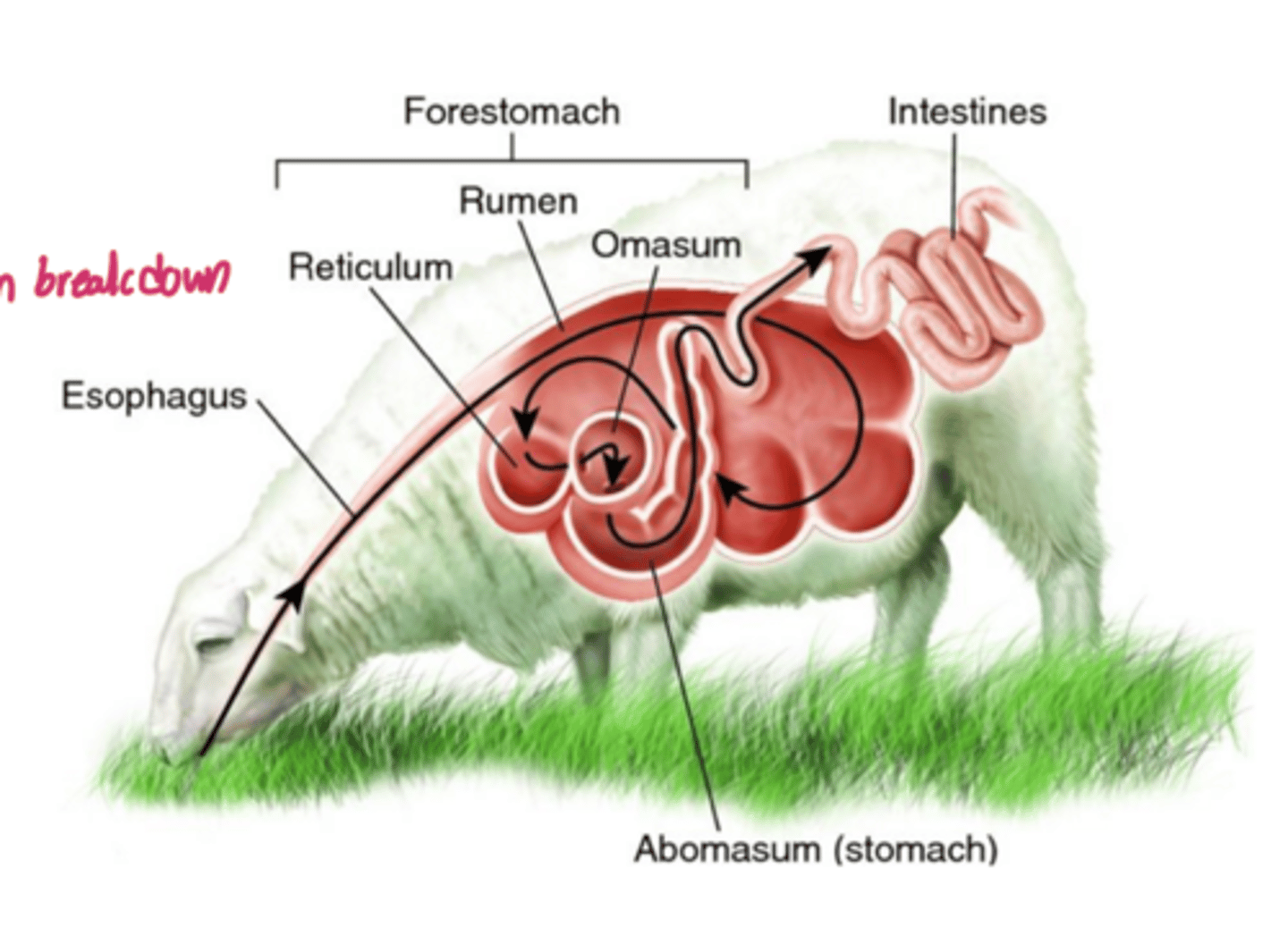
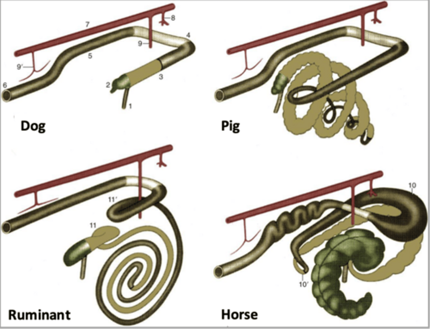
"fore-gut fermenters"
while equine are considered "hind-gut fermenters", ruminants are considered ____
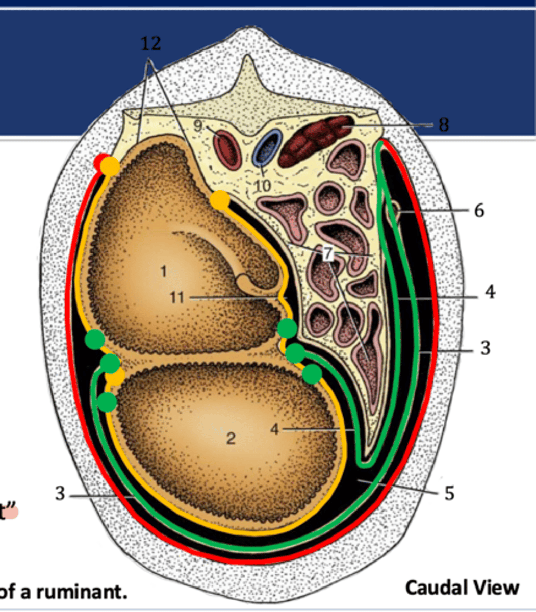
1. rumen
2. reticulum
3. omasum
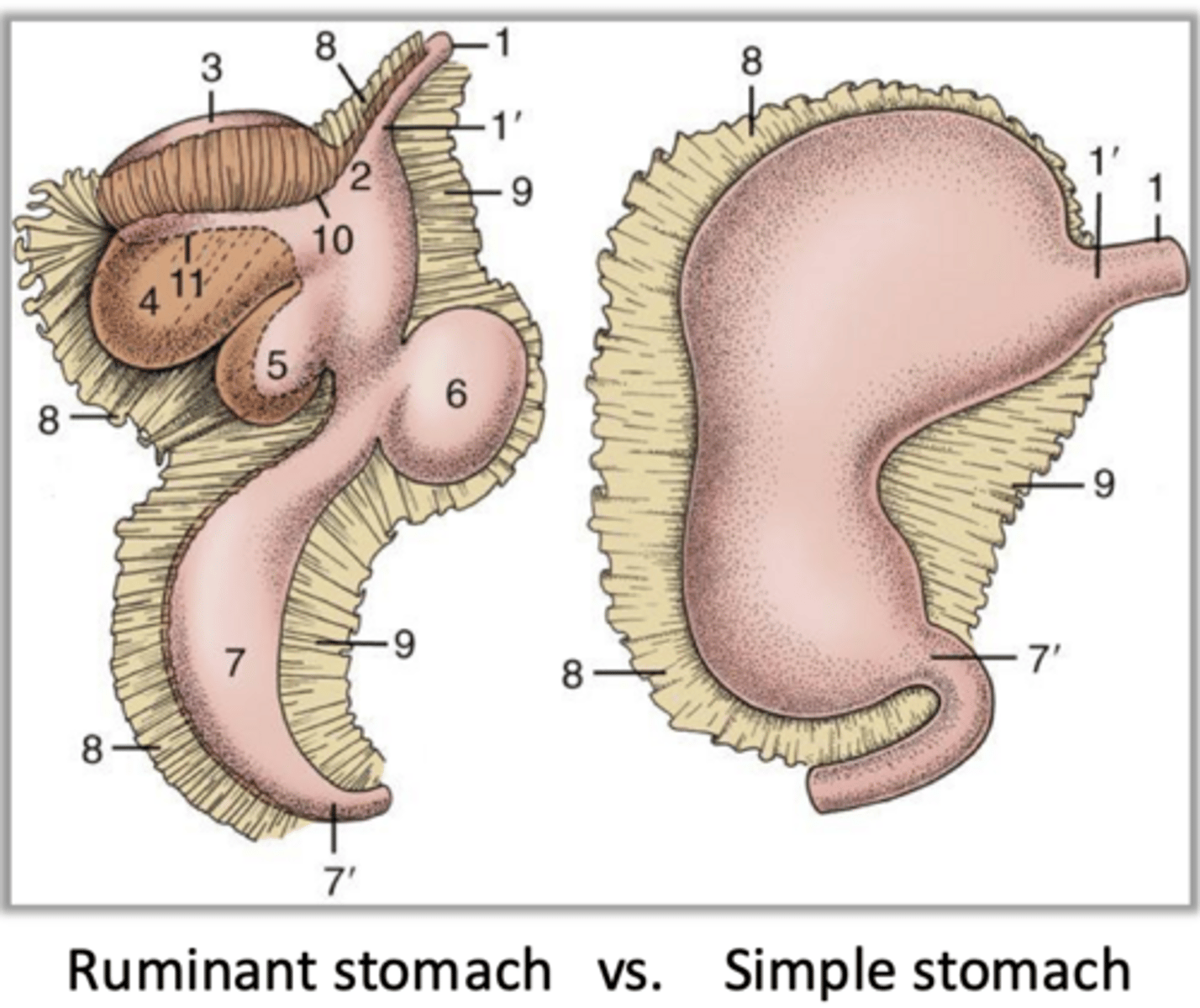
what are the three chambers of the forestomach/proventriculus
keratinized stratified squamous epithelium
the forestomach/proventriculus is filled with ______ epithelium to assist with mechanical mixing of food and absorption
microbial fermentation to produce VFAs (acetate, propionate, butyrate) to serve as the primary energy source for the animal
what process occurs within the rumen/reticulum of the forestomach/proventriculus

concentration of ingesta via water/electrolyte/and remaining VFA absorption
what process occurs within the omasum of the forestomach/proventriculus

abomasum
what chamber makes up the "true" stomach/ventriculus
1. contain glandular epithelium to secrete HCL and pepsin
2. location of chemical/enzymatic digestion of proteins in preparation SI absorption
significance of the abomasum of the ruminant stomach
reticulum
1

rumen
4
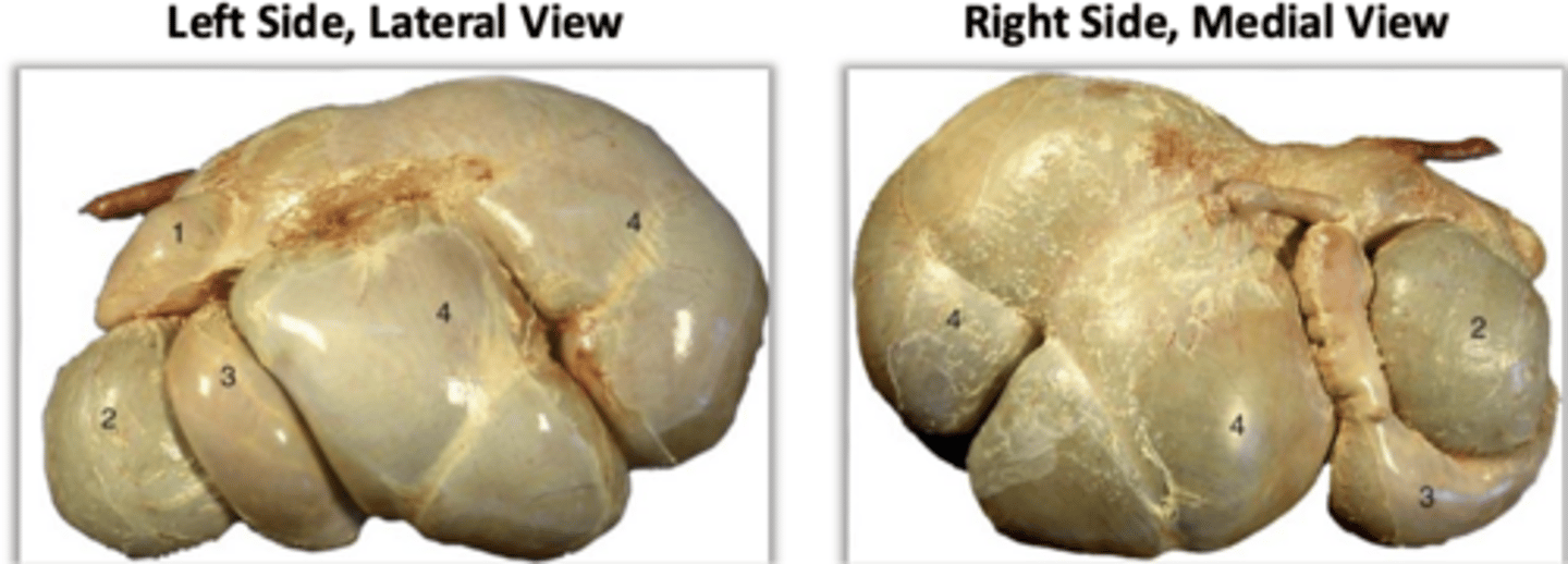
omasum
2

abomasum
3

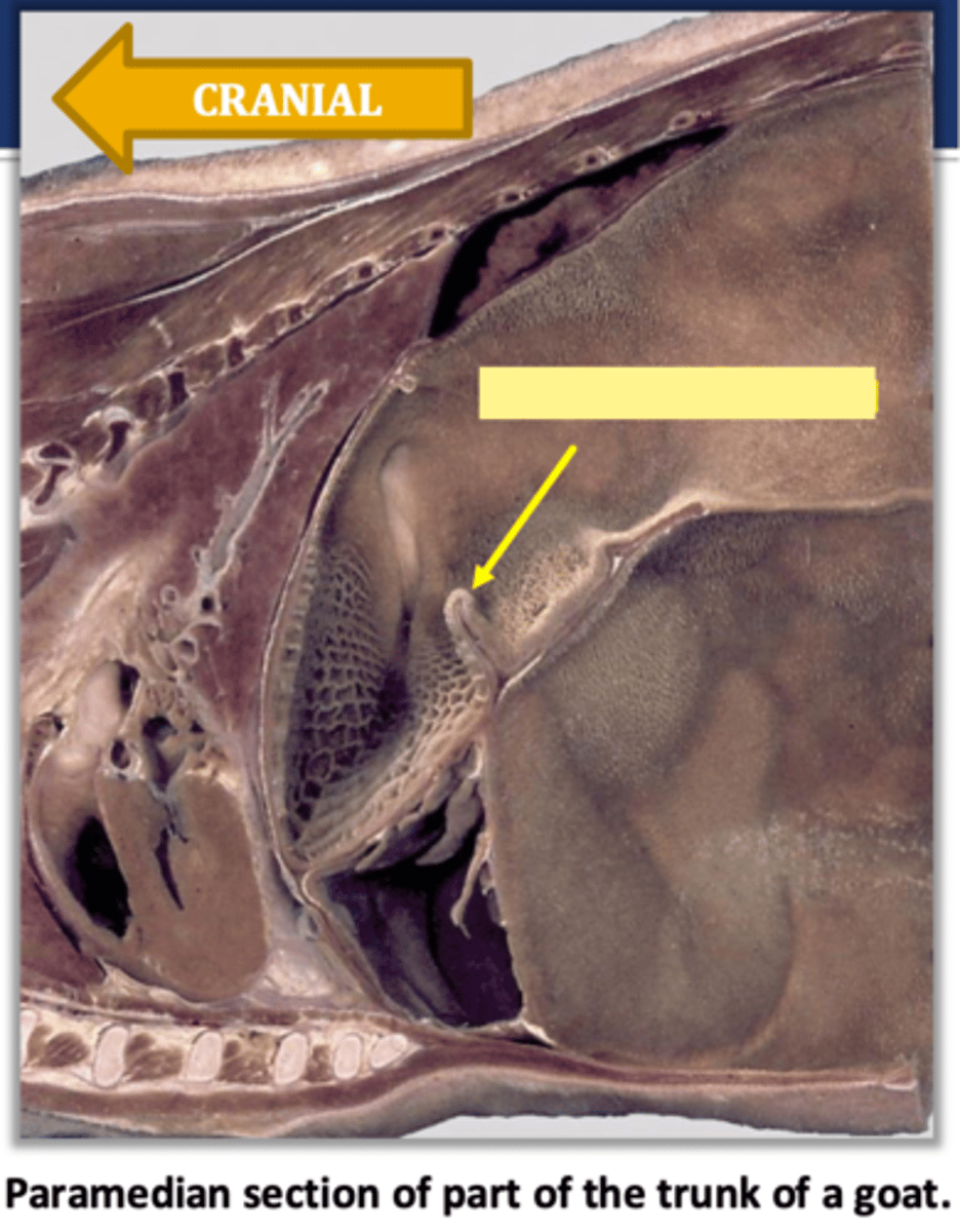
reticulum...a week functional closure allowing frequent regurge/eructation
what part of the stomach is the esophagus/cardia found in and what is its function
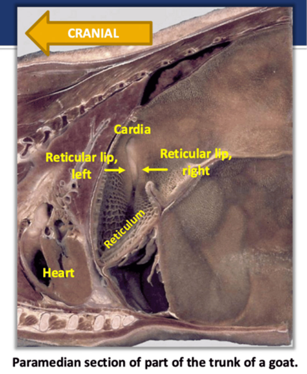
reticular groove
what structure indicated in the image is the continuation of the cardia/esophagus within the reticulum and it bounded by the reticular lips
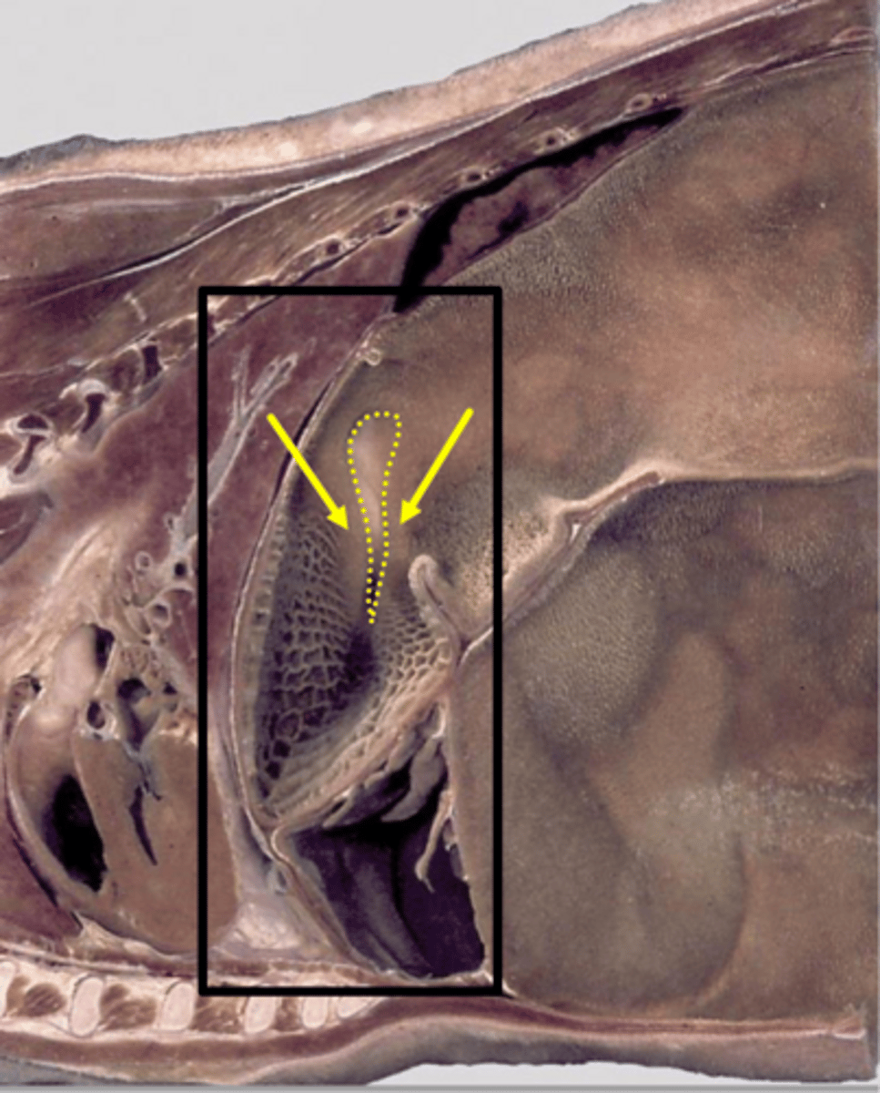
reticulo-omasal orifice
by what structure does the reticular groove open into the omasum

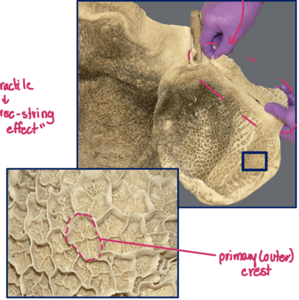
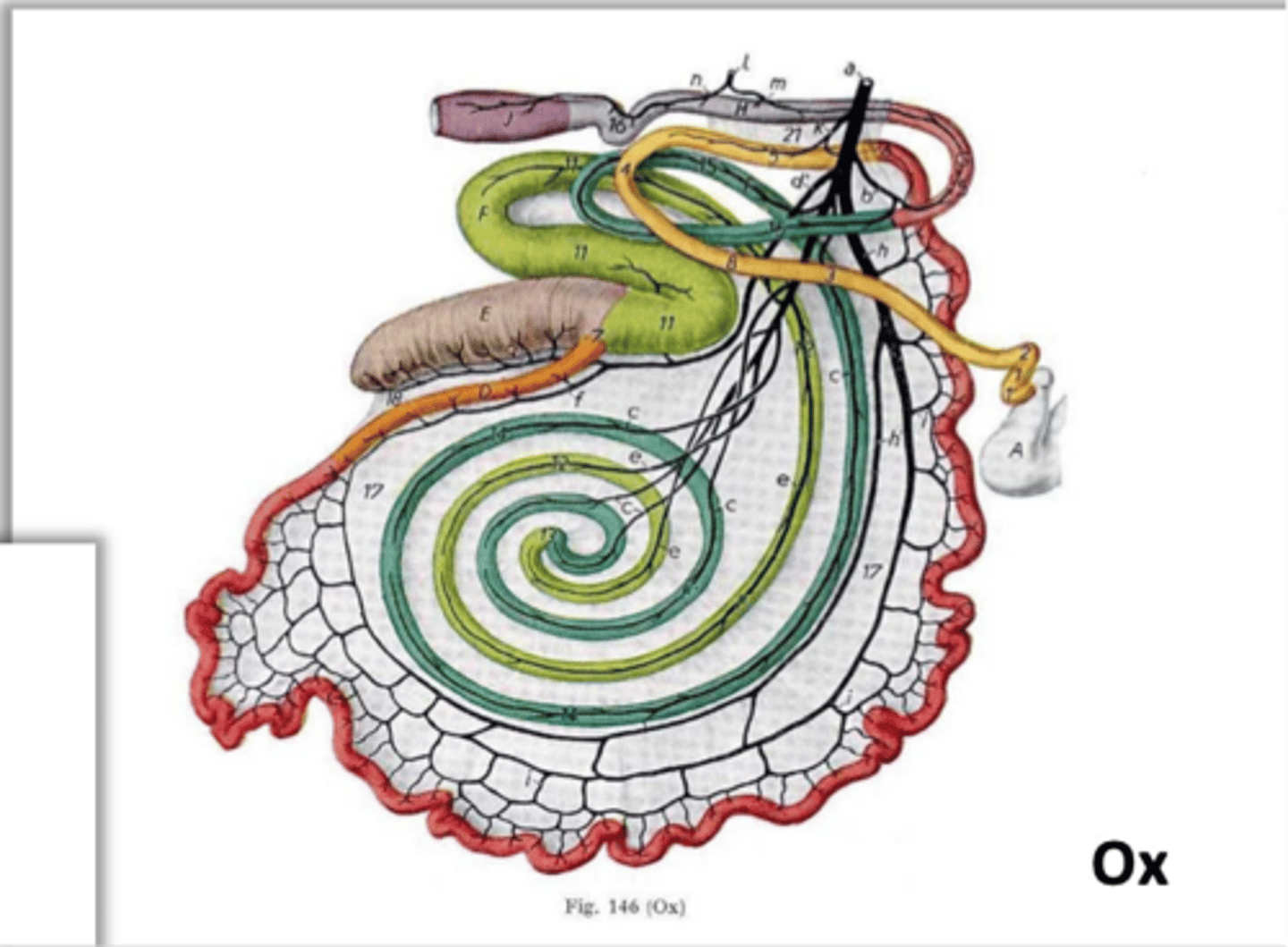
honeycomb
the reticulum is said to have a _____ mucosal pattern formed by the reticular crests which filters particles
....
outer crests have smooth mm. core with small corial papillae
ruminoreticular orifice
what structure within the ruminoreticular permits unrestricted exchange of ingesta between the rumen and reticulum
ruminoreticular fold
what structure indicated here separates the rumen from the reticulum internally
ruminoreticular groove
what structure indicated here externally corresponds with the ruminoreticular fold
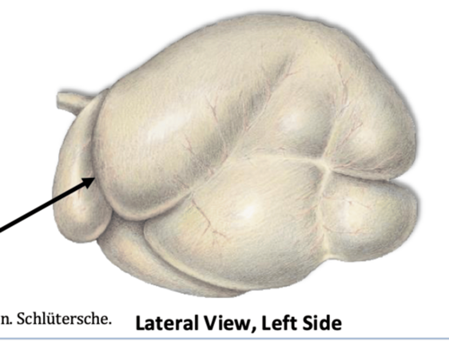
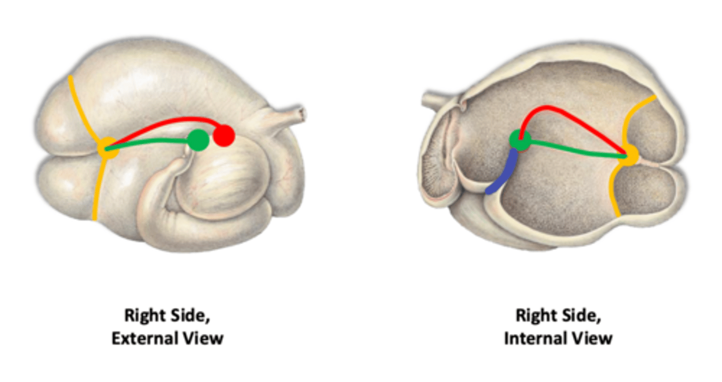
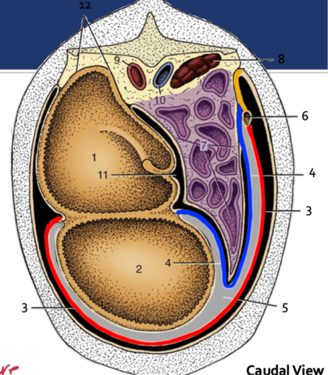
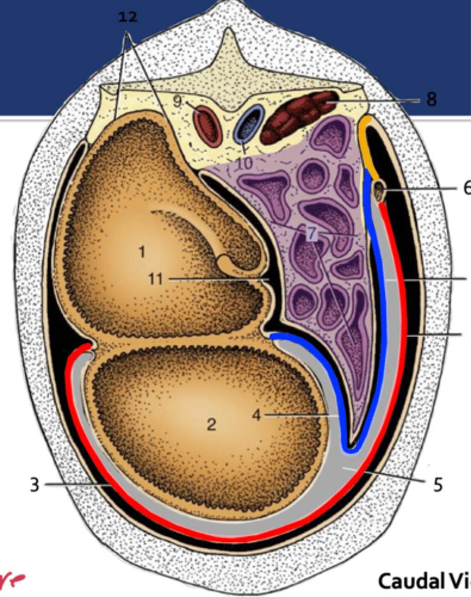
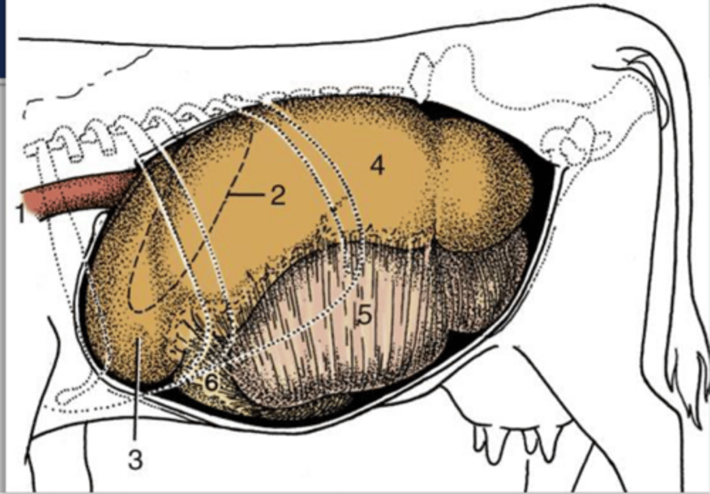
longitudinal groove-->divide to L and R
which external ruminal groove is represented by the green line and how do they divide the rumen

cranial and caudal grooves
which external ruminal groove is represented by the black line and how do they divide the rumen

coronary grooves-->dorsal and ventral on L and R sides
which external ruminal groove is represented by the orange line and how do they divide the rumen
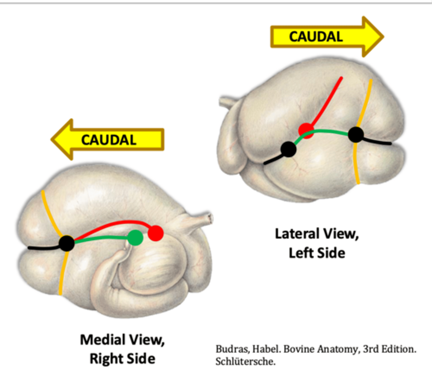
accessory grooves-->extend dorsally on L side and caudally on R side
which external ruminal groove is represented by the ref line and how do they divide the rumen

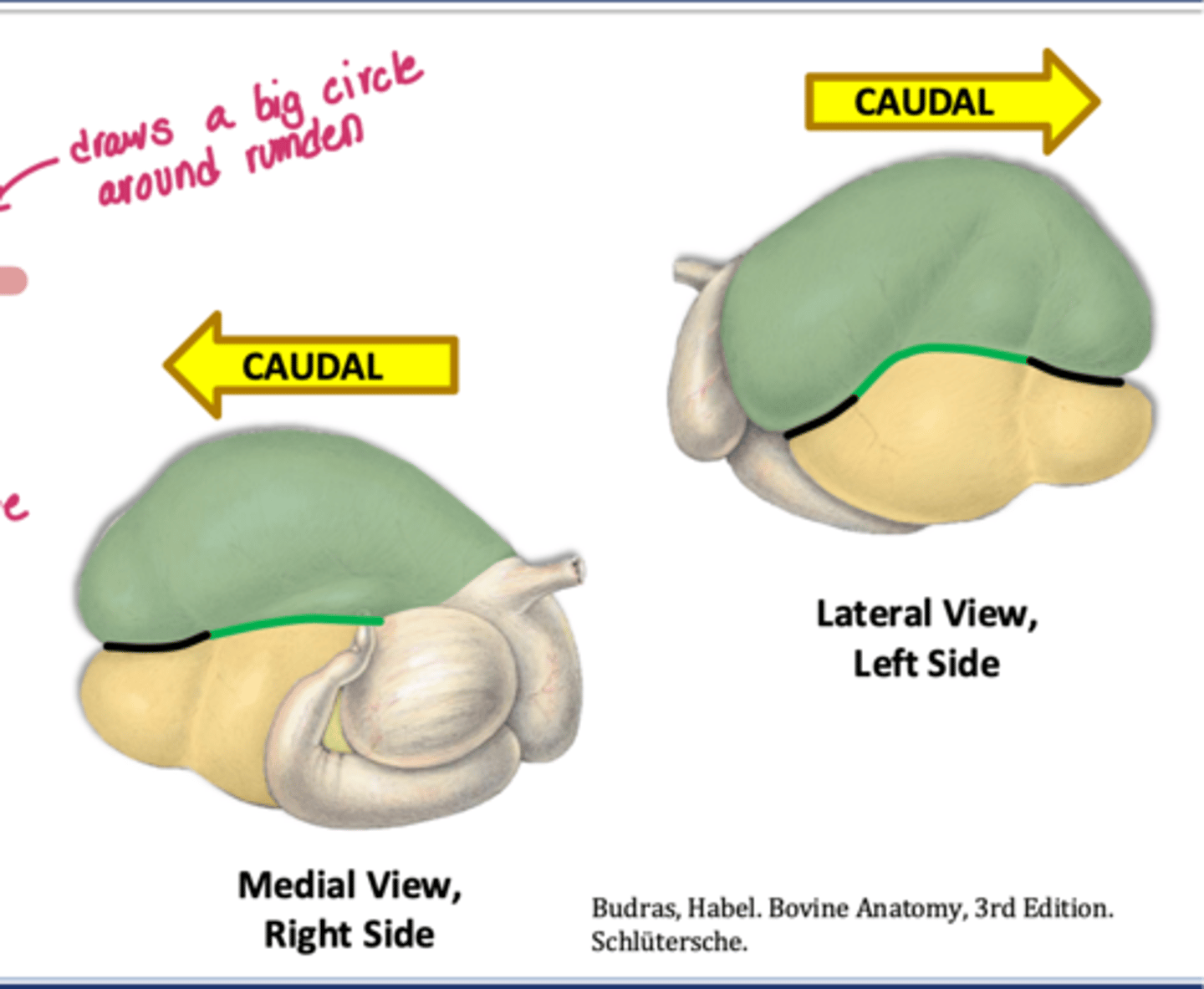
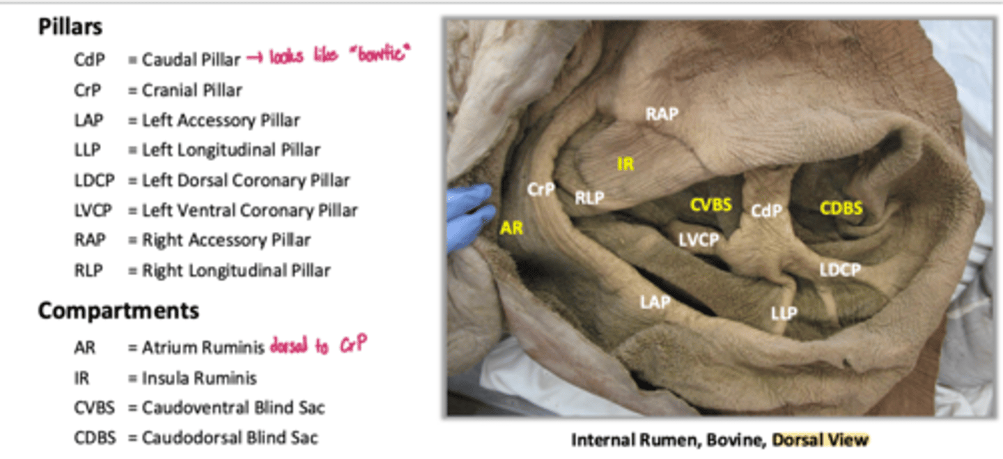
dorsal and ventral sacs
what ruminal sacs are indicated here which are divided by longitudinal and cranial and caudal groove
atrium ruminis-AR
part of the dorsal sac at the cranial-most extent
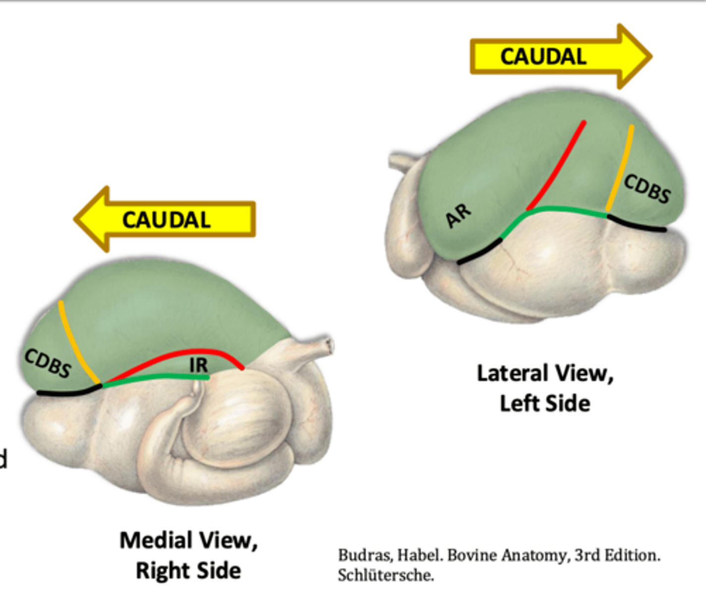
caudodorsal blind sac-CDBS
part of the dorsal sac at the caudal-most extent

right sided dorsal sac located between R accessory groove and R longitudinal groove
how would you describe the location of the insula ruminis-IR

ruminal recess-RR
part of the ventral sac at the cranial most extent
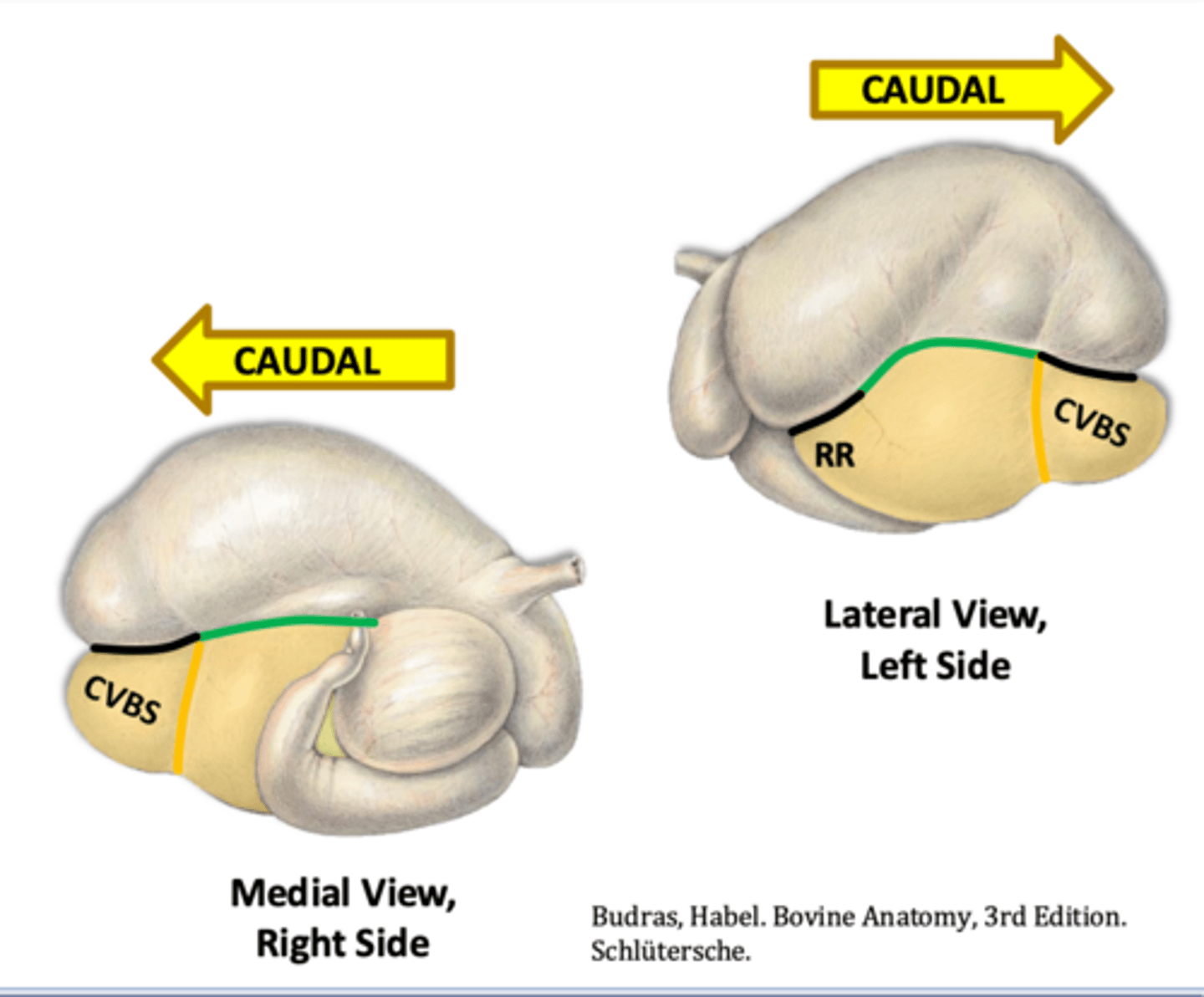
caudoventral blind sac-CVBS
part of the ventral sac at the caudal most extent

R accessory pillar
what internal pillar is shown in red that is associated with the R accessory groove

R longitudinal pillar
what internal pillar is shown in green that is associated with the R longitudinal groove
R coronary pillars
what pillars are shown in yellow that are associated with the R coronary grooves

R cranial pillar
what pillar is shown in blue that is associated with the R cranial groove
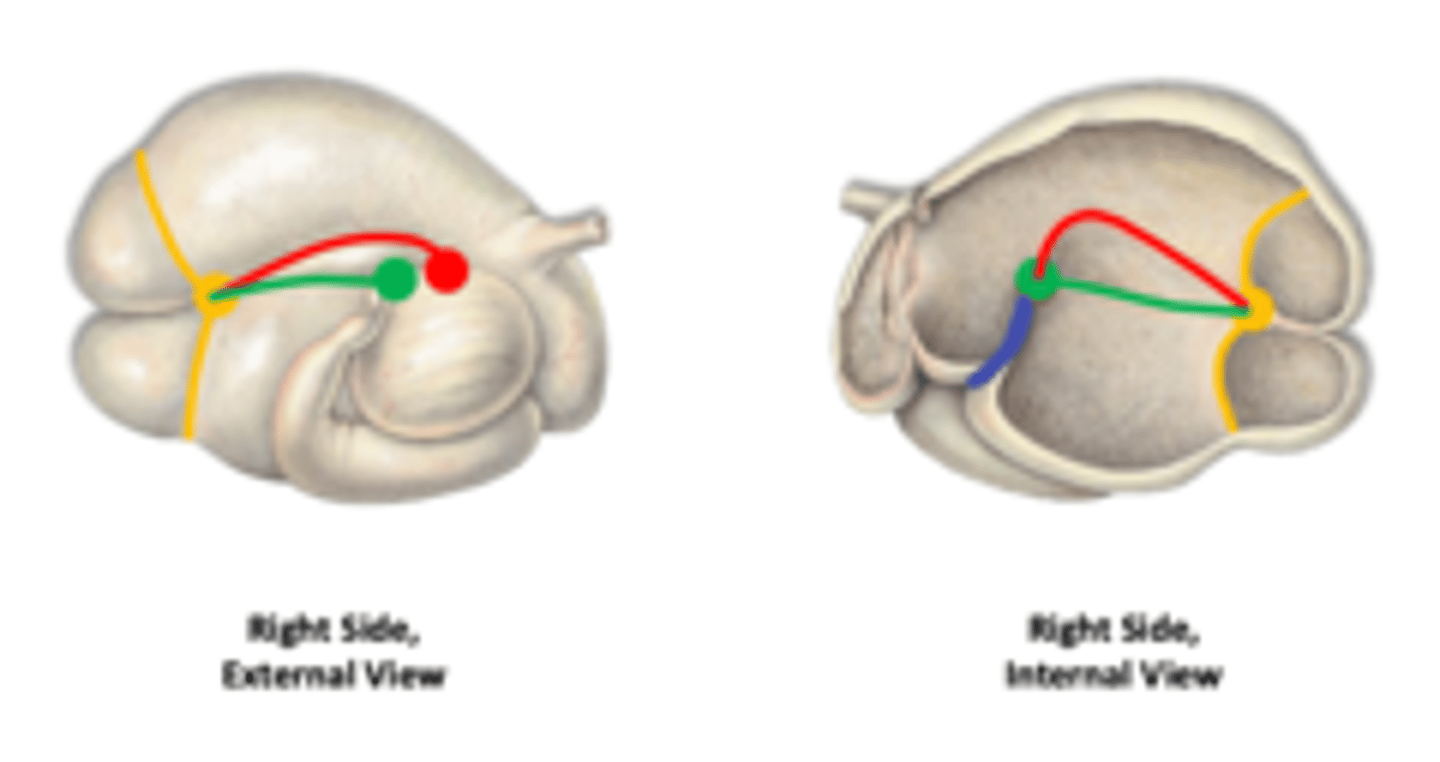
caudal pillar-CdP
which caudal ruminal pillar is said to resemble a bow tie and which abbreviation is it in this image
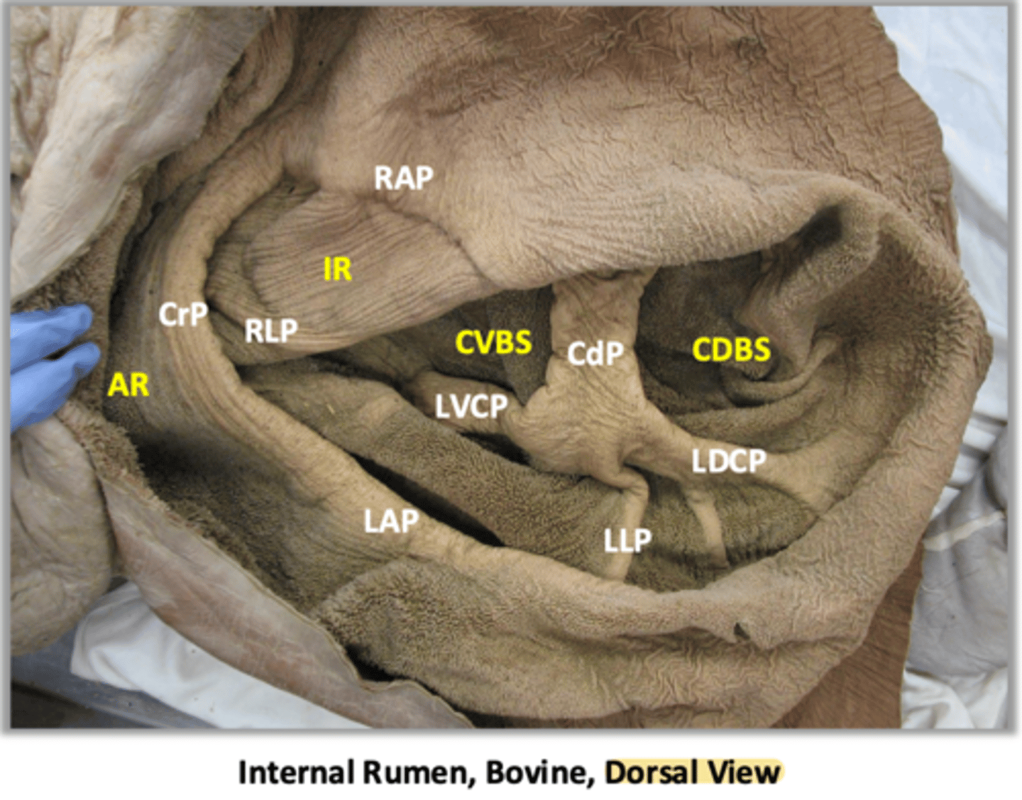
reviewed
review this image of the ruminal pillars
1. mucosal papillae to increase SA for VFA absorption
2. dvlp of papillae reflects diet, age, and functional region of rumen
characteristics of internal ruminal features

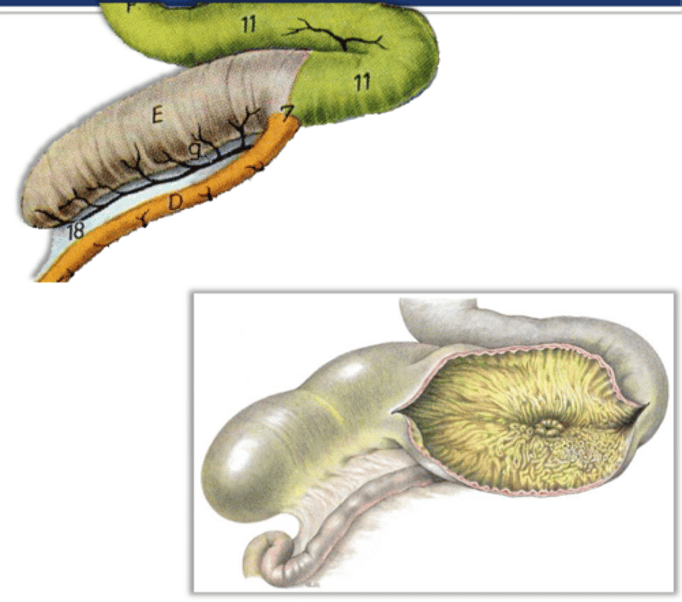
crescent shaped lamine resembling "book pages" with interlaminar recesses in between
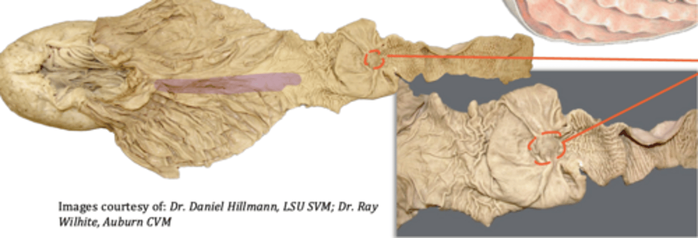
how would you describe the internal features of the omasum
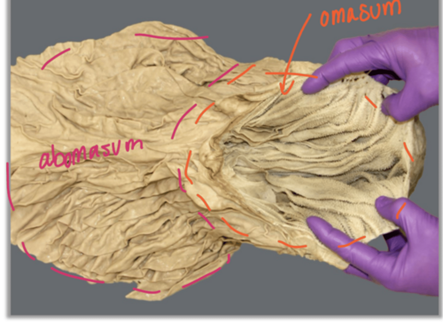
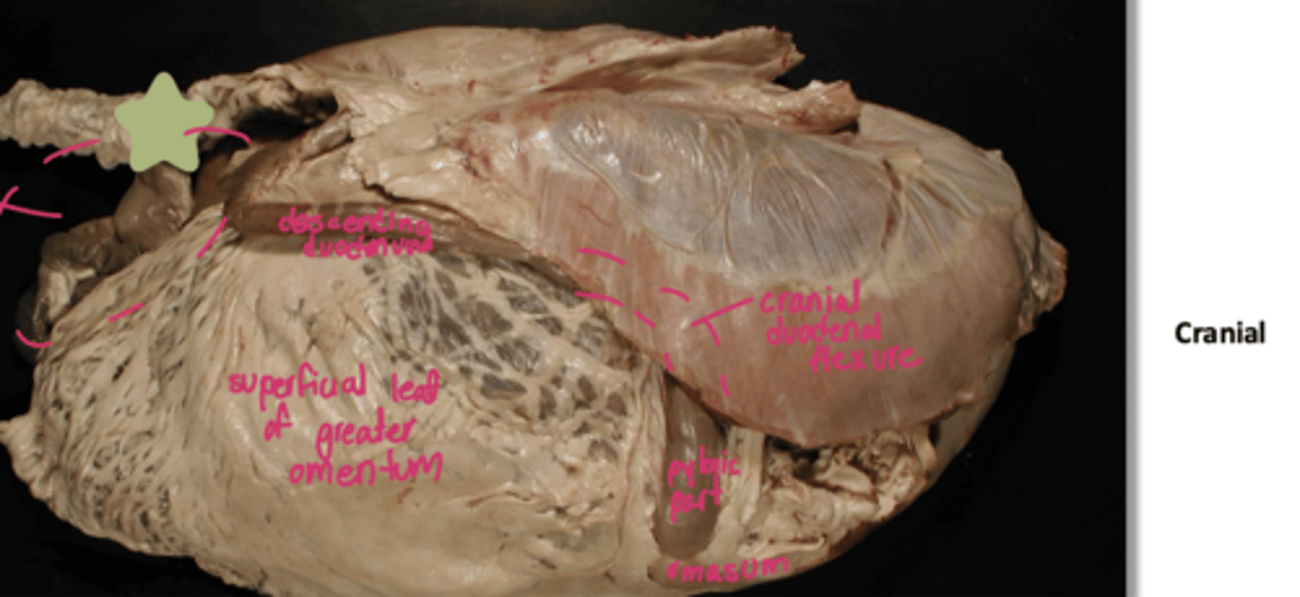
pylorus
what part of the abomasum is indicated by the star
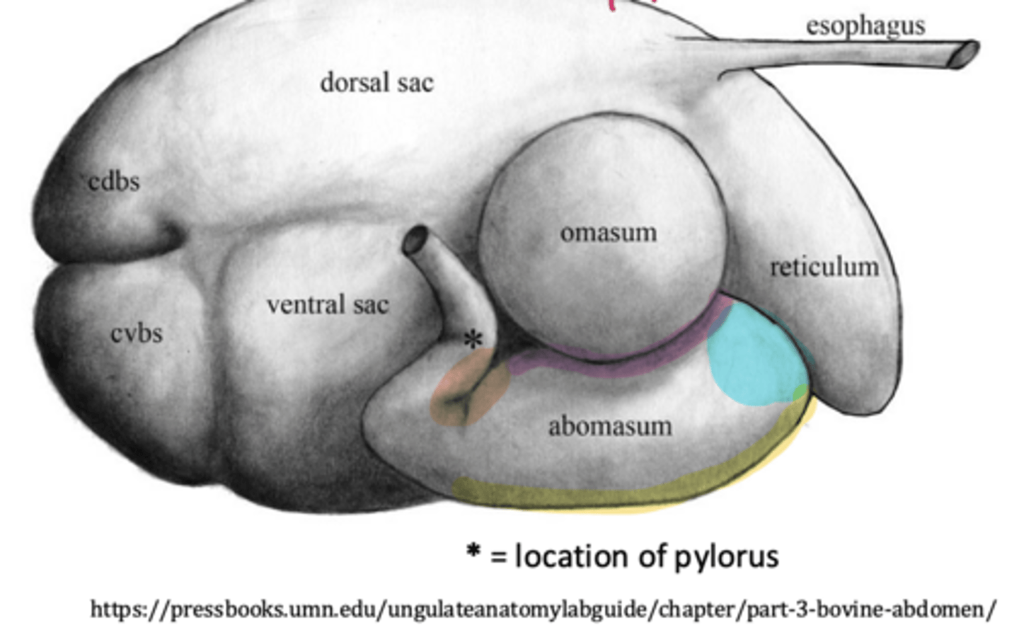
1. permanent spiral folds covered in glandular epi
2. abomasal groove (purple line) as a continuation of omasal groove
3. torus pyloricus near the pylorus
what are some of the internal features of the abomasum
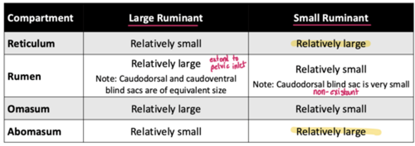
of equivalent size
the rumen of the large ruminant is relatively large and the caudodorsal and caudoventral blind sace are...
caudodorsal blind sac
the rumen of the small ruminant is relatively small ad the ____ blind sac is very small
1. large ruminant: rumen and omasum and relatively large
2. small ruminant: reticulum and abomasum are relatively large
review and recognize which parts of the stomach are largest for the large ruminant vs small ruminant
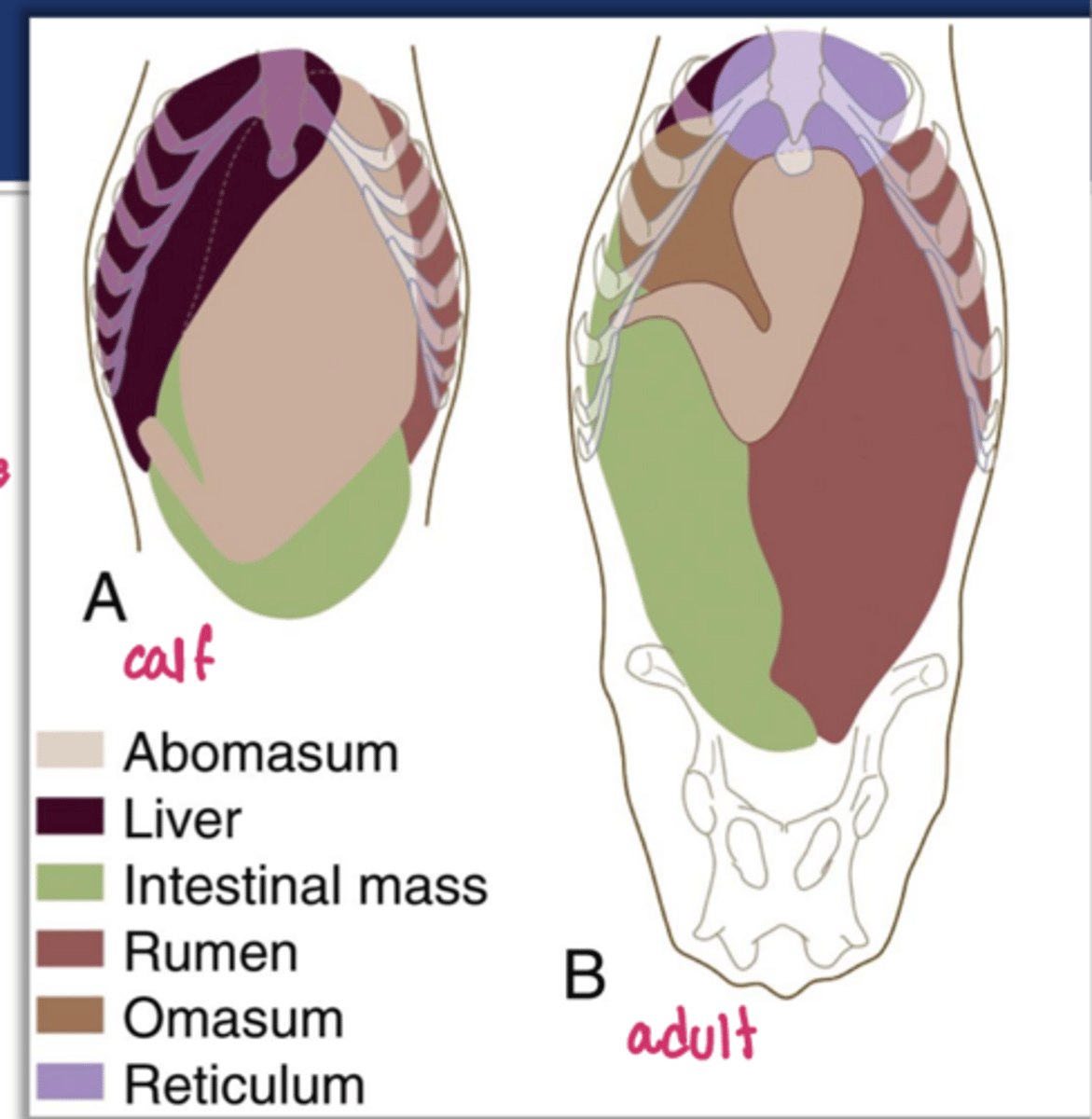
>50%
the abomasum of the calf has ____ capacity and occupies almost the entire floor of the abdominal cavity
ventricular groove/NAV/sulcus ventriculi
the ___ is the continuity of the reticular groove + omasal groove + abomasal groove and allows direct passage to the abomasum in the calf via closure of reticular lips
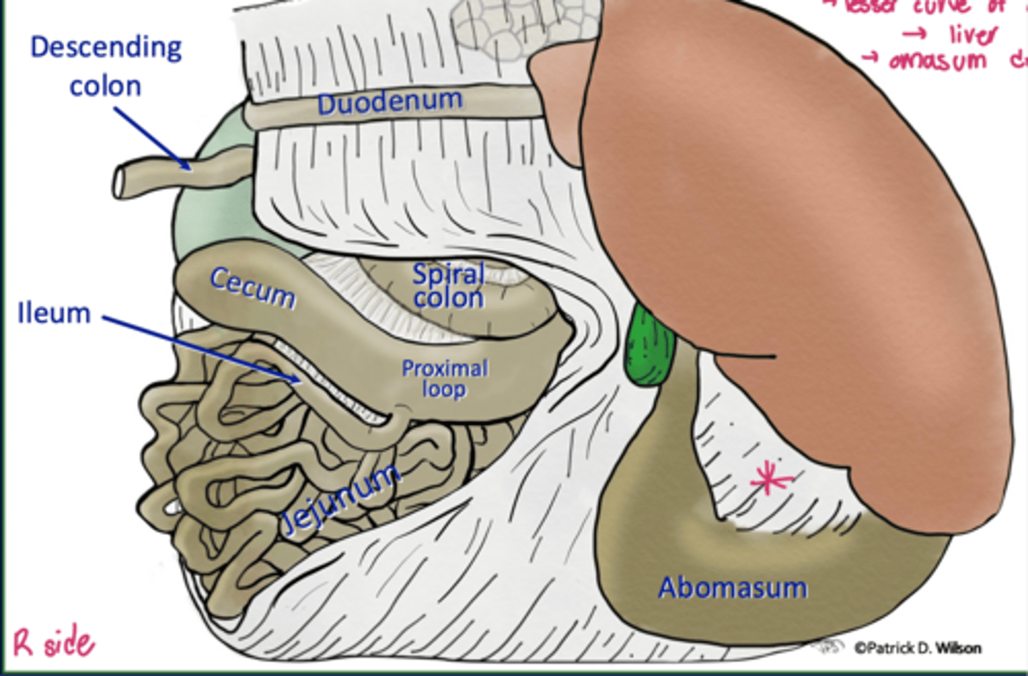
jejunum
in the ruminant, the ____ encircles the spiral colon/ascending colon
discrete cecocolic orifice
though ruminants possess the ileocecal fold, ileal orifice, and ileal papilla...they do not possess a ____ _____ ____ and the cecum and ascending colon communicate freely
1. cecum and origin of ascending colon are R of ROM
2. transverse colon passess cranial to ROM
3. descending colon is to the left of the ROM
what are some of the LI consistency associated with the ROM/root of mesentery in ruminants
s-shaped proximal loop
A

spiral loop w/ centripetal (in) and centrifugal (out) gyri
B

central flexure
C
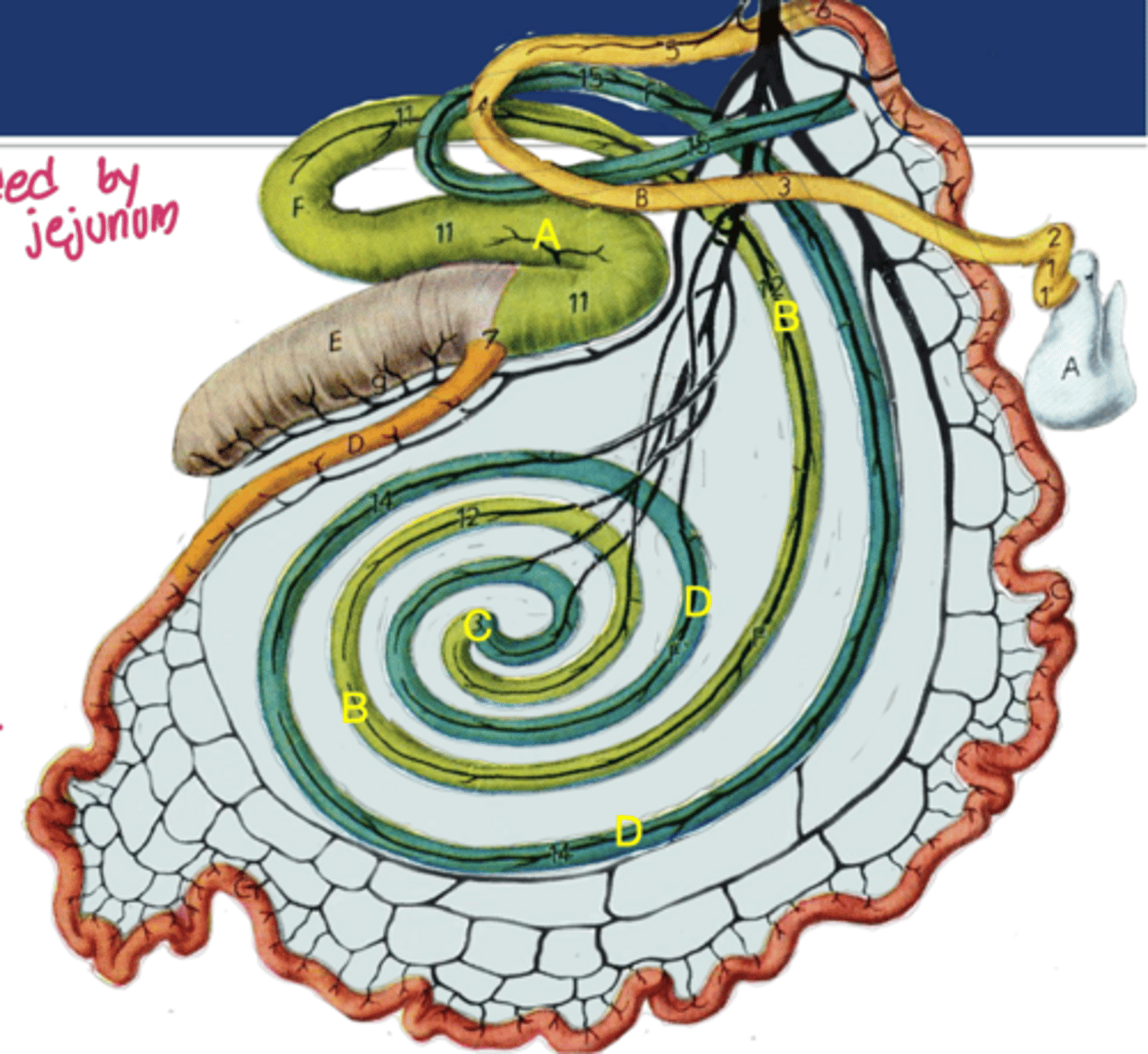
distal loop
D

jejunal flange
what aspect of the bovine GI indicated is susceptible to volvulus
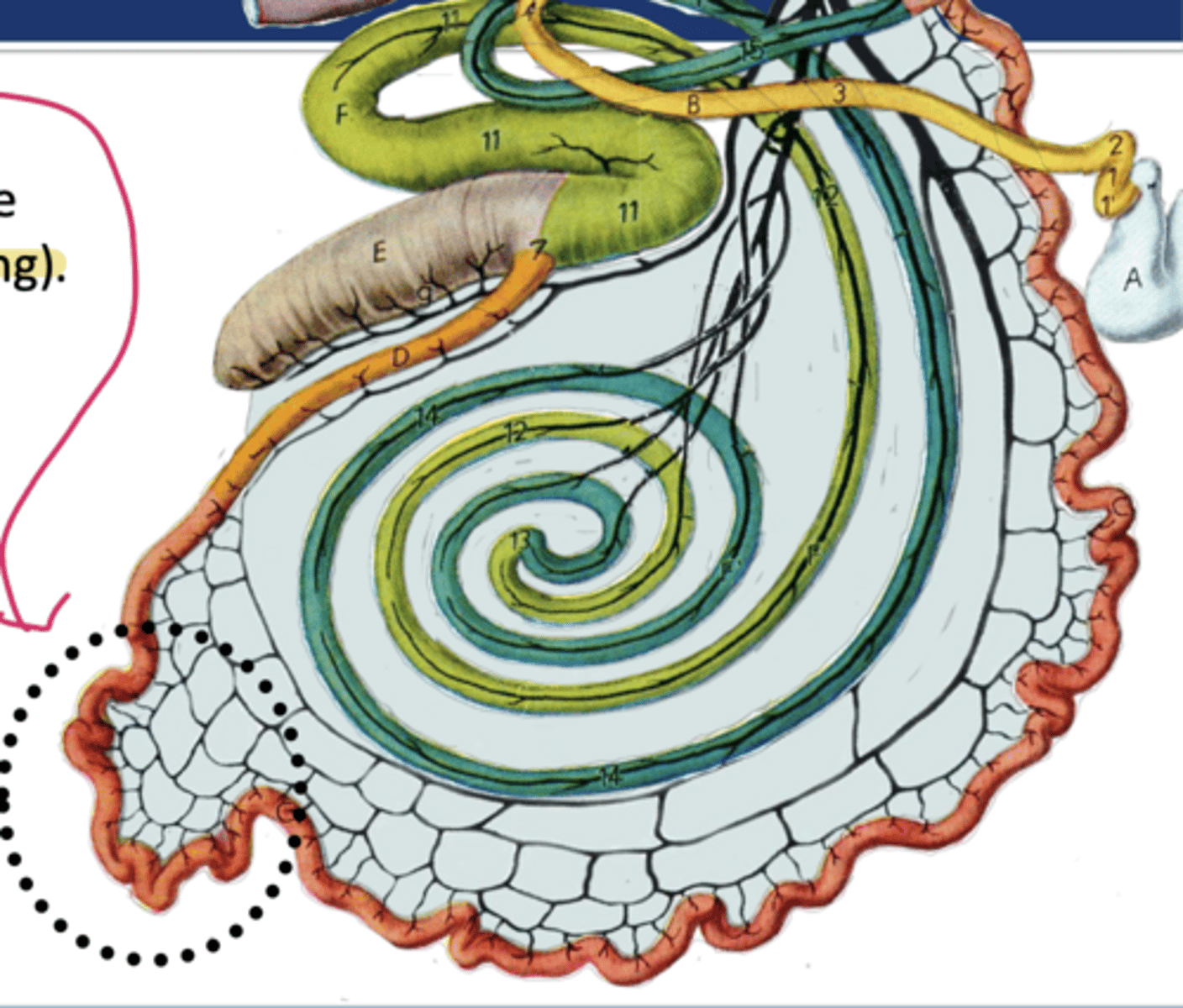
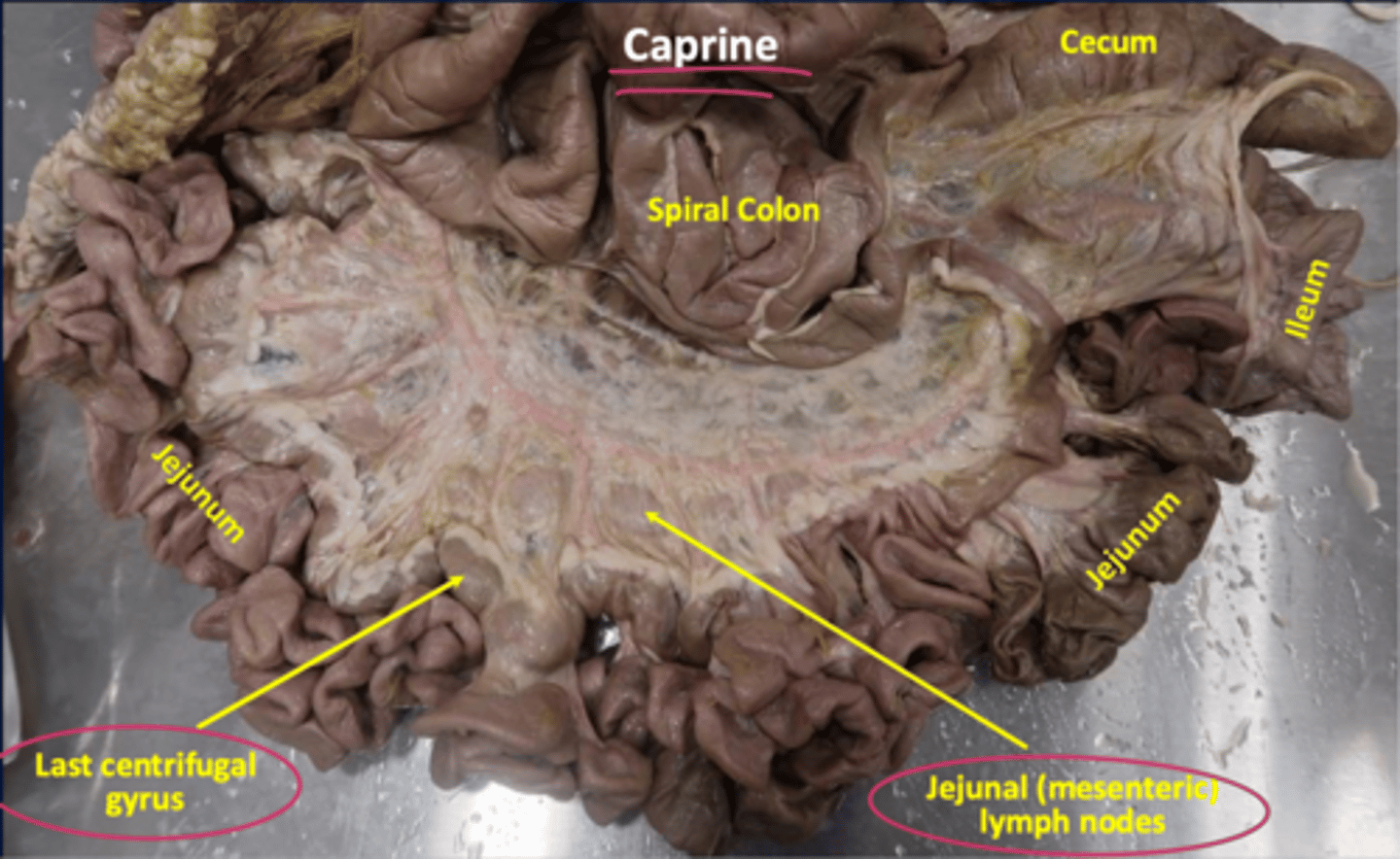
between last centrifugal gyrus and spiral colon
where are the jejunal LN of caprine located
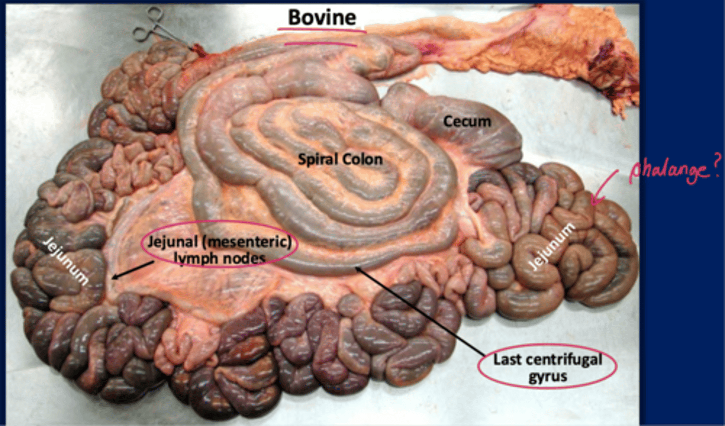
between jejunum and spiral colon
where are the jejunal Ln of bovine located
mesos-connection to body wall
greater/lesser omentum (gastrium), duodenum, jejunum, ileum, and colon are considered which connecting peritoneium
ligaments/folds-viscera to viscera connections
gastrosplenic/rumino splenic (spleen-->dorsal sac of rumen) ligament, ileocecal fold, cecocolic fold, duodenocolic fold are considered which connecting peritoneum

nephrosplenic lig.
which connecting ligament do ruminants not possess that equine do
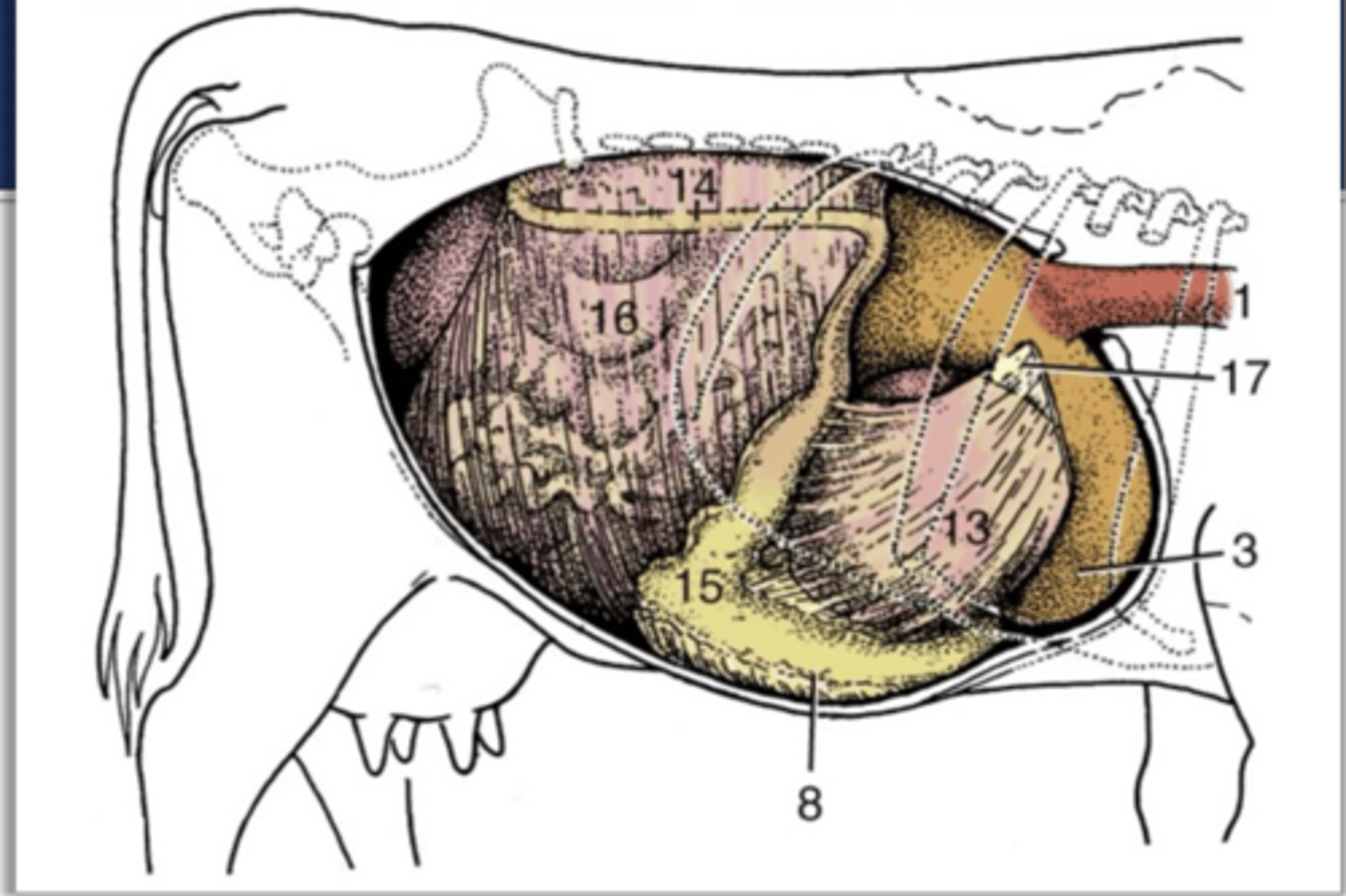
dorsal mesogastrium/greater omentum attaching to dorsal surfaces like greater curvature of stomach/abomasum, dorsal and ventral ruminal sac, and reticulum
8

ventral mesogastrium/lesser omentum attaching to ventral surfaces like lesser curvature of stomach/abomasum and omasum
9
L longitudinal groove of rumen to duodenum
the superficial leaf of the greater omentum indicated here by the red line extends from...

R longitudinal groove of rumen to mesoduodenum
the deep leaf of the greater omentum indicated here by the blue line extends from...
supraomental recess
the deep lead of the greater omentum forms a "sling" for the intestinal mass called the...
ventral sac of the rumen
what is located within the omental bursa

intestinal mass at the supraomental recess
what is indicated by the starred area
lesser omentum
what does the small star in this image represent
1. cupular at 6th ICS
2. caudal extent at upper part of 11th/12th ICS
3. much steeper slope than in horses
describe the topography of the diaphragm
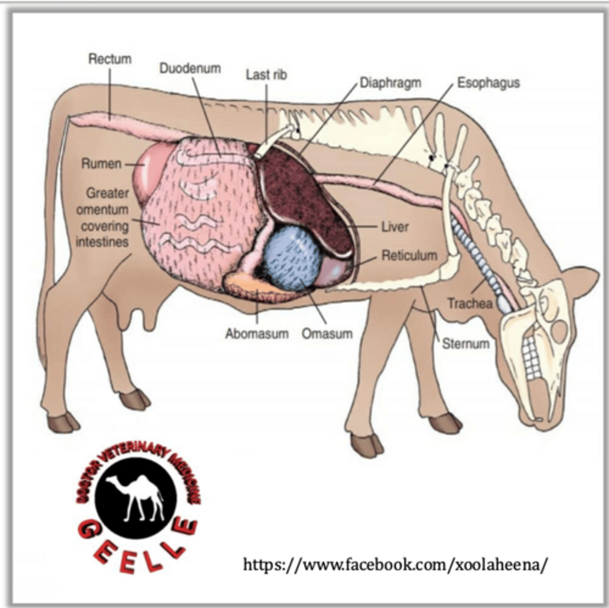
~8th ICS on the L side
where is the cardia of the stomach found---at the star

reticulum---abuts diaphragm at 6th rib
3
rumen
4,5

L side but does cross midline and extend caudally
where is the rumen (3) primarily found

rumen
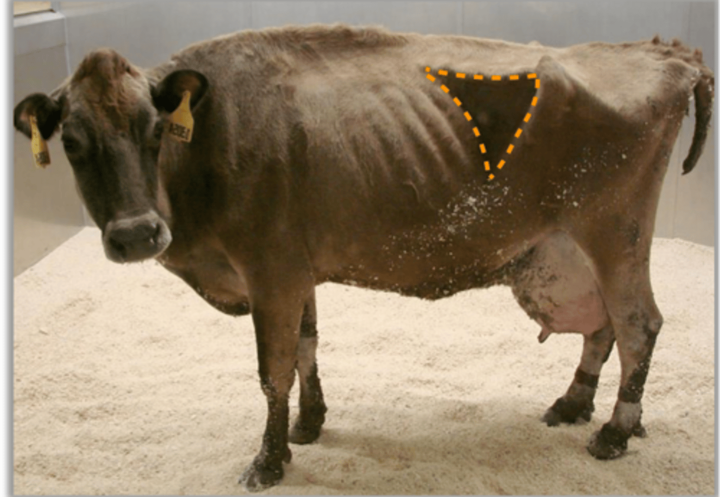
what part of the GIT is accessed by the L paralumbar fossa
R side at the 7th-11th ICS covered by the (13) lesser omentum
where is the omasum found
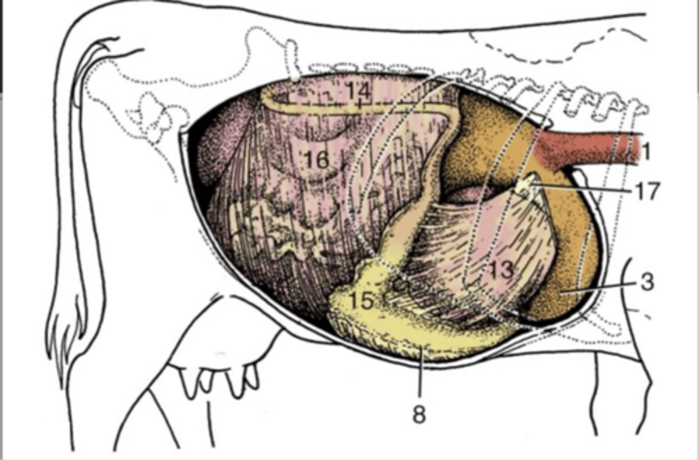
fundus of the abomasum
6

body of abomasum
8

found ventrally with the fundus protruding to the L btwn ruminoreticular groove and the (15) pylorus on the R
where is the abomasum found

cecum and intestinal mass w/in supraomental recess on the R side
what aspect of the GIT is accessed via the R paralumbar fossa
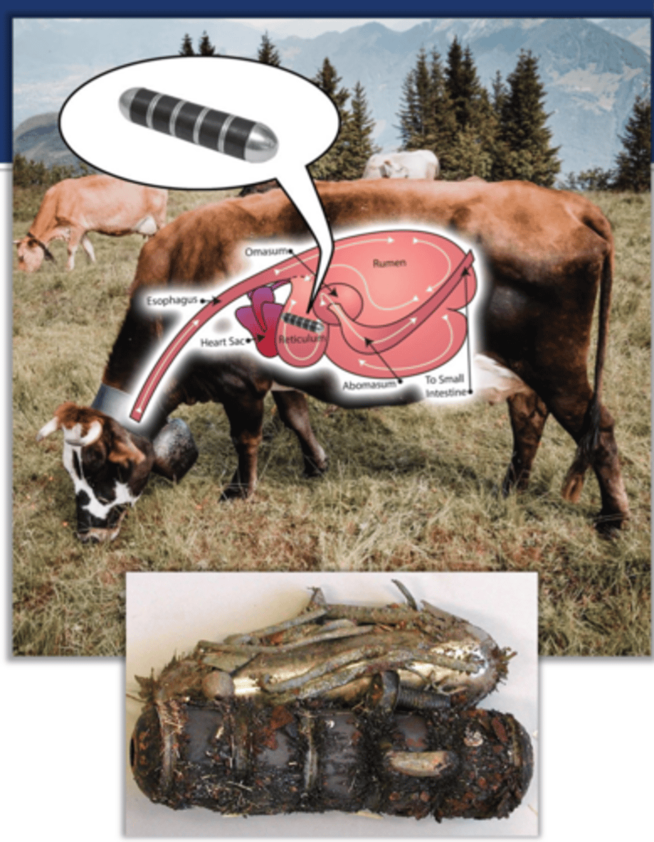
"hardware disease" w/in reticulum which can penetrate to the heart...prevention with prophylactic magnet in the reticulum
explain the significance of the position of the reticulum bieng located just caudal to the heart
1. ruminal tympany-bloat
2. gravid uterus-prgnancy
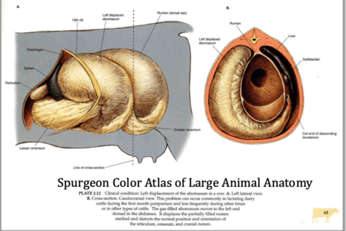
2. LDA-left displaced abomasum
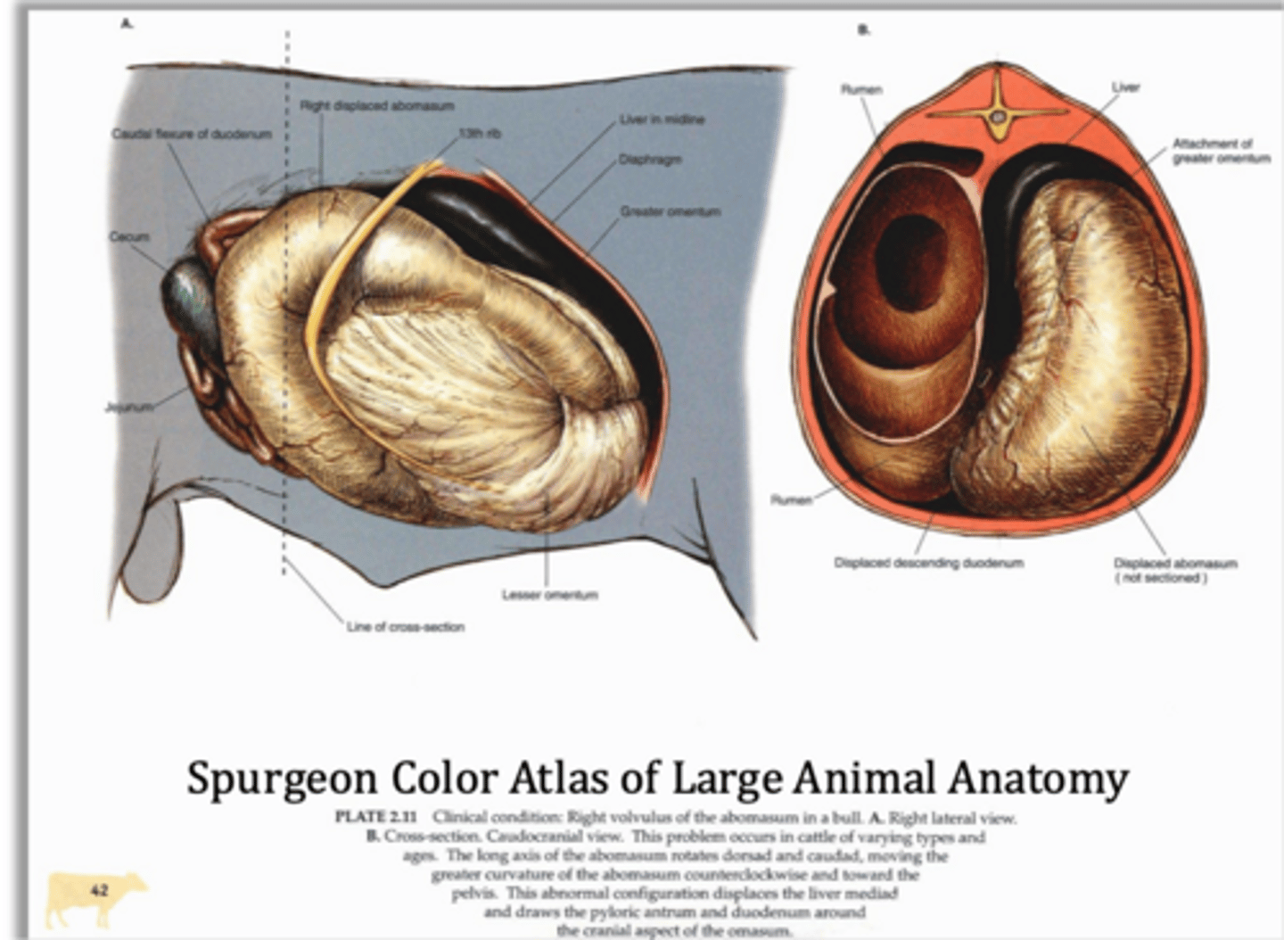
3. RDA- right displaced abomasum
what are some of the common causes of GIT organ displacements
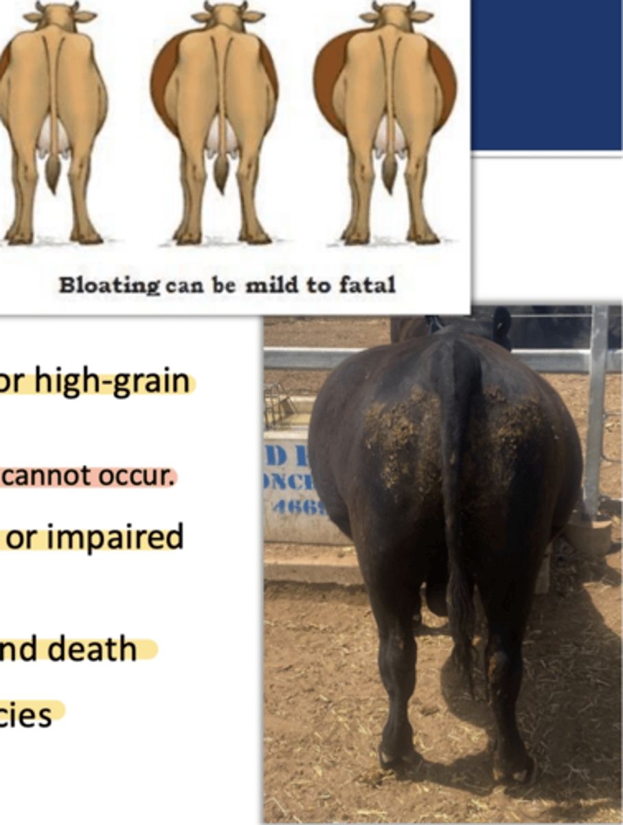
1. accumulation of gas in the rumen leading the distention of left abdomen which can compress diaphragm
2. frothy bloat associated with lush legumes or high grain diet preventing formation of free gas cap
3. free-gas bloat caused by esophageal obstruction or impaired rumen motility/vagal indigestion
4. relief via stomach tube or trochar
significance of ruminal tympany/bloat
rumen dorsally, intestinal mass cranially and dorsally, and abomasum cranially
a gravid uterus/pregnancy will occupy ventral and R abdominal spaces leading to displacement of...
LDA-left displaced abomasum
what is the most common abomasal displacement (typically in dairy cattle first few weeks postpartum) but does not usually compromise blood supply
RDA- right displaced abomasum
this form of displacement is less common but is more clinically serious as the long axis of the abomasum rotates dorsally and caudally creating a high risk of volvulus and blood supply compromise