Histology - Respiratory
1/104
There's no tags or description
Looks like no tags are added yet.
Name | Mastery | Learn | Test | Matching | Spaced |
|---|
No study sessions yet.
105 Terms
Ciliated pseudostratified epithelium
Identify the type of epithelium of #2.

Nasal Cavity
What are the spaces found immediately lateral to #2?

Nasal Turbinates (conchae)
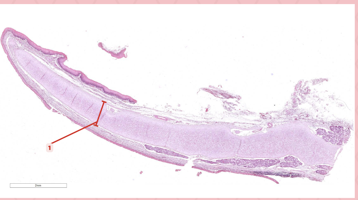
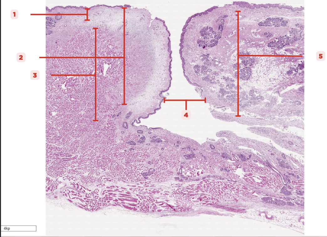
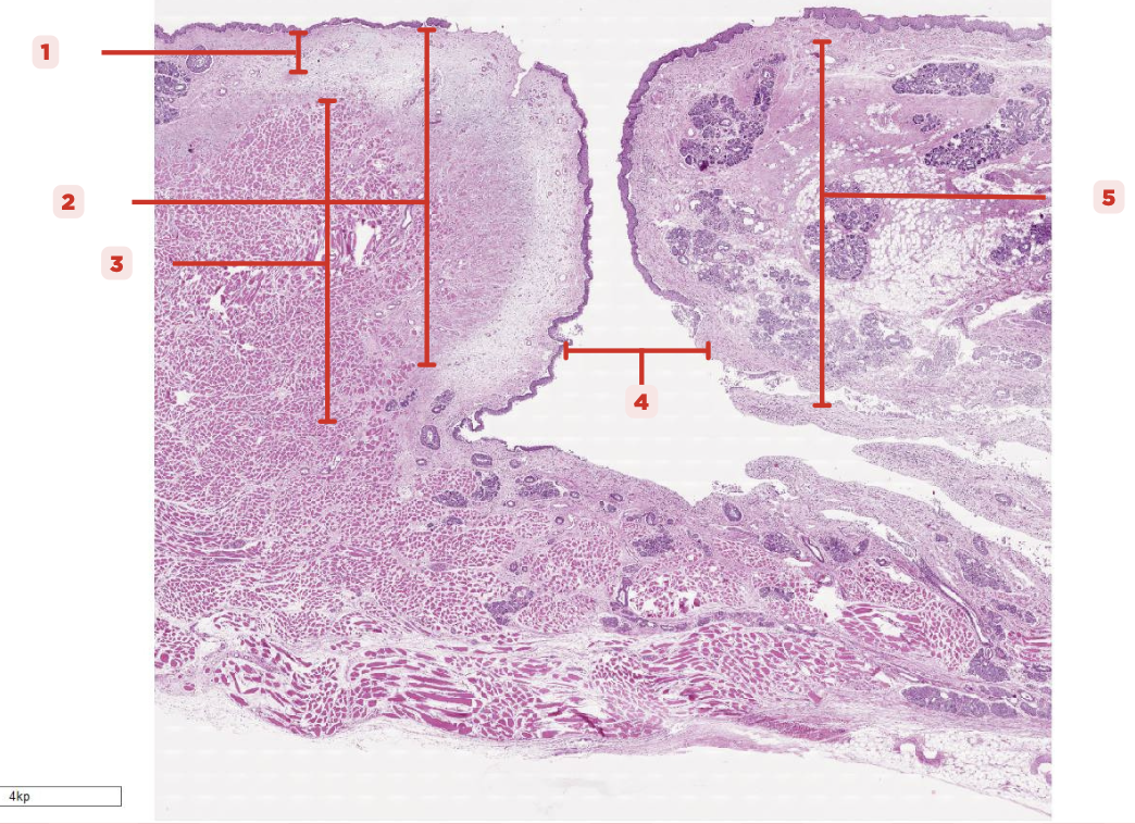
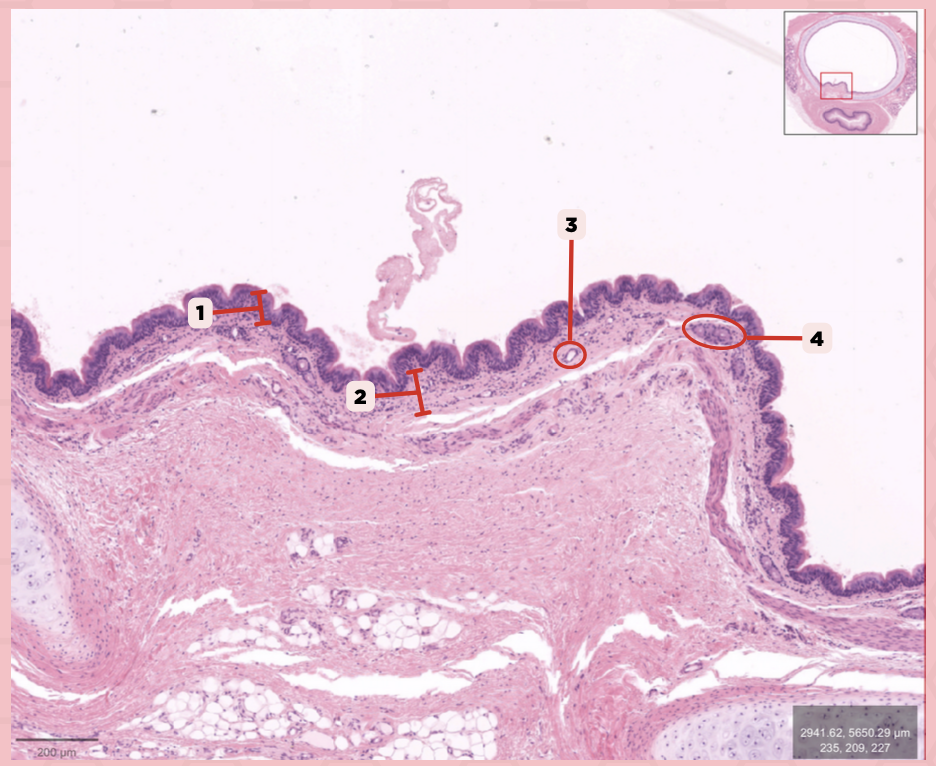
Identify the structure labeled as 1.

Nasal septum
Identify the structure labeled as 2.
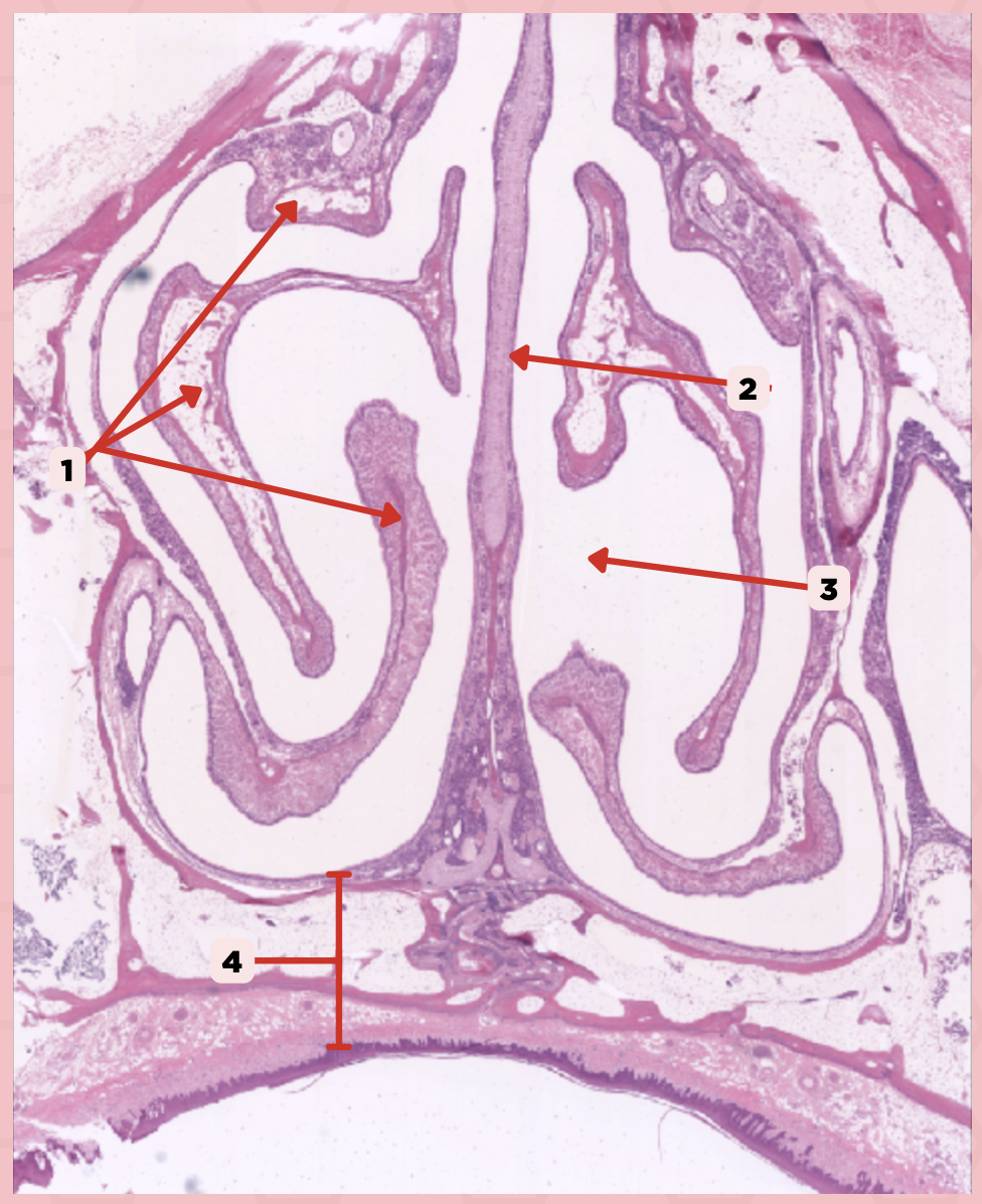
Nasal cavity
Identify the structure labeled as 3.

Hard palate
Identify the structure labeled as 4.

Mucosal Associated Lymphoid Tissue (MALT)
What time of lymphocyte is numerous in the nasal cavity?

Loose Connective Tissue
What type of cells make up #1?

Lamina Propria
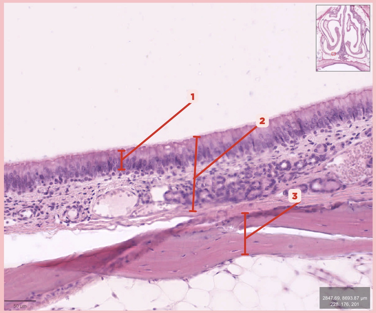
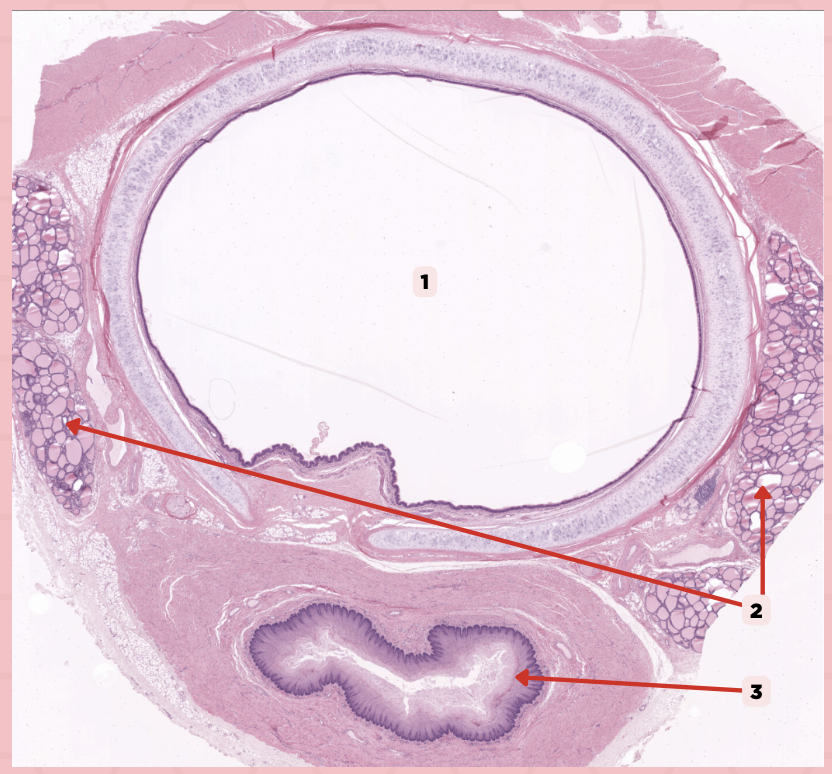
Identify the structure labeled as 1.

Hyaline Cartilage
Identify the structure labeled as 2.

Respiratory Epithelium
Identify the structure labeled as 3.

Nasal Cavity
Identify the structure labeled as 4.

Basal Cells
Identify the structure labeled as 5.

Goblet Cells
Identify the structure labeled as 6.
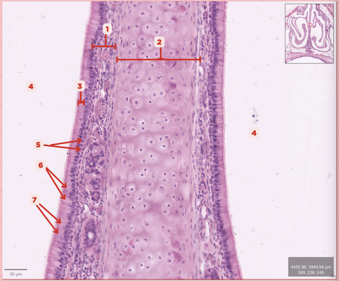
Ciliated Columnar Cells
Identify the structure labeled as 7.

Adipose tissue
What type of tissue is sometimes seen between the bony spicules?

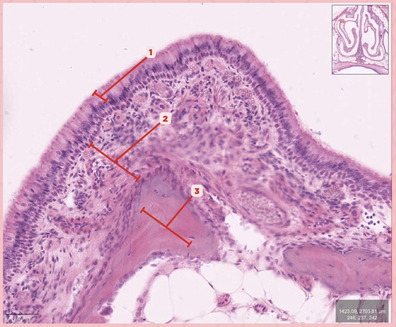
Serous and Mucous Glands
What glands are present in the Lamina Propria?

Respiratory Epithelium
Identify the structure labeled as 1.

Lamina Propria
Identify the structure labeled as 2.

Bony spicule
Identify the structure labeled as 3.
Respiratory Epithelium
What epithelium lines this structure?

Ciliated pseudostratified epithelium
What type of epithelium lines this structure?

Respiratory Epithelium
Identify the structure labeled as 1.
Mucosa
Identify the structure labeled as 2.

Bone
Identify the structure labeled as 3.

Keratinized stratified squamous epithelium
What’s the lining epithelium at #3?

No
Are Glands present at #1?
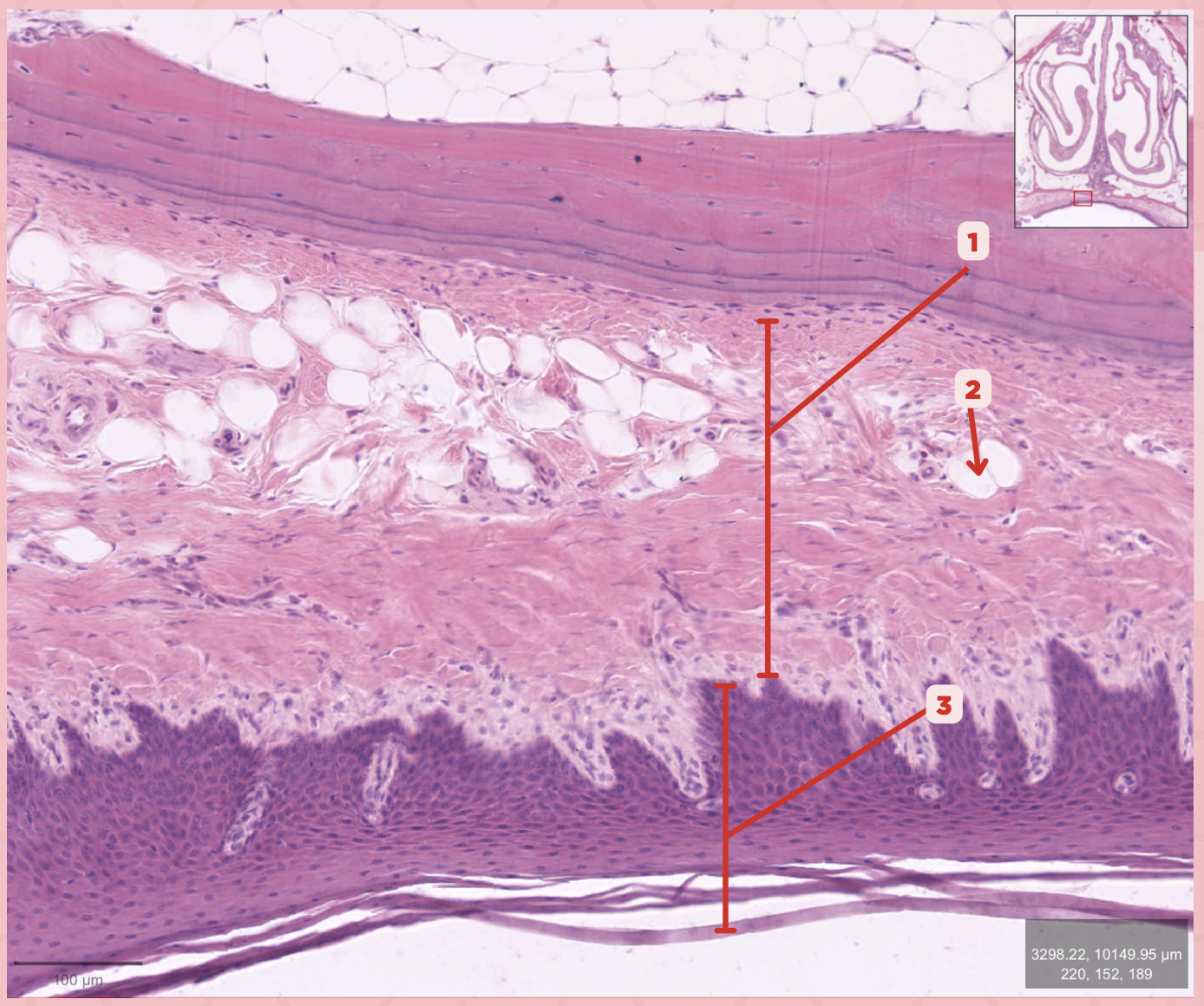
Lamina Propria
Identify the structure labeled as 1.

Adipocytes
Identify the structure labeled as 2.

Keratinized stratified squamous epithelium
Identify the structure labeled as 3.

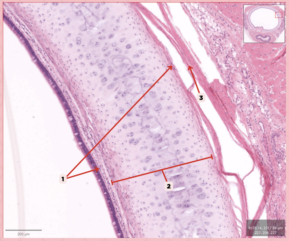
Unpaired cartilage
What is the type of elastic cartilage at #1, paired, or unpaired?
Olfactory Epithelium
Type of Epithelium that lines the roof of the nasal cavity and its adjacent areas

Epiglottic Elastic Cartilage
Identify the structure labeled as 1.

Serous Gland
What is the gland at #2?
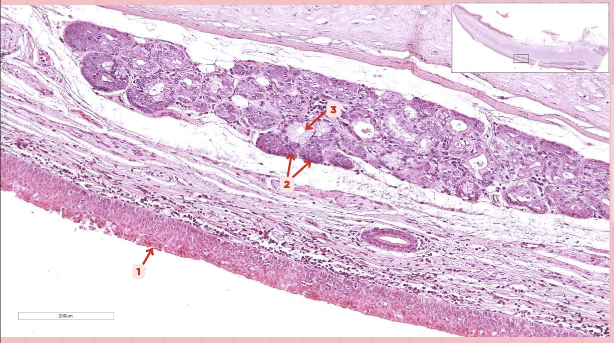
Mucous Glands
What is the gland at #3?

Respiratory Epithelium
Identify the structure labeled as 1.

Laryngeal Ventricle
Space that separates the true and false vocal cords (#4)

True Vocal Cord
The #2 consists of a lamina propria that includes the vocalis ligament and a group of skeletal muscles known as the vocalis muscle.
Vocalis Ligament
Identify the structure labeled as 1.

True Vocal Cord
Identify the structure labeled as 2.

Vocalis Muscle
Identify the structure labeled as 3.

Laryngeal Ventricle
Identify the structure labeled as 4.

False Vocal Cord
Identify the structure labeled as 5.
Lumen
It is a large ovoid space that occupies the center of the trachea.

Thyroid gland
This consist of cystic structures that contain highly eosinophilic material on either side of the trachea.
Lumen
Identify the structure labeled as 1.

Thyroid Gland
Identify the structure labeled as 2.

Esophagus
Identify the structure labeled as 3.

Ciliated Pseudostratified Columnar Epithelium
What is the epithelium of no. 3?

MALT
What type of lymphoid tissue supplies no. 3?

Lamina Propria
Identify the structure labeled as 1.

Basement Membrane
Identify the structure labeled as 2.
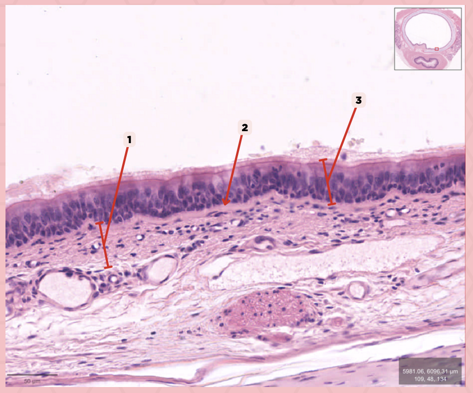
Respiratory Epithelium
Identify the structure labeled as 3.

Tracheal glands
What glands are found in no. 2?

Mixed Tubuloalveolar Glands
Specify the type of glands are tracheal glands.
Mucosa
Identify the structure labeled as 1.

Submucosa
Identify the structure labeled as 2.

Mucous gland
Identify the structure labeled as 3.

Serous gland
Identify the structure labeled as 4.

Loose connective tissue
What type of connective tissue comprise no. 3?

Adipocytes
Together with blood vessels and nerves, these cells are abundant in no. 3.

Perichondrium
Identify the structure labeled as 1.

Hyaline Cartilage
Identify the structure labeled as 2.

Adventitia
Identify the structure labeled as 3.
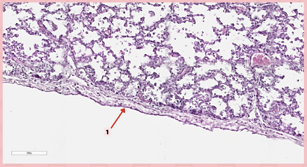
Simple squamous epithelium (Mesothelium)
The surface related to the pleural cavity is in line with what type of epithelium?

Lumens and Interalveolar septa
Name the irregular spaces and the fine threads of tissue that separate them.

Visceral pleura
Identify the structure labeled as 1.
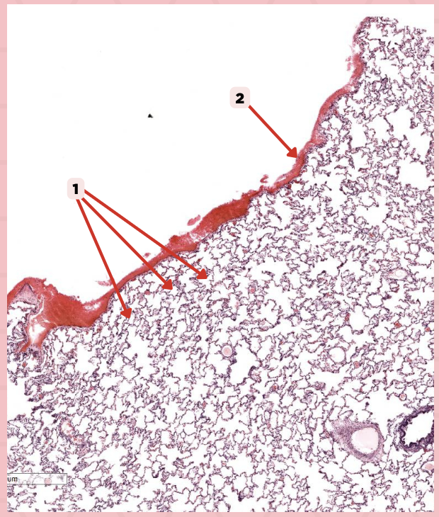
Alveoli
Identify the structure labeled as 2.

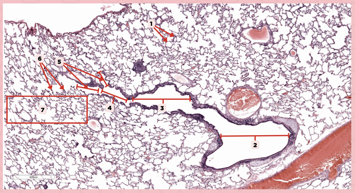
Bigger and smaller intrapulmonary bronchi
What are the two types of intrapulmonary bronchi?

Pseudostratified epithelium (bigger bronchioles), Simple columnar or cuboidal (smaller)
What are the types of epithelium in No. 3?

Interalveolar septa
Identify the structure labeled as 1.

Intrapulmonary bronchi
Identify the structure labeled as 2.

Bronchiole
Identify the structure labeled as 3.

Terminal bronchiole
Identify the structure labeled as 4.

Respiratory bronchiole
Identify the structure labeled as 5.

Alveolar ducts
Identify the structure labeled as 6.

Alveolar sacs
Identify the structure labeled as 7.
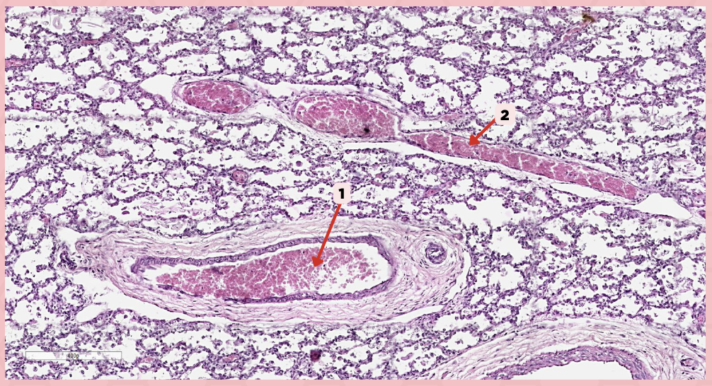
Deoxygenated blood
What type of blood does vessel No. 1 carry?
Connective tissue septae
Where would No. 2 be located?

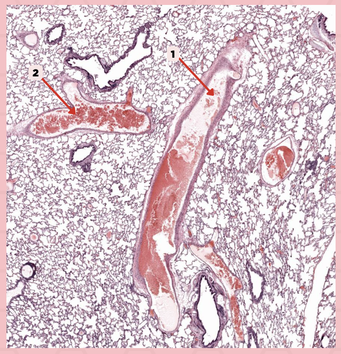
Pulmonary artery
Identify the structure labeled as 1.

Tributary of pulmonary vein
Identify the structure labeled as 2.

Oxygenated blood
What type of blood does this vessel supply?

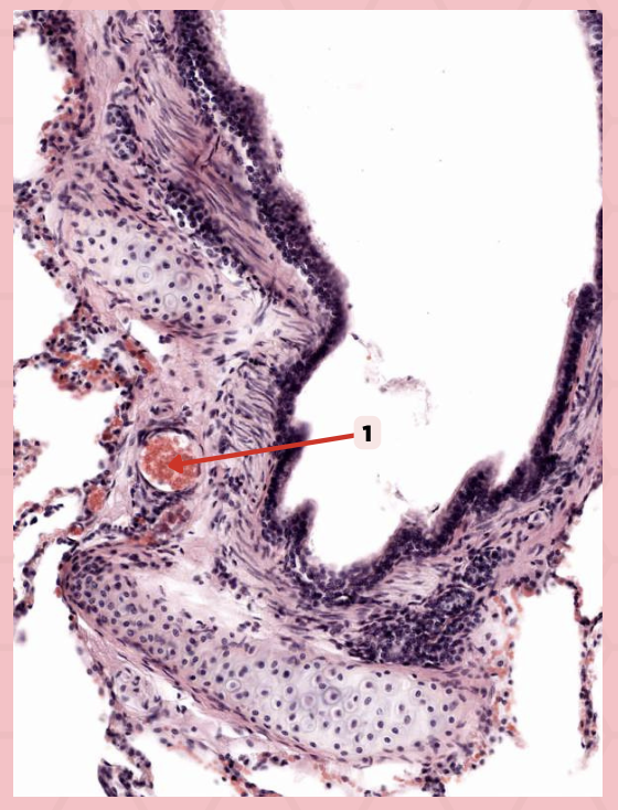
Bronchial artery
Identify the structure labeled as 1.
Type 1 and Type 2 Alveolar Cells
What are the 2 cell types for alveoli?

Simple squamous epithelium
What kind of epithelium are these alveolar cells?

Interalveolar septum
Identify the structure labeled as 1.
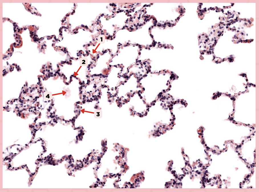
Type I alveolar cell
Identify the structure labeled as 2.

Type II alveolar cell
Identify the structure labeled as 3.

Pulmonary Alveolar Macrophages (Dust Cells)
Identify the structure labeled as 4.

Pulmonary Alveolar Macrophages (Dust Cells)
What are the most numerous cells inside the alveoli?

Pneumocyte Type I
Which of the following labeled structures covers 95% of the alveolar surface?

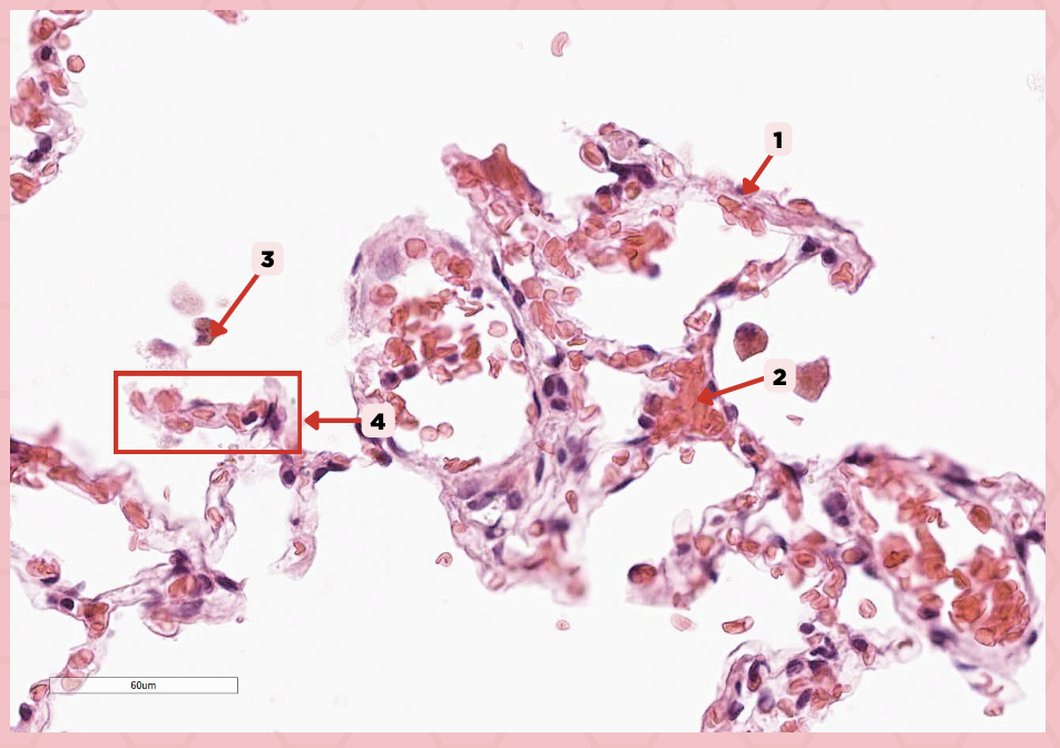
Type I alveolar cell
Identify the structure labeled as 1.

Type II alveolar cell
Identify the structure labeled as 2.

Pulmonary Alveolar Macrophages (Dust Cells)
Identify the structure labeled as 3.

Endothelial Cells
Identify the structure labeled as 4.
Visceral Pleura
What type of pleura is a thin layer of connective tissues that borders on the outside by mesothelium?

Simple Squamous Epithelium
What epithelium lines the red arrow?
Mesothelium
Identify the structure labeled as 1.

Pulmonary Capillaries
Oxygenated blood is collected by structure #2 from what source/structure?

Pulmonary Trunk
From what other structure does structure #1 arise from?