Muscle Tissue
1/55
There's no tags or description
Looks like no tags are added yet.
Name | Mastery | Learn | Test | Matching | Spaced | Call with Kai |
|---|
No analytics yet
Send a link to your students to track their progress
56 Terms
Pericytes
Found wrapped around tiny blood vessels (capillaries and venules).
Act like “support cells/stem cells” that help control blood flow, keep vessels strong, and help in healing.
Found outside capillaries
Myofibroblast
Special cells that appear when there’s injury or wound healing.
They contract (pull the tissue together) to help close wounds.
Think of them as “the body’s stitchers.”
Myoepithelial Cells
Found in external surface of glands (like sweat, salivary, or mammary glands).
They squeeze the glands to push out the fluid (like milk, sweat, saliva).
Think of them as “tiny squeezers in the glands.”
Muscle Cells
The actual cells that make up muscles (skeletal, smooth, cardiac).
They contract to help us move, pump blood, or push food in the gut.
Think of them as the “engines of movement.”
Muscle Tissue
Demonstrates the greatest degree of contractility
Due to muscle cells (muscle fiber)
Highly cellular tissue
Supported and bound together by intercellular material
Consists of connective tissue
Responsible for locomotion and movement
Muscle Fiber/Muscle Cells
Elongated Cell
Called a “fiber” because it’s long, thin, and thread-like
From Mesoderm EXCEPT iris (Ectoderm)
Enveloped by Basal Lamina (Made of reticular tissue)
Cell membrane of Muscle Fiber/Tissue
Sarcolemma
Cytoplasm of Muscle Fiber/Tissue
Sarcoplasm
Smooth Endoplasmic Reticulum of Muscle Fiber/Tissue
Sarcoplasmic Reticulum
Mitochondria of Muscle Fiber/Tissue
Sarcosome
Actin & Myosin
Inside: Filled with special proteins _____ that allow contraction.
Myofibrils & Sarcomeres
Internal unit: Contains _____ → which are made up of repeating units called _____ (the contractile units).
Skeletal Muscle
Striated (striped) due to Sarcomere → has light and dark bands under the microscope.
Contraction → Quick & Forceful
Forms Mouse Shaped organs → Muscles or Skeletal Muscles
Voluntary → meaning we can control it consciously (EX: Limbs, Body wall, Face) (NOT AN EX: Pharynx and upper part of Esophagus)
It is typically attached at either end by a DENSE CONNECTIVE TISSUE called Tendon to a part of the SKELETAL SYSTEM called Bone or Cartilage. The attachment are referred to as Origin and Insertion.
Tendon
To what DENSE CONNECTIVE TISSUE is the Skeletal Muscle attached?
Bone or Cartilage
Skeletal muscle is attached to Tendon to a part of the SKELETAL SYSTEM called?
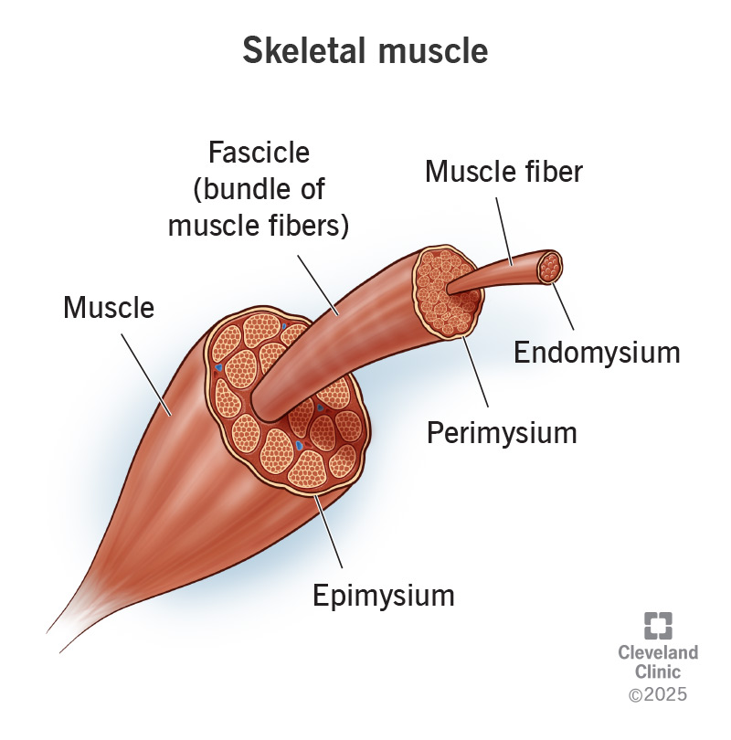
Muscle Fiber
The basic structural unit of skeletal muscle.
Long, cylindrical, multinucleated cells that contain myofibrils.
Myofibrils are made up of sarcomeres (the contractile units with thin and thick filaments).
Fascicle
A bundle of muscle fibers grouped together.
Surrounded by connective tissue called perimysium.
Fascicles give muscles their visible grain or striated appearance.
Muscle
The whole skeletal muscle is formed by bundles of fascicles.
Surrounded by a connective tissue covering called epimysium.
Muscles work together with tendons to produce movement by contracting and pulling on bones
Endomysium
Made of: Loose connective tissue (mainly reticular fibers and some collagen) that surround each individual muscle fiber.
Contains capillaries and nerves that supply the muscle cells.
Provides support and helps transmit force from one fiber to the next.
Perimysium
Made of: Dense irregular connective tissue sheath that surrounds a bundle of muscle fibers, called a fascicle.
Provides pathways for blood vessels and nerves to reach the muscle fibers inside.
Also contributes to the strength of muscle by binding fibers together.
Epimysium
Made of: Dense irregular connective tissue (rich in collagen fibers, mainly reticular fibers and some collagen) that surrounds the entire muscle.
It merges into tendons, which attach muscles to bones.
Protects the muscle from friction and helps distribute force generated during contraction.
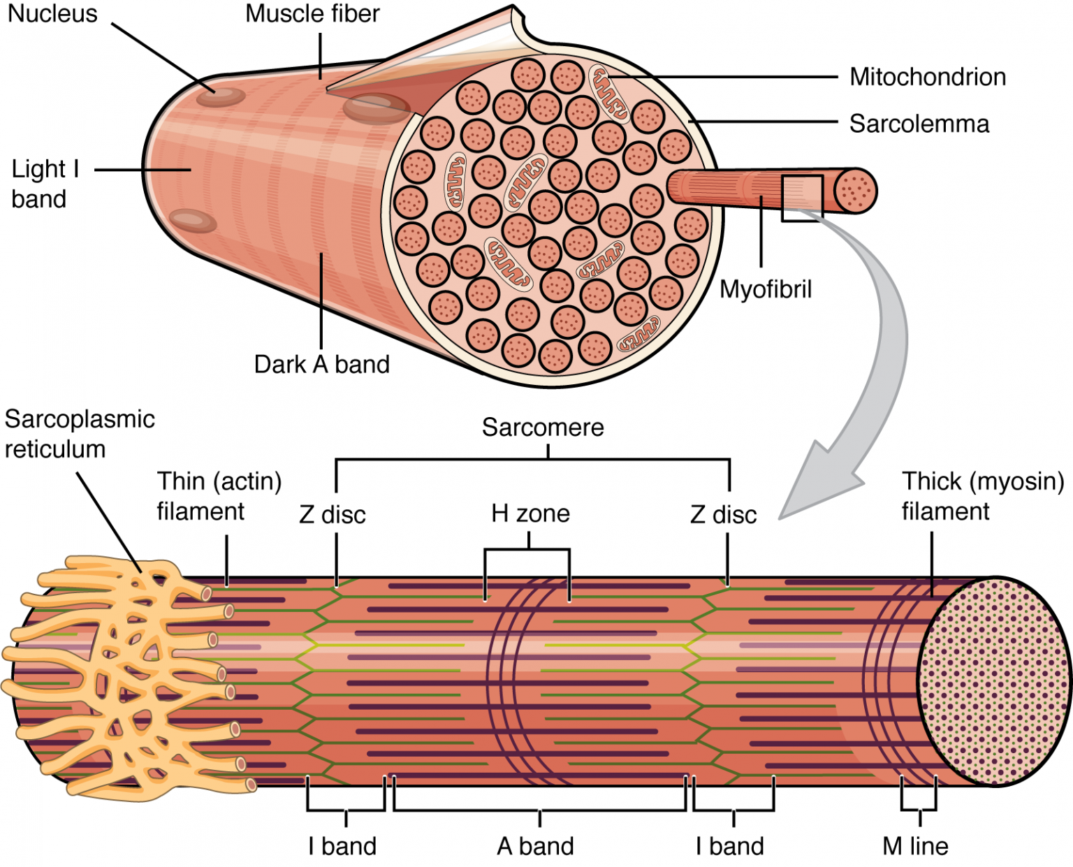
Skeletal Muscle Fiber
Instead, it is long, cylindrical, and multinucleated, meaning it has many nuclei scattered along its length.
Composed of Myofibrils (made up of sarcomeres)
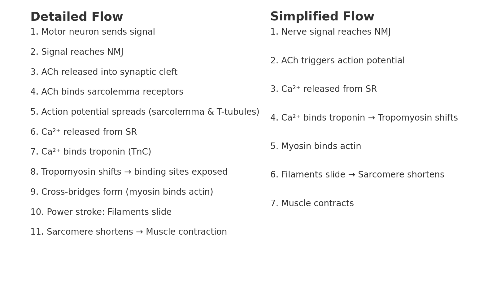
Contract when stimulated by motor neurons at the neuromuscular junction (NMJ).
Myofibrils
Long protein structures inside the fiber, made of repeating sarcomeres (the contractile units).
Sarcoplasmic Reticulum (SR)
Store and release calcium ions (Ca²⁺) needed for muscle contraction.
T-tubules
Carry electrical signals deep inside the fiber.
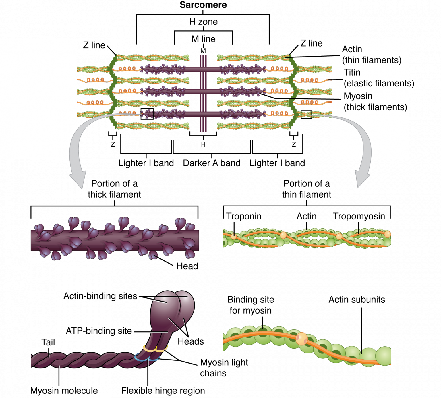
Sarcomere
Made up of Microfilaments.
Thin Filaments: Actin, Tropomyosin, and Troponin
Contraction occurs through the sliding filament model, where actin and myosin filaments slide past each other, shortening the sarcomere. When sarcomeres shorten, the muscle contracts.
It is the region of a myofibril between two Z-discs (Z-lines).
The principal component of thin filaments is F-Actin which consist of 2 strands of Globular and Soluble Actin (G-Actin).
One end of each thin filament is attached to a Z-Line while the other end is free
Thick Filament: Myosin
Occupy the middle zone of a sarcomere
Among the myofilaments, actin and myosin are the most abundant, accounting for about 60% of total muscle for protein.
Red Muscle Fibers
Appearance: Dark red (due to high myoglobin and many mitochondria). Smaller and have richer blood supply than white muscle fibers
Contraction: Slow but sustained; resist fatigue. “Slow-twitch” muscle fibers
Sarcoplasm has more mitochondria, glycogen granules and myoglobulin
White Muscle Fibers
Appearance: Pale/white (low myoglobin, fewer mitochondria).
Contraction: Fast and forceful than Red, but fatigue quickly.
“Fast-switch” muscle fibers
Intermediate Muscle Fibers
Appearance: Pinkish (moderate myoglobin and mitochondria).
Contraction: Faster than red fibers but more resistant to fatigue than white fibers.
Morphology and characteristics are between Red and White
General Sensory Receptors
Simple nerve endings
Vater-Pacinian Corpuscles
Ruffini’s Corpuscles
Proprioceptors
Simple nerve endings
Neuromuscular spindles
Golgi tendon organs
Neuromuscular Spindle
Encapsulated fusiform structure that has several striated muscle fibers (intrafusal fibers)
Present in all skeletal muscles
Embedded in Endomysium and Perimysium
A stretch receptor that detects the degree and velocity stretch applied to a muscle (Prevents over stretching)
Example: When a doctor taps your knee, the muscle spindle detects the sudden stretch → sends a signal → causes the quadriceps to contract.
2 kinds of Intrafusal Fibers
1. Nuclear Bag Fibers
2. Nuclear Chain Fibers
Nuclear Bag Fibers
Structure: The central region of the fiber is swollen and filled with nuclei that “bunch up” like a bag.
Function:
Detect dynamic changes in muscle length (speed and rate of stretch).
Important for sensing when the muscle is being stretched quickly.
Nuclear Chain Fibers
Structure: The nuclei are arranged in a straight chain/row along the length of the fiber.
Function:
Detect static muscle length (steady or maintained stretch).
Important for sensing muscle position rather than speed.
Golgi Tendon Organ
Location: In tendons, near where the muscle fibers connect to the tendon.
Small, one mm-long structures that attach skeletal muscles to their insertions and origins
Measures the tension that is generated by muscle contraction
Sensitive to muscle contraction than stretch
Cardiac Muscle
Location: Found only in the walls of the heart (myocardium).
Striations: Yes, like skeletal muscle, because of sarcomeres (thin and thick filaments).
Control: Involuntary – you cannot consciously control it.
Nucleus: Usually one nucleus per cell (sometimes two).
Branching
Cell junctions demonstrate intercalated disc
Cardiac Muscle Fiber
Cylindrical and much shorter than skeletal muscle cells, branches at their ends unlike skeletal muscle
Contains only 1 to 2 nuclei, centrally located
Sarcoplasm is more abundant and mitochondnia more numerous and larger
With myofibrils with cross striations that are not as prominent as that of skeletal muscle cell
Contraction of cardiac muscle
Similar to the contraction of skeletal muscle, except that inside - sarcoplasmic reticulum
Calcium ions also came from the outside of cell
Cells contract without neural stimulation
The impulse that initiates the contractions is generated by the sinoatrial node
Intercalated Disest
Unique to cardiac muscle
Appear as dark, transverse lines that occur at irregular intervals
Purkinje Fibers
Non-contractile cardiac muscle cells specialized to initiate and conduct electrical impulse that controls cardiac contraction
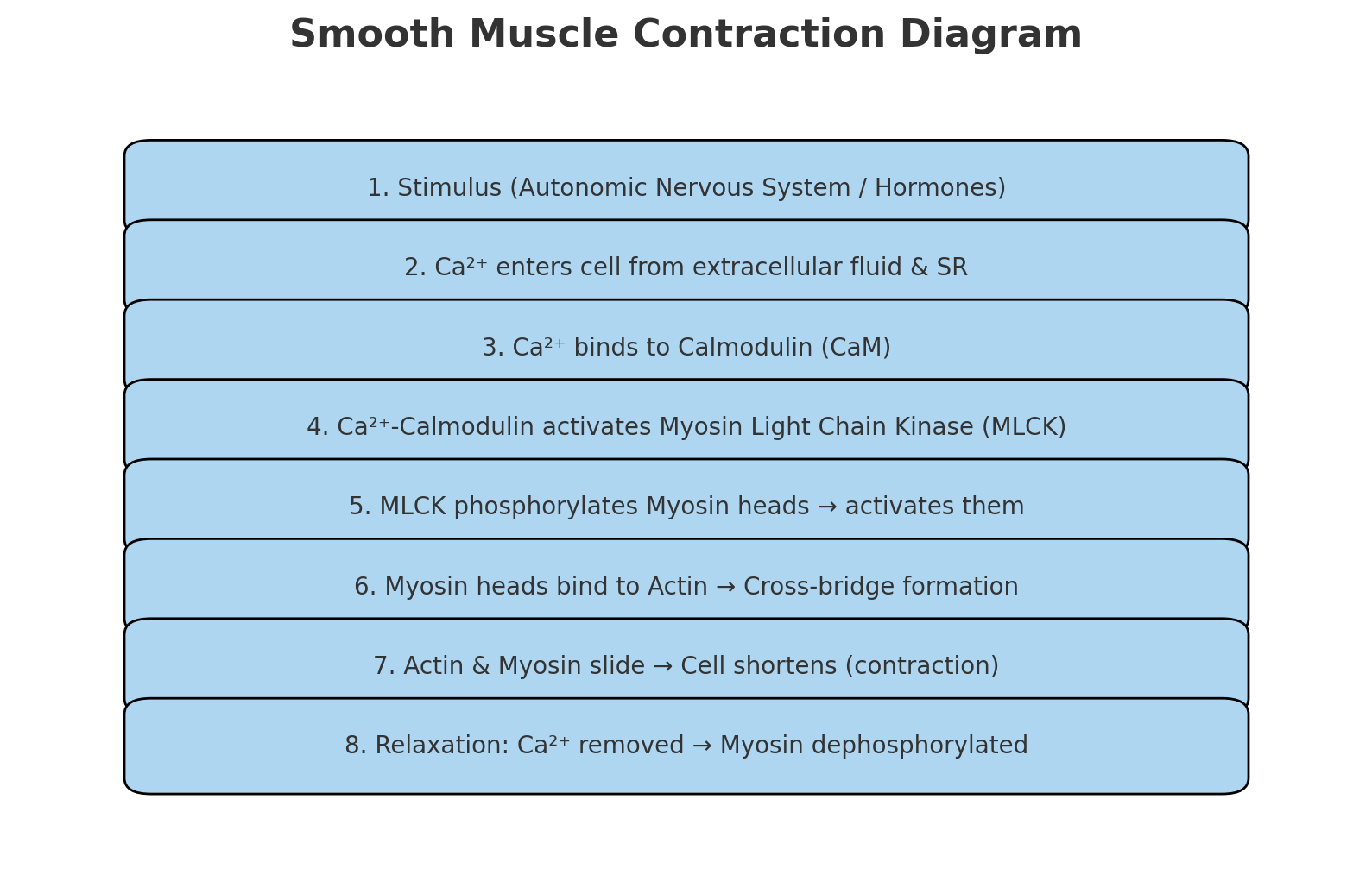
Smooth muscle
Also known as visceral muscle
Involuntary muscles
Slower and less forceful than striated muscles
No striations (unlike skeletal & cardiac muscles) because actin and myosin are arranged differently.
Smooth muscle cell
Fusiform
Single nucleus
Fusiforn, broad at middle, and tapering at both ends
Contains a single, oval nucleus that is located in the thick part of the cell.
Acidophilic sarcoplasm, filled with myofilaments
May be mistaken for collagen fibers
Organization of Smooth Muscle
Fascicles are enveloped by endomysium
Desmosomes and gap junctions attach the sarcolemma of adjacent cells
Skeletal musc
Repair and Regeneration: Skeletal Muscle Tissue
Capable of regeneration despite its inability to undergo cell division.
Satellite cells (myeloblast - like stem cells) - source of new skeletal muscle cells
Repair and Regeneration: Smooth Muscle Tissue
Can regenerate from a type of stem cell called a “Pericyte”, which is found in some blood vessels. Pericytes allow smooth muscle cells to regenerate and repair much more readily than skeletal and cardiac muscle tissue.
Repair and Regeneration: Cardiac Muscle Cells
Have very limited to no regenerative capabilities
Smooth Muscle Contracyion
2 strands of Globular and Soluble Actin (G-Actin)
Coiled around each other much like the fibers of a rope
Neuromuscular junction (NMJ)
Where Skeletal muscle fibers contract when stimulated by motor neurons