Ap Bio Unit 2: Cells
Cytology- the study of cells
Cell Theory
All living things are made of cells
Cell is the basic unit of life
All cells come from pre-existing cells
Cytology Techniques
Light Microscopy- up to 1000x magnification
Electron microscopy- up to 10,000,000x magnification
Cell fractionation- isolate different components of cells for a detailed study
Types of Cells
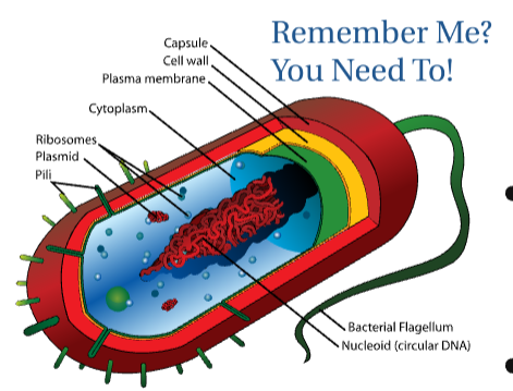
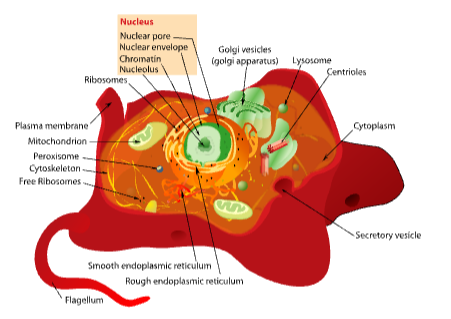
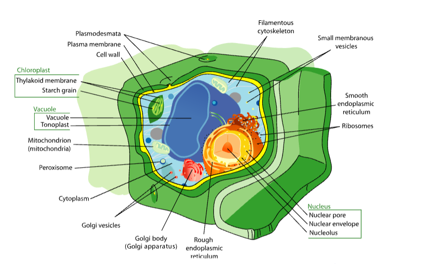
Prokaryotic- Simpler, smaller cells that are more abundant Unicellular Ex) | Eukaryotic- Lots of membrane-bound organelles BOTH multicellular and Unicellular Unicellular: protists, yeast Multicellular: plant, animal, mushrooms+ |
No nucleus No membrane-bound organelles Can have: Cell wall, plasma membrane, capsule, Nuceloid (circular DNA), ribosomes, cytoplasm, plasmid, pili, flagella | ALL: Plasma membrane, cytoplasm, nucelus, smooth/rough ER, ribosomes, cytoskeleton, golgi body, plasma membrane mitochondria, vesicles Plant ONLY: cell wall, vacuole, chloroplast Animal ONLY: centrioles, lysosomes, extracellular matrix (ECM), can have flagella |
 |   |
The Utility of Membrane-Bound Organelles
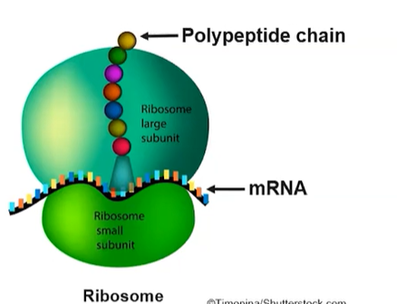
Nucelus -Site of DNA storage and replication—relays info to ribosomes -Nuclear Envelope- Double membrane surrounds the nucelus with protein pore channels 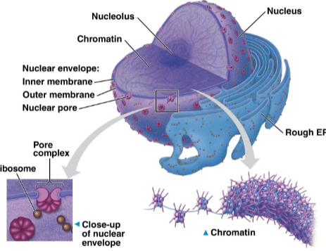 Nuceleolus -Region of the nucelus where ribosomal RNA genes are concentrated | Ribosomes -Site of protein synthesis using RNA transcript of a gene -Complex of RNA + proteins with 2 subunits (large + small) 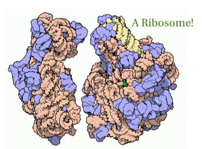 Types of Ribosomes: based on PROTEINS made
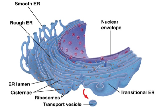
| Endoplasmic Reticulum- netwoek of membrane channels attached to a nucelar membrane Rough ER:
Smooth ER:
 |
Vesicles -Small compartment surrounded by membrane Many functions: | Golgi Apparatus -Modifies, sorts, + packages proteins & lipids for delivery “UPS of the cell” -Series of flattened, membrane-bound sacs 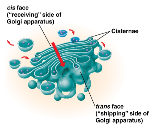 | Plasma Membrane -Controls movement of materials in/out the cell + communication between cell/environment -Phospholipids bilayer with embedded proteins 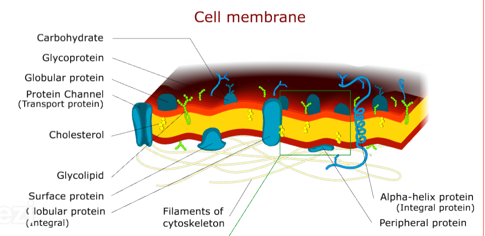 Membrane-protein functions:
|
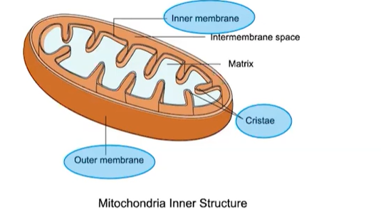
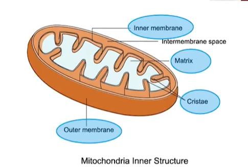
Mitochondria -Converts glucose to ATP energy through aerobic cellular respiration, in charge of apoptosis (programmed cell death) Structure:
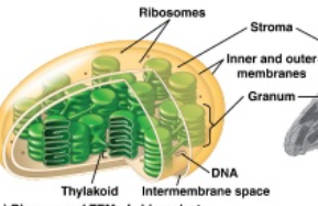
*Reproduce independently of the cell 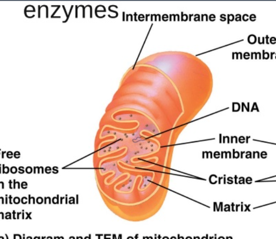 | Chloroplasts -Photosynthesis: building (anabolism) of sugar from ATP, CO2 and light with O2 byproduct -Structure:
 | Lysosomes -Digestion of waste materials, damaged cell parts, large molecules, sometimes apoptosis -Sac full of digestive (hydrolytic) enzymes 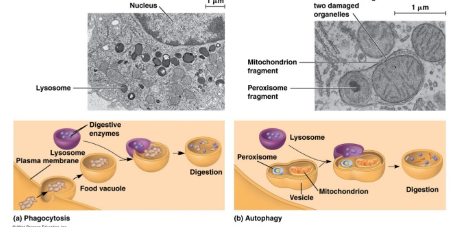 Lysosomal Storage diseases: -Lysosome picks up molecules but cnanot digest → grow larger until it disrupts cell/organ function -Often fatal |
Peroxisomes -Digestive sac that breaks down fatty acids -Detoxifies poisons like alcohol -Produces peroxide (H2O2) | Vacuoles -Storage
-Membraneous sac full of storage materials | Cytoskeleton Functions:
-Network of structural proteins extending throughout the cytoplasm assembled from protein subunits
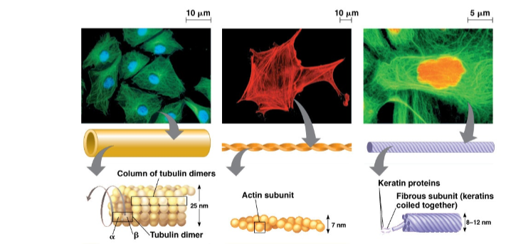 Cilia + Flagella: -Motility (movement) related extension of cytoskeletal proteins Centrosome: -Microtube-oganizing center ONLY in animal cells 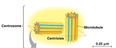 |
Cell Wall -Provide structural support -Cross-linked netwoek of structural polysaccharides
| Extracellular Matrix (ECM) -Cell anchorage/cell communication ONLY in animal cells -Network of connective proteins and proteoglycan molecules outside the cell membrane 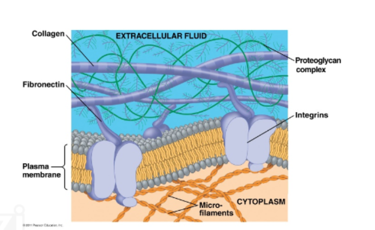 | Intercellular Junctions* -Proteins that connect cells to other cells Open Junctions: ALLOW communication and exchange of materials
Closed Junctions: PREVENT movement of substances between cells
|
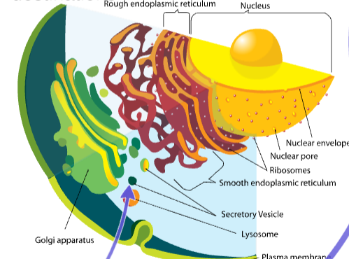
The Endomembrane System- how eukaryotic cells send proteins from ribosomes → destinations
Organelles involved: Ribosomes, Endoplasmic reticulum, Nucelus, Golgi apparatus, Vesicle
Ex. Pathway of processing/Packaging a secretory protein: Secretory vesicles → rough ER → Golgi body → Membrane
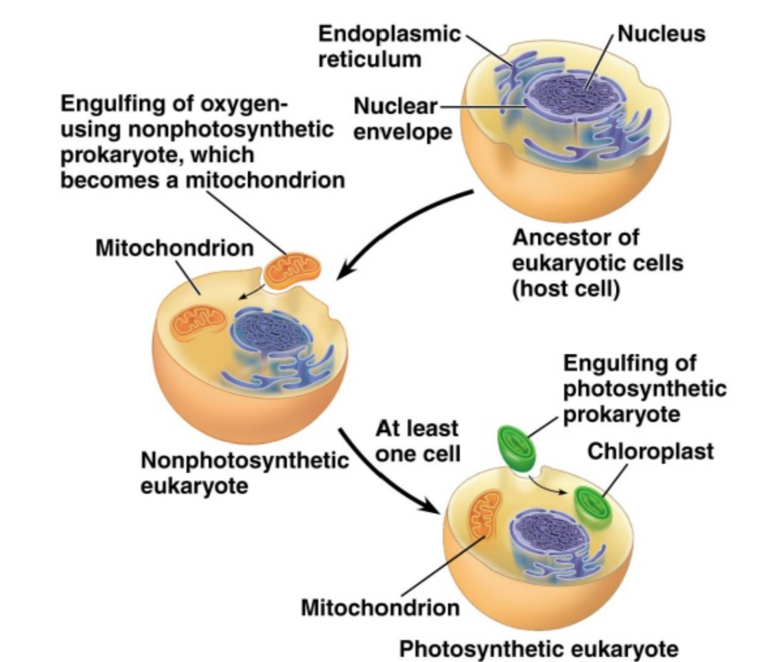
Endosymbiotic Theory- Explains the origins of eukaryotes
Chloroplasts & Mitochondria arose from endosymbiosis
Membrane-bound organelles evolved from once free-living prokaryotes (aerobic bacteria and photosynthetic bacteria) engulfed by a host cell w(ancestor of eukaryotic cells)A
Evidence:
Both have their own DNA, DOUBLE membrane, ribosomes, enzymes
Both divide independently
Compartimentalization:
Refers to way eukaryotic cells are divided into membrane-bound organelles with specialized functions
Purpose: More efficient movement of nutrients/materials through cell through increasing surface area + increased metabolic efficiency
Cells are small to maintain a LARGER/HIGHER surface area : volume ratio
Compartimentalization helps bigger cells like eukaryotes to get nutrients to the center more efficiently
AP Classroom Videos
2.1 Subcellular Components
ALL living cells contain a genomse and ribosomes—reflect the common ancestry of all life
Ribosomes synthesize protein acording to the mRNA sequence and the instructions are encoded in that mRNA sequence originate from the genome of the cell—free floating not membrane-bound
Consists of TWO subunits not membrane enclosed
Made of ribosomal RNA (rRNA) and proteins
Endoplastic Reticulum- network of membrane tubes withn the cytoplasm of eukaryotic cells
Rough ER
Ribosomes attached to membrane
Compartimentalizes cell b/c associated with packaging the newly synthesized proteins made by attached ribosomes for possible export from cell
Smooth ER
No ribosomes attached
Detoxification and lipid synthesis
Golgi Apparatus
Series of flattened membrane-bound sacs in eukaryotes
Correct folding + chemical modification of newly synthesized proteins and packaging for protein trafficking
Mitochondria
Double membrane—outer and inner
Outer is smooth—inner is highly convoluted forming folds called cristae
Functions in production of ATP energy
Lysosome
Membrane-enclosed sacs found in some eukaryotic cells with hydrolytic enzymes
Used to digest a variety of materials like damaged cells or macromolecules
Vacuoles
Membrane-bound sacs in eukaryotes
Variety of roles from storage to release of waste
Chloroplasts
In eukaryotic cells like plants/algae
Double outer membrane
Captures energy from the sun and producing sugar through photosynthesis
2.2 Cell Structure and Function
Chloroplasts- photosynthesis
Thylakoid
Highly folded membrane compartments organized in stacks called grana
Contain chlorophyll pigments that comprise the photosystems and electron transport proteins found between the photosystems embedded in the thylakoid membrane
LIGHT-DEPENDENT reactions occur here
*Folding of membranes increases efficiency of reactiosn
Stroma
Fluid between inner chloroplast membrane and outer thylakoids
Carbon fixation (Calvin-Benson cycle) reactions occur here
Mitochondria- metabolic reactions
Double membrane provides compartments for different metabolic reactions
Krebs Cycle reactions occur in the MATRIX of the mitochondria
Electron transport/ATP synthesis occur in the inner mitochondrial membrane
Folding of inner membrane increases surface area enabling more ATP production
Vacuole
In plants vacuoles aid in retention of water for turgor pressure
Turgor Pressure- Internal cellular force caused by water pushing up against membrane and cell wall
Lysosome
Intracellular digestion
Recycling of organic material
Programmed cell death (apoptosis)
Endoplasmic Reticulum
Mechanical support
Intracelullar transport
Rough ER protein synthesis on the bound ribosomes
2.3 Cell Size
Cells are typically small
Moving materials in and out of cells gets more difficult the larger a cell is—smaller cells more efficient
Smaller cells have higher surface area to volume ratios thus more efficient with exchange of materials—needed for demands like exchanging oxygen, removing waste, taking in nutrients
As cells increase in volume relative SA decrease making it larger for larger cells to meet the demand for internal resources
Complex structures used to increase efficiency
Folded membranes increase surface area
Root hairs on plant root surface increases surface area → more absorption of water and nutrients
Ex. Small Intestine
Membrane folding increases surface area
Outer lining is highly folded containing infger-like projections called villi
Surface of each villi has additional microscopic projections called microvilli further increase surface area
As organisms increase in size their SA:V ration decreases, affecting properties like rate of heat exchange with the evironment
Elephant having large flat ears to dissipate more thermal energy as blood flows closer to the surface
Organisms evolve highly efficient strategies to obtain nutrients and eliminate wastes
Stomatal openings of leaes obtain molecules from and release molecules into the environment
When stomata are open CO2 eneters and O2 and H2O can be released into the atmosphere
Topic 2.4 Plasma Membranes
Cell embranes provide a boundary between the interior of the cell and outside environment
Control transport of materials in and out of cell
Phospholipids are amphipathic—Hydrophilic head hodrophobic tail
Spontaneously form a bi-layer in an aqueous environment
Tails located inside the bilayer
Heads exposed to the aqueous envrionment outside
Peripheral proteins
Losslely bound to the surface of the membrane
Hydrophilic with charged and polar side groups
Intergral proteins
Span the membrane
Hydrophilic with charged and polar side groups
Hydrophobic with nonpolar side grousp that penetrate hydrophobic interior of bilayer
Ex. Transmembrane proteins
Membrane protein functions:
Transport
Cell-Cell recognition
Enzymatic activity
Signal transduction
Intercellular joining
Attachment for extracellular matrix/cytoskeleton
Structure—a mosaic of protein molecules in a fluid bilayer of phospholipids
Held together primarily by hydrophobic interactions—weaker than covalent bonds
Most lipids and some proteins can shift and flow along the surface of the membrane or across the bilayer
Cholestrol (steroid) is randomly distributed/wedged between phospholipids in the membrane of eukaryotes
Regulates bilayer fluidity under different environmental conditions
Carbohydrates- diversity and location of carbodydrates and lipids enable them to function as markers/identifiers
Glycoproteins- one or more carbohydrates attached to a membrane protein
Glycolipids- lipid with one or more carbohydrates attached
Topic 2.5 Membrane Permeability
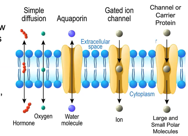
Selective permeability is a consequence of membrane structure
Smaller molecules pass freely
Hydrophilic substances such as large polar molecules and ions CANNOT freely move across the membrane
Hydrophilic substances move through transport proteins
Channel Proteins- a hydrophilic tunnel spanning the membrane that allows specific target molecules to pass through
Carrier Proteins- Spans the membrane and change shape to move a target molecule from one side to another
Small polar molecules like H2O can pass through directly in small amounts
Cell Walls act as a structural boundary:
Protects and maintains the shape of the cell
Prevents against cellular rupture when internal water pressure is high
helps plants stand up against the force of gravity
Cell Walls act as a permeable boundary
Plasmodesmata- small holes between plant cells allow the transfer of nutrients, waste, ions
Cell Wall- comprised of complex carbohydrates
Plants- Cellulose (Polysaccharide)
Fungi- Chitlin (Polysaccharide)
Prokaryotes- peptidoglycan (polymer consisting of sugar and amino acids)
Topic 2.6 Membrane Transport
Concentration gradient
When a solute is more concentrated in one area than another
Membrane separates two different concentrations of molecules
Passive transport:
Net movement of molecules from high to low without metabolic energy like ATP needed
Primary role in import of materials and export of wastes
Active Transport:
Direct input of energy to move molecules from low to high concentration
Endoctosis requires ENERGY to take in molecules to the cell
Exocytosis- internal vesicles use energy ot fuse with the plasma membrane and secrete large macromolecuels out of cell
Includes: Proteins like signaling proteins, hormones, waste
Topic 2.7 Facilitated Diffusion
Facilitated Diffusion- movement of molecules from high → low concentration through transport proteins
Large and small polar molecules
Large quantities of water pass through aquaporins
Charged ions (NA+ and K+) require channel proteins
Active Transport- moves molecules AGAINST concentration gradient (low → high)
Require carrier proteins called protein pumps
Require metabolic energy (ATP)
Establish and maintain concentration gradients
Cotransport- secondary active transport uses energy fom electrochemical gradient to transport two DIFFERENT ions across the membrane through a protein
Symport- two different ions transported in the SAME direction
Antiport- two different ions transported in the OPPOSITE direction
Cell membrane allows for formation of gradients
Electrochemical gradient
Type of concentration gradient
Membrane potential: electrical potential difference (voltage) across the membrane
Membranes may become POLARIZED by the movement of ions across

Ex. Sodium-Potassium (Na+/K+) Pump contributes to the maintenance of the membrane potential
3 sodium ions pumped for every 2 potassium ions pumped to establish concentration gradient
Topic 2.8 Tonicity and Osmoregulation
Osmosis- diffusion of free water across a selectively permeable membrane
Move larger quantities of water via aquaporins
Osmolarity- total solute concentration in a solution
Water has high solvency
Solute- being dissolved
Solvent- dissolves a solute
Solution- uniformed mixture of one of more solutes dissolvd in a solvent
Tonicity- measurement of the relative concentration of solute between two solutions (in and out of the cell)
Internal cellular environments can be hypotonic, hypertonic, or isotonic to external environments
Hypertonic- MORE solute, less solvent
Isotonic- equal concentrations
Hypotonic- less solute, more solvent
Water moves by osmosis into area with higher solute concentration
Water concentration and solute concentrations are inversely related
Water would diffuse OUT of a hypotonic environment (less solutes) into a hypertonic one (more solutes) OR high water potential to low water potential
Solutes diffuse along their own concentration gradients from hypertonic environment to hypotonic
When a cell is in an isotonic environment a dynamic equilibrium exists with equal amounts of water oving in and out of the cell at equal rates—no net movement of water
Osmoregulation
In plant cells it maintain water balance and allows control of internal solute composition/water potential
Environmental hypertonicity- less cellular solute and more cellular water → Plasmolysis- water leaves the cell
Isotonic- Equal solute and water → Flaccid plant cell
Environmental hypotonicity- more cellular solute and less cellular water → Turgid
Turgidity- The optimum state for plant cells
Cell wall helps maintain homeostasis for plant in environmental hypotonicity
Osmotic pressure high outside of the plant cell (hypotonicity)
Water flows into the plant vacuoles via osmosis → vacuoles expand and press against cell wall
Cell wall expands until it exerts pressure back on the cell → Turgor Pressure
Osmoregulation in animal cells maintains water balance and allows control of internal solute composition/water potential
Environmental hypertonicity- Less cellular solute more cellular water → shriveled
Isotonic solution- equal solute and water → normal state
Environmental hypotonicity- more cellular solute less cellular water → lyse (bursting)
Graphing
Characteristics:
Title- experiment details and what is measured
Labeled axes with units
Scaling—unifrom intervals; scale large enough to analyze data and scale numbers on grid lines
Identifiable lines/bars
Trend line- line of best fit shows overall direction/pattern of data
Line graph
Reveals trends or progress over time for multiple groups/treatments
Tracks changes over time, concentrations, etc.
Scatterplot (X Y Graph)
Determine relationships between two different things
Compare two variables that may/may not have linear relationship
Histogram
How values in data are distributed across equal intervals
Explore relationships between two or more variables
Bar graphs
Comparing multiple groups/treatments
Box and Whisker
Shows variability in sample
Compare distributions in relation to the mean
Dual Y*
Represent relationship between two dependent variables
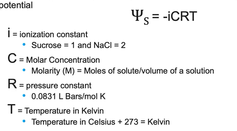
Water Potential
Water potential measures TENDENCY of water to move via osmosis
Calculated from Pressure Potential and Solute Potential
Water potential = pressure potential + solute potential (measured in bars)
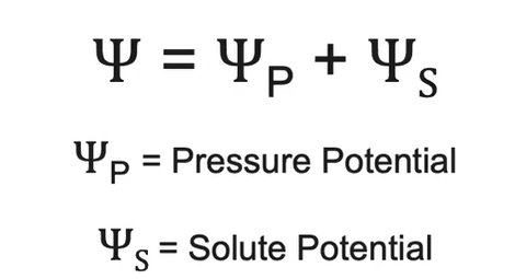
Moves from area of HIGH → LOW water potential areas
More negative/lower water potential = more likely water moves INTO the area
Water potential of PURE water has a value of ZERO in an open container
Osmoregulation and Water Potential
Increasing amount of solute = Decrease in solute potential / decrease in water potential
Increasing water potential = Increase in PRESSURE potential
Decreasing pressure potential = Decrease in water potential*
In an open system, pressure potential is zero, so water potential is equal to the solute potential
Topic 2.9 Mechanisms of Transport
Diffusion- Movement of molecules from high → low concentration
Small nonpolar molecules pass freely (O2, CO2, N2)
Small amounts of very small polar molecules also diffuse
Facilitated Diffusion- movement of molecules from high concentration to low concentration through transport proteins
Large and small polar molecules
Charged ions (Na+, K+) require channel proteins
Large quantities of water move via aquaporins
Differences in relative solute concentrations facilitates osmosis
Active transport- move molecules/ions against concentration gradient from low → high concentration
Protein pumps are carrier proteins in active transport
Require metabolic energy (ATP)
Establishes and maintains concentration gradients
Endocytosis- Cell uses energy to TAKE IN macromolecules/particulate matter by forming new VESCICLES derived from the plasma membrane
Phagocytosis- Cell engulfs/eats large particles then fuses with lysosomes to produce digestive enzymes to break down materials; common in immune cells
Pinocytosis- drinking/uptake of extracellular fluid with dissolved substances
Receptor-mediated endocytosis- Selective—receptor proteins on the membrane capture specific target molecules
Exocytosis- internal vescicles use energy to FUSE with plasma membrane and secrete macromolecules OUT of the cell
Topic 2.10 Compartimentalization
Cells have a plasma membrane that allows them to establish and maintain internal environments different from external environments
Eukaryotic cells have additional internal membranes/membrane-bound organelles that compartmentalize the cell
Cellular compartments allow for various metabolic processes and specific enzymatic reactions ot occur simultaneously → increased cell efficiency
Membrane minimizes competing interactions
Example:
Hydrolytic enzymes of lysosome function at an acidic environment
With this compartmentalization, inside of lysosome can maintain a more acidic pH and allow for efficient hydrolysis to occur while the rest of the cytoplasm can remain a more neutral environment
May lead to cell damage/death if membranes around lysosomes were to burst as it would lead to the release of hydrolytic enzymes released into the cytoplasm that would digest important cellular materials/molecules
Mitochondria membrane folding maximizes surface area for metabolic reactions to occur
Electron transport and ATP synthesis occur in mitochondrial membrane
Folding of inner membrane increases surface area allowing MORE ATP to be made
Chloroplasts membrane folding maximizes surface area for metabolic reactions to occur
Thylakoids highly folded membrane compartments that increase the efficiency of light dependent reactions in the chloroplast
Topic 2.11 Origins of Cell Compartmentalization
Both eukaryotes and prokaryotes have a plasma membrane that separates their internal environment from their surroundings
Prokaryotic cells have an internal nucleoid region containing its genetic material while eukaryotic cells store genetic material in a membrane-bound nucelus
Nucelus and other internal membranes (ER) theorize to have formed from the infoldings of the plasma membrane
Mitochondria evolved from previously free living prokaryotes via endosymbiosis
A free living aerobic prokaryote engulfed by anaerobic cell
The prokaryotic cell did not get digested but eventually formed a symbiotic arrangement
Over time engulfed cell lost some of its independent functionality and became the mitochondria
Chloroplast evolved similarly
Free-living photosynthesic prokaryote engulfed by another cell and formed a mutually beneficial arrangement then lost its independent functionality over time to become the chloroplast
Similarities
Both have a double membrane-regulate passage of materials into and out of cell to maintain a stable internal environment
Both have their own circular DNA encoding genetic information and can reproduce similarly to prokaryotes
Both contain their own ribosomes that synthesize proteins