Lec 9 Circulatory System: Biol 302 Histology Midterm #2
1/55
There's no tags or description
Looks like no tags are added yet.
Name | Mastery | Learn | Test | Matching | Spaced | Call with Kai |
|---|
No analytics yet
Send a link to your students to track their progress
56 Terms
Name the 4 functions and relationships of Circu Sys
1) O2 & nutrients in the blood distributed by the heart.
2) De-O2 blood to the lungs
3) Waste products (i.e. kidney & liver)
4) Transport system
Simple Squamous Epithelium in the Circu Sys is called what?
Endothelium = lines bv + Squamous for diff and transport.
Main Components of Circu Sys
1) Heart - pumps
2) Vascular System - arterial = blood away from heart, venous = blood to heart
3) Lymphatic System - drains fluid back to venous
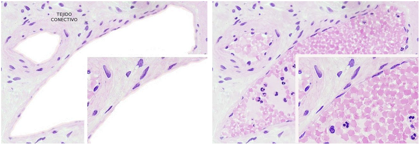
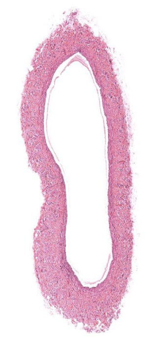
What system is this? And what are its features?
Lymphatic Sys: collects fluid form CT, bv w/ Endothelium, no RBCs = pink
What system is this?
Lymphatic Sys: collects fluid form CT, bv w/ Endothelium, no RBCs = pink
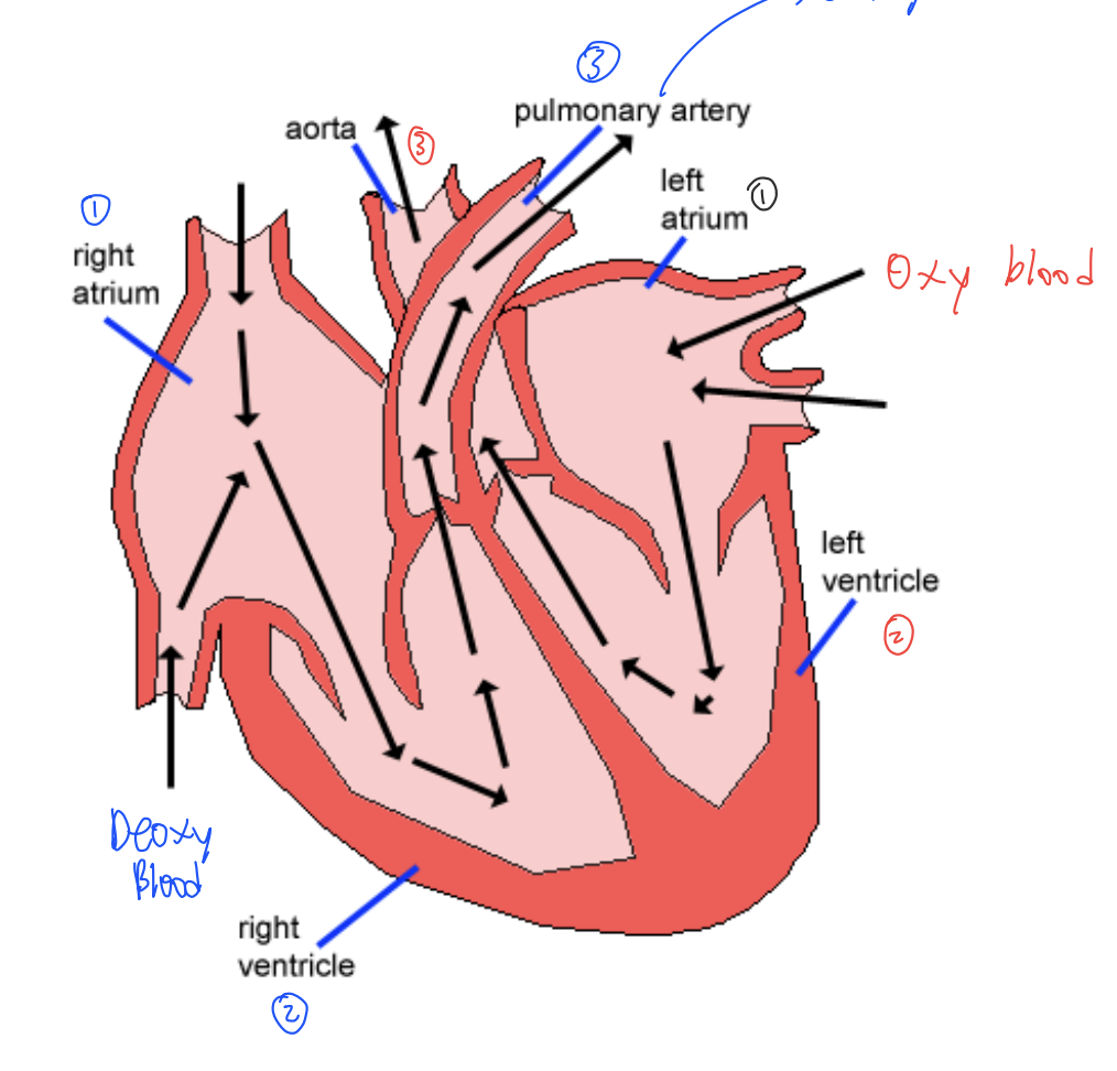
What are the Components of the Heart/which way does blood flow?
Deoxy blood → RA → RV → PA → Lungs
Oxy blood → LA → LV → Aorta
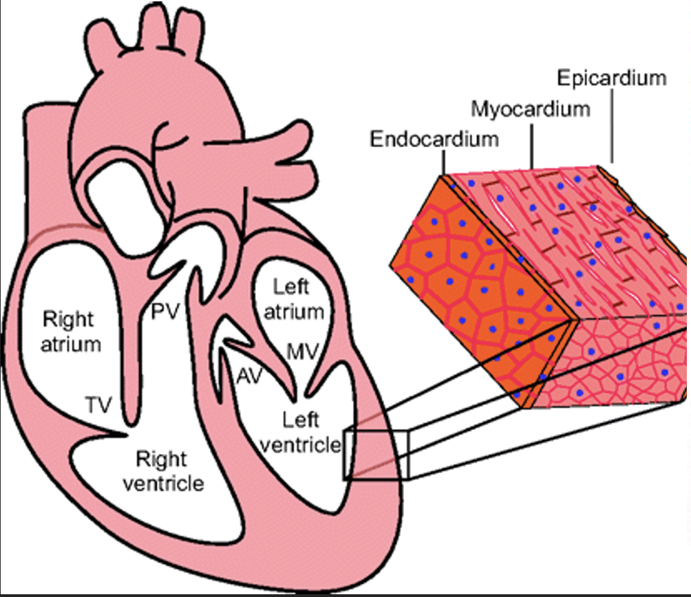
What is the Heart Partitioning?
Endocardium (in), Myocardium (middle), Epicardium (out, and gets ripped off sometimes)
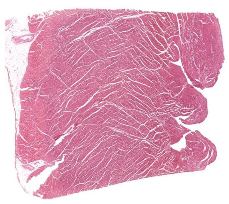
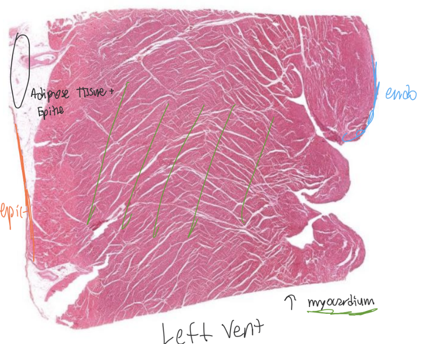
Which ventricle is this?
LV
What are functions of LV?
Gets oxy blood from lungs
Pumps into systemic circu
BV close to heart have characteristics that match its F
Which ventricle is this?
RV
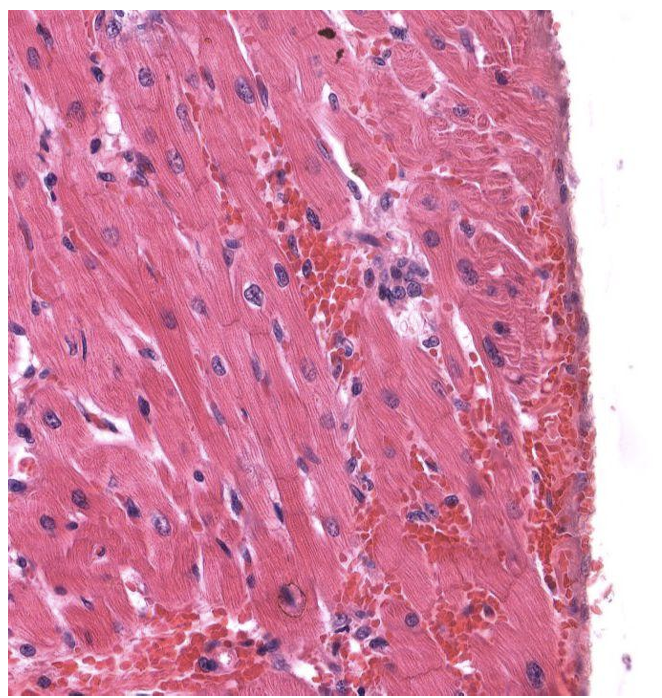
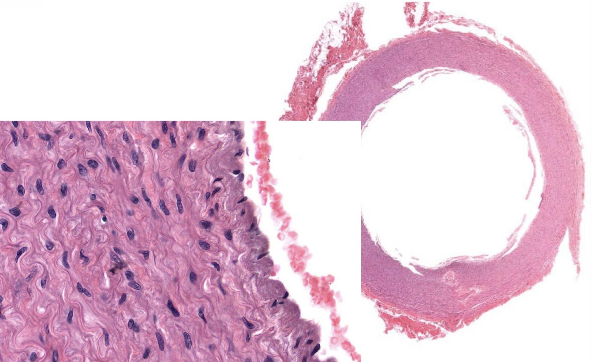
Identify Which partition this is and Where?
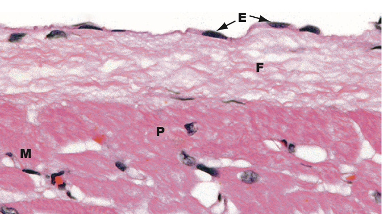
I = Endocardium in Ventricle.
Has simple squam epit and is supported by few fibres of CT.
Identify Which partition this is and Where?
I = Endocardium in Atrium.
Has simple squam epit and is supported by thin layer of DICT.
Identify Which partition this is and Where?
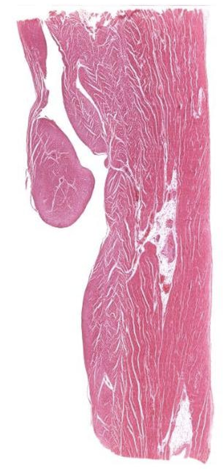
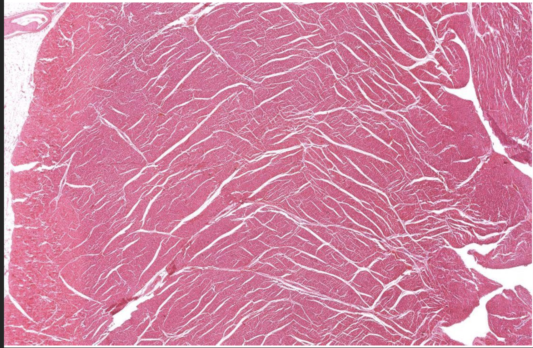
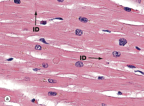
I = Myocardium in Ventricle.
(Char = Cardiac M. Is the thickest layer v/ ventricular folds called trabecular carnae)
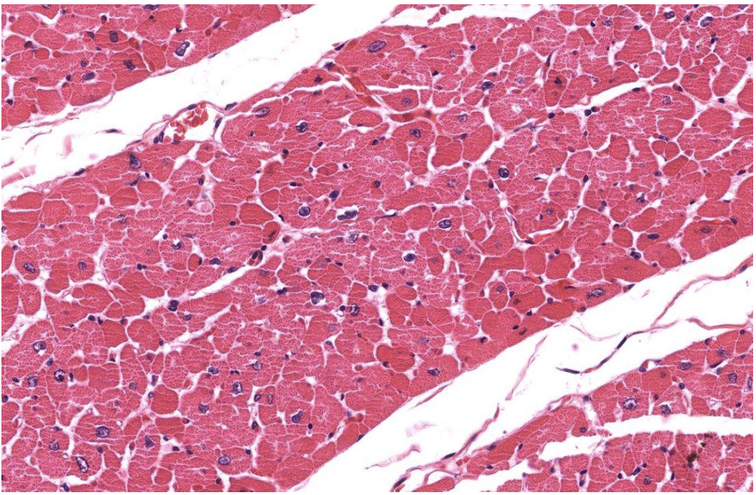
Identify Which partition this is?
I = Myocardium in Ventricle.
(Char = Cardiac M. Is the thickest layer v/ ventricular folds called trabecular carnae)
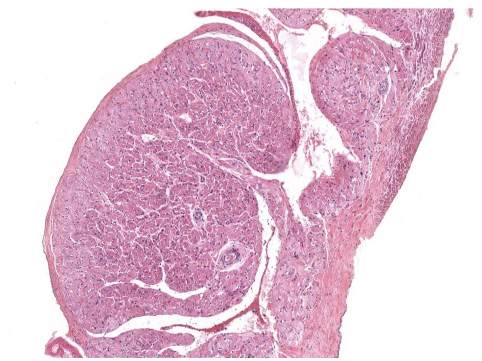
Identify Which partition this is and Where?
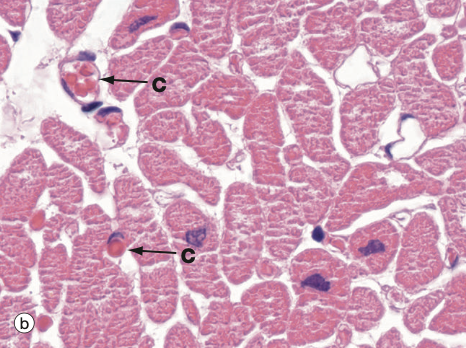
I = Myocardium in Atrium.
Has large abundance of Cardiac T
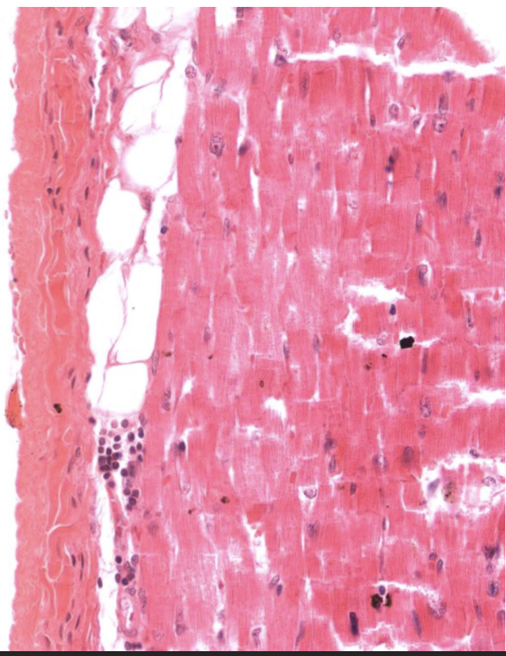
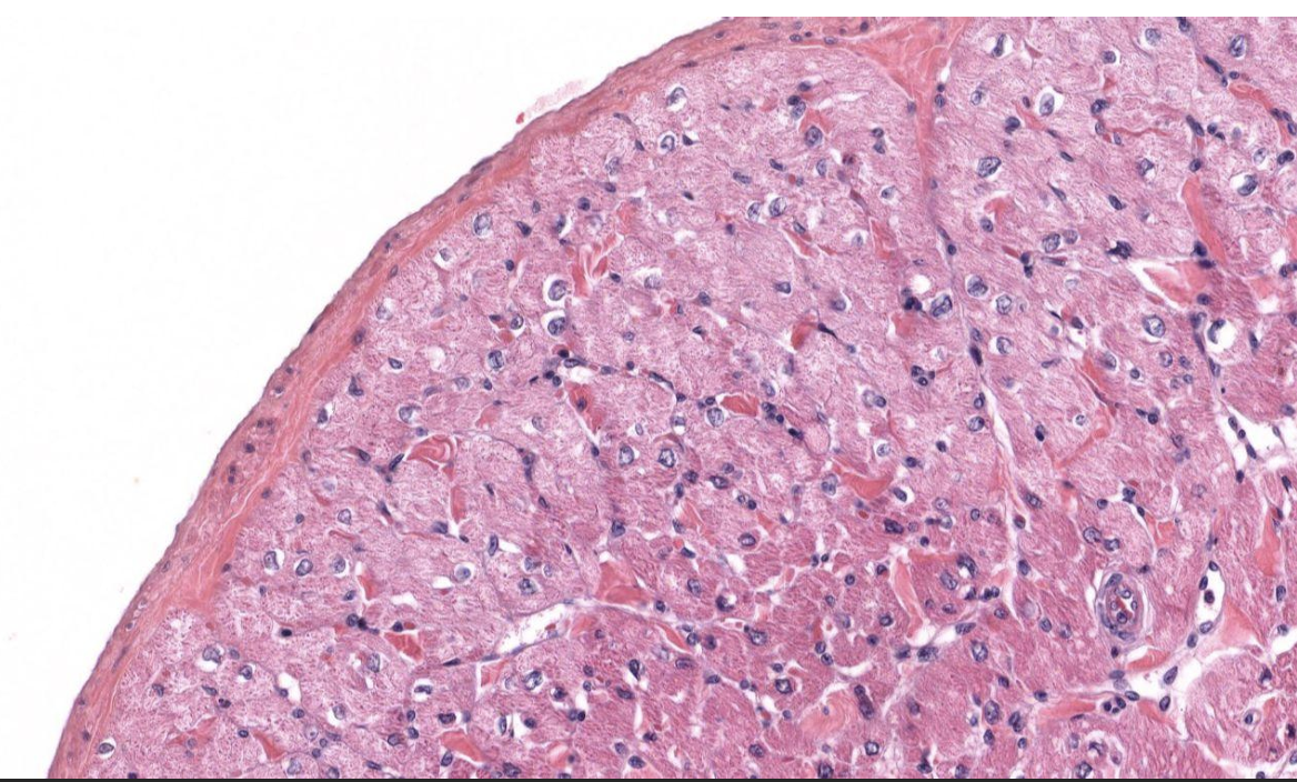
Identify Which partition this is and Where?
I = Epicardium in Ventricle.
Outer mesothelial (simple squam epit) cells missing, supported by DICT on outside and has Loose Unilocular Adipose CT.
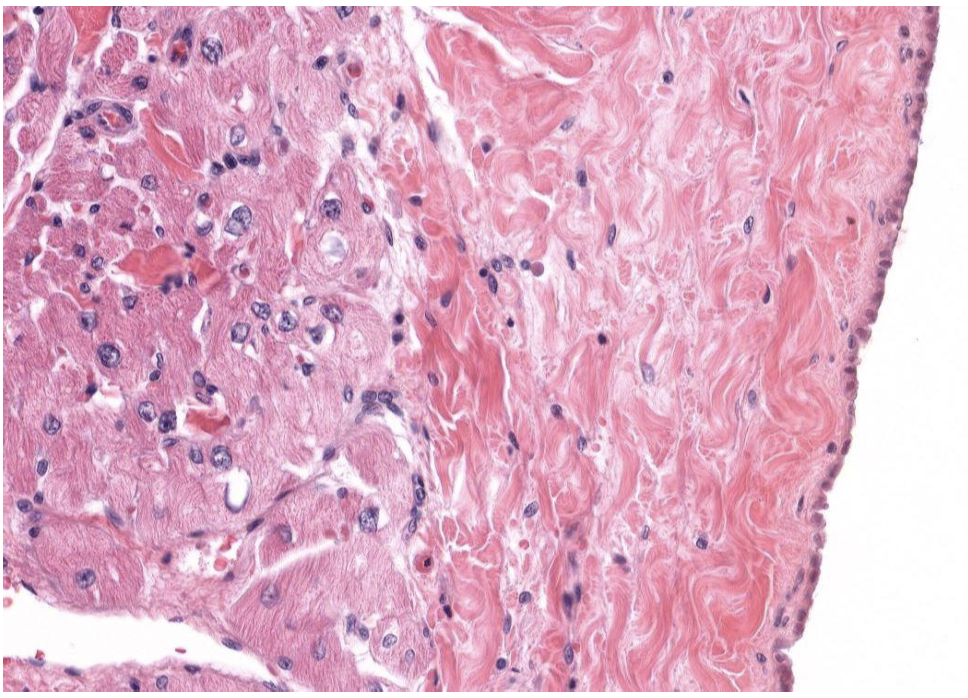
Identify Which partition this is and Where?
I = Epicardium (right side) and Myocardium (Left) in Atrium.
Outer mesothelial (simple squam epit), supported by DICT.
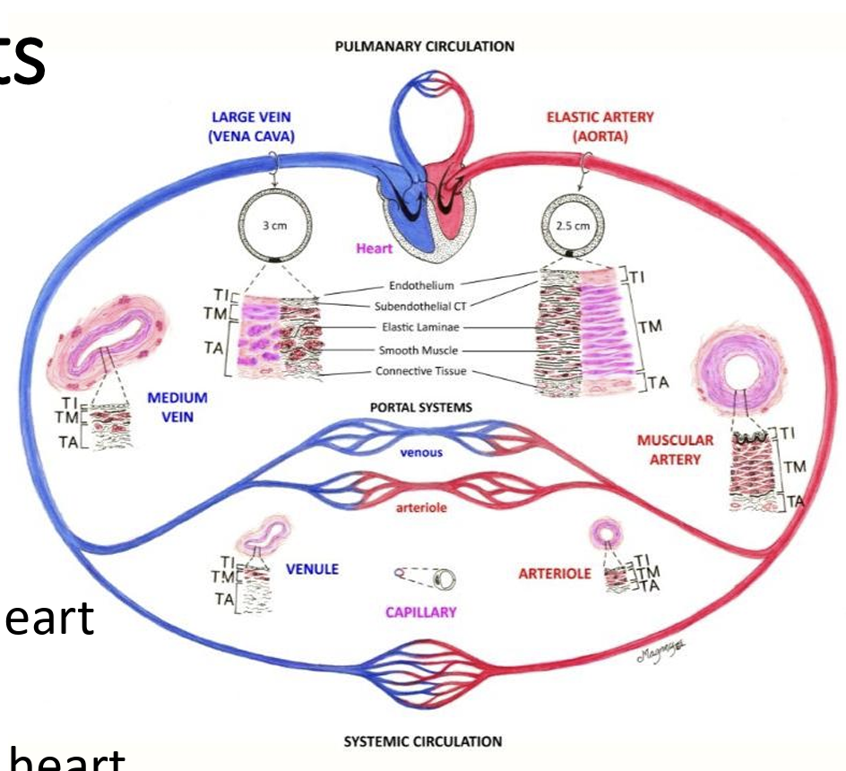
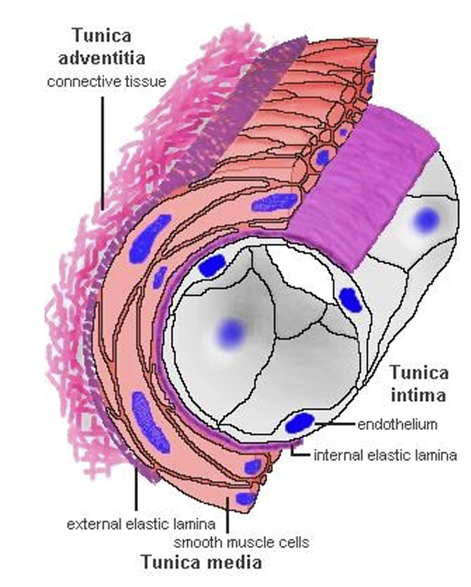
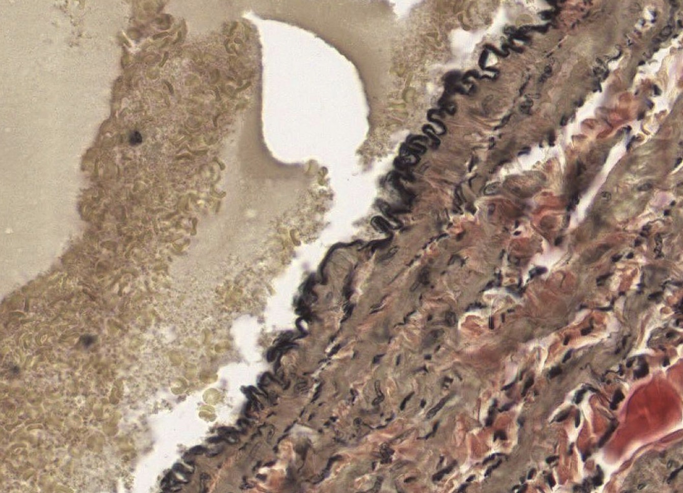
Name the Bv Nomenclature for Layers
Tunica intima (inside, endothelium)
Tunica media (middle, smooth M)
Tunica adventitia (area away from lumen/outside, DICT)
Walls ate 1 layer of M, all same direction and not oblique.
What is pictured?
Neuromuscular bundle = vein, artery, and nerve tgth
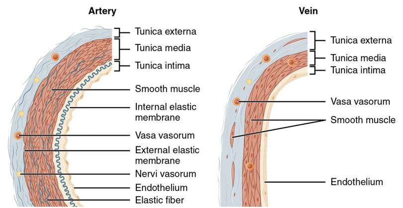
What are Layers in Veins & Arteries
Tunica externa, media, and intima.
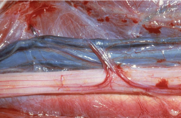
What is pictured?
Neuromuscular bundle = vein (blue), artery (red line), and nerve (white) tgth
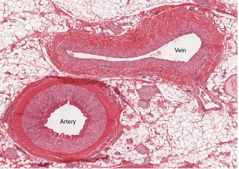
Identify what is pictured?
Vein (big Te)
Identify what is pictured?
Artery (big Tm)
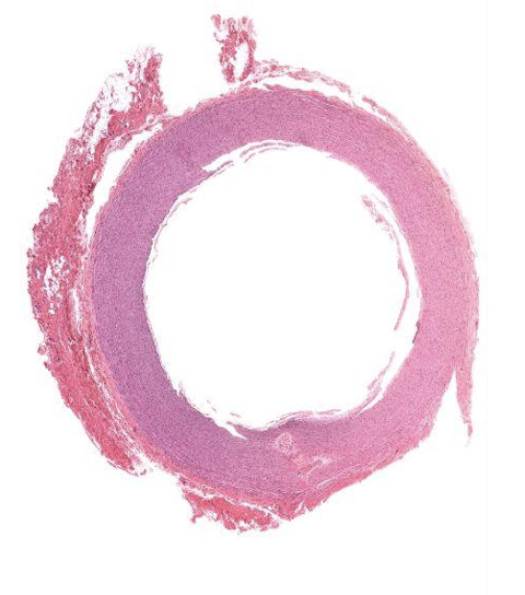
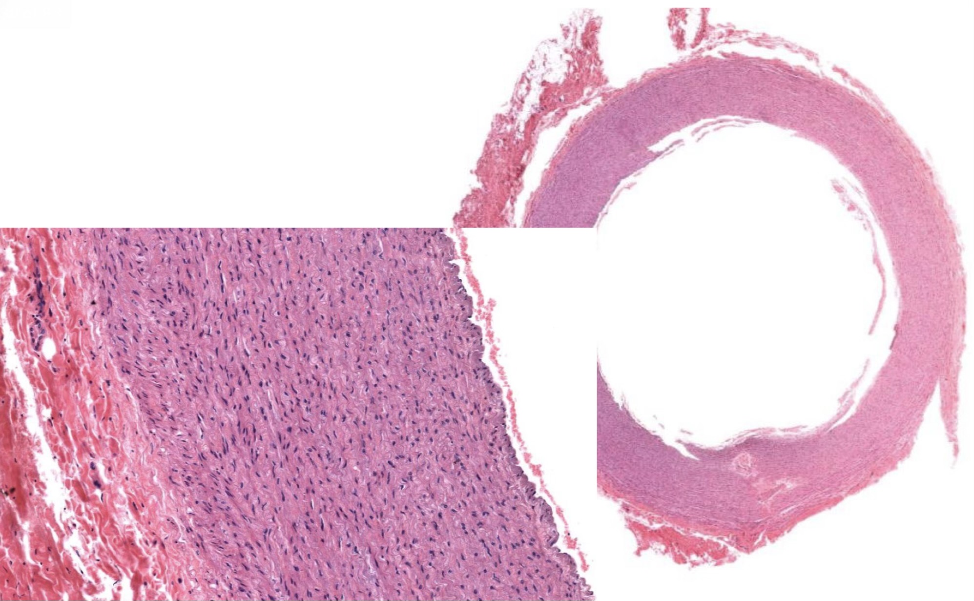
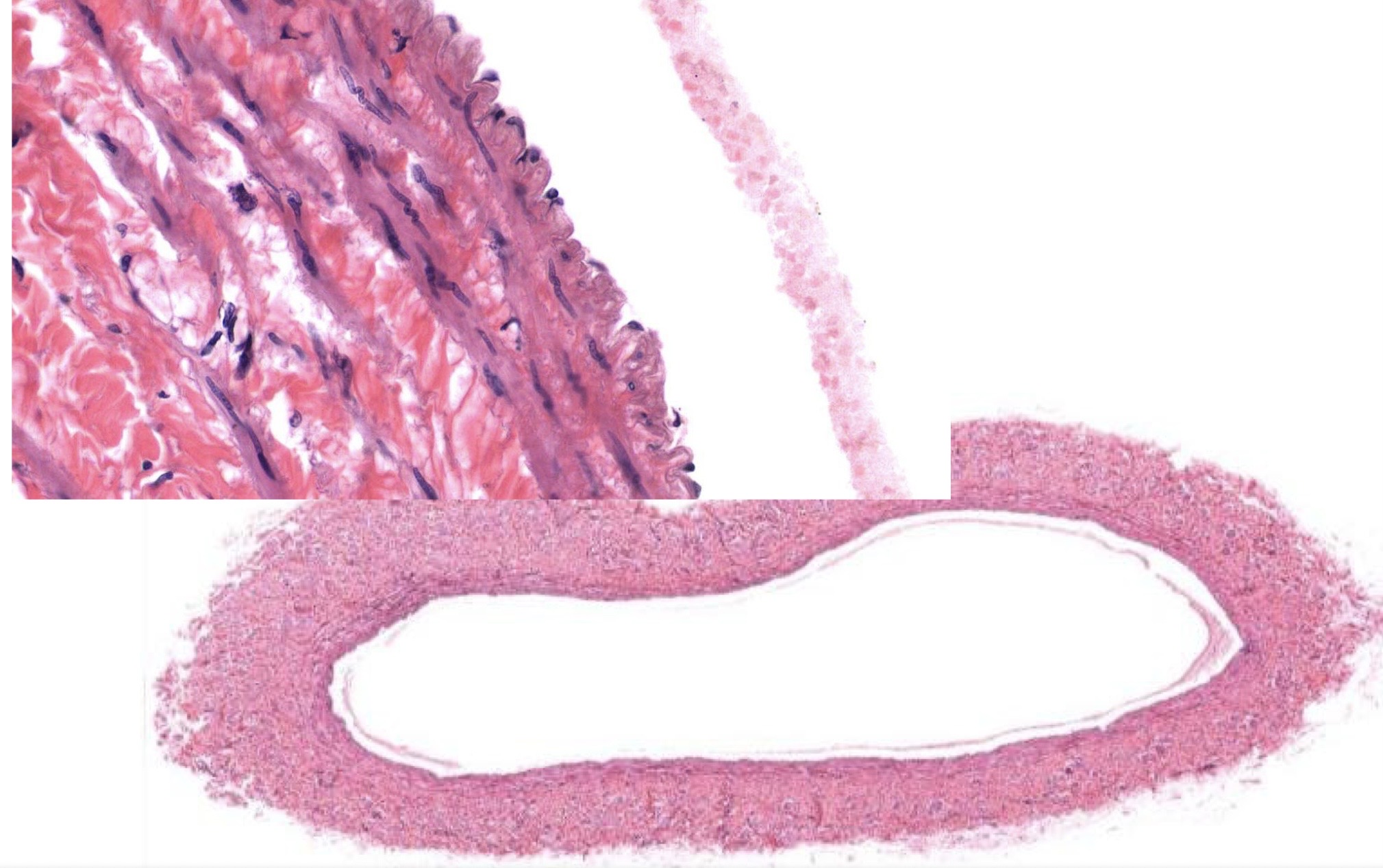
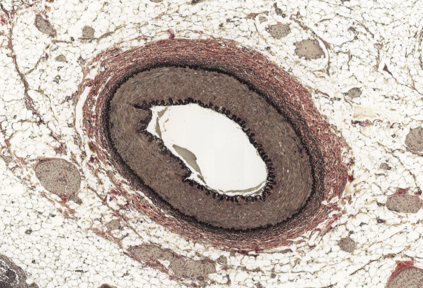
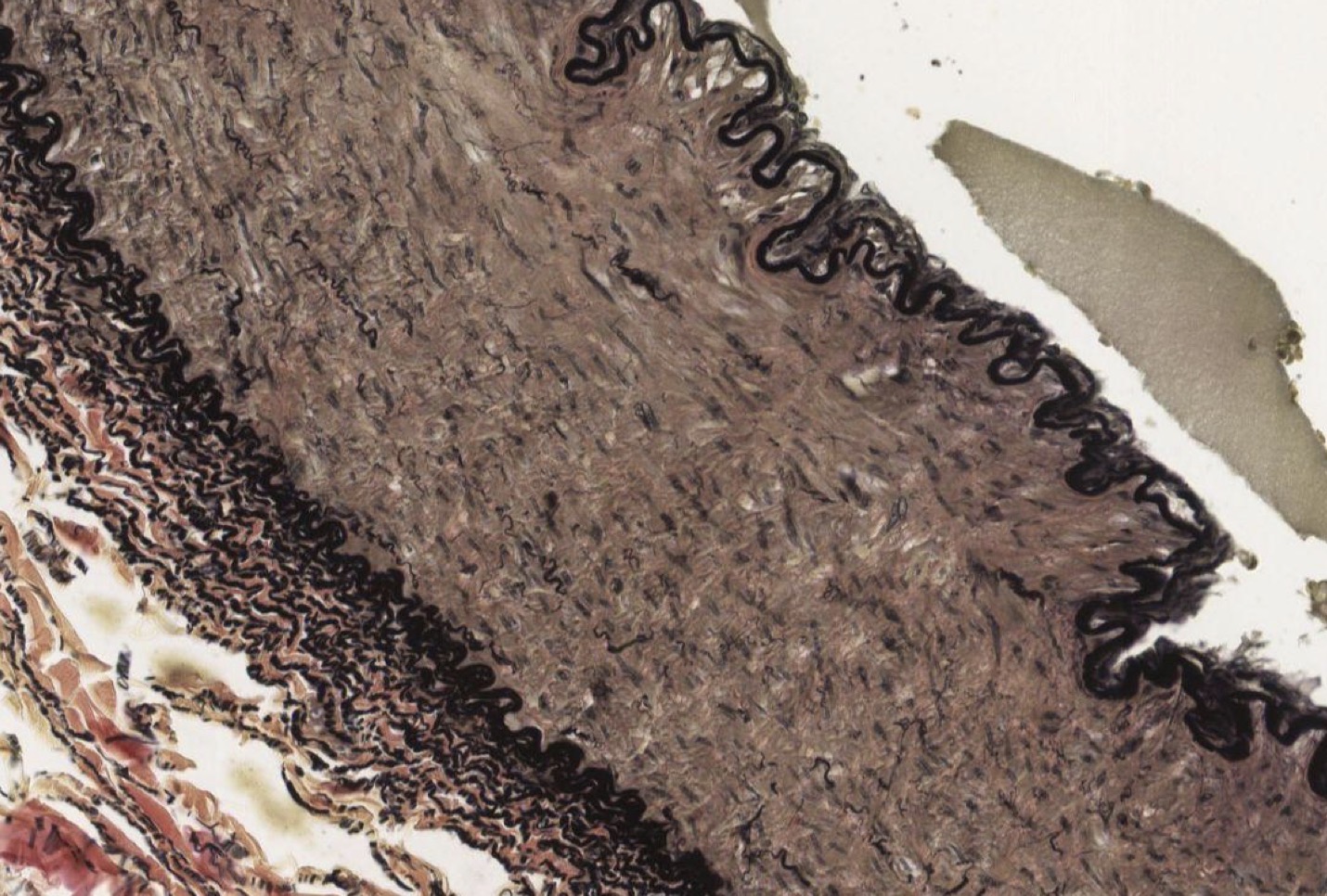
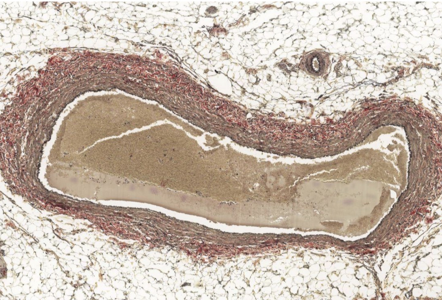
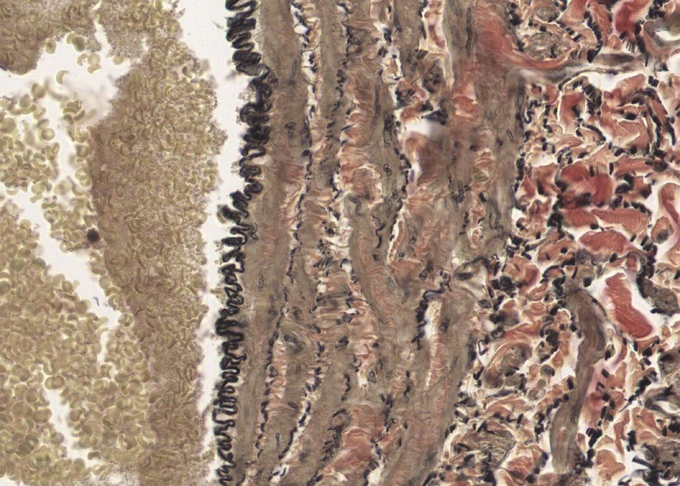
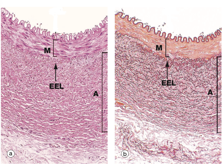
Idenitfy the Vessel and what are the Properties?
Large/Elastic Artery
close to heart
Ti made of endot + elastin + collagen
Tm made of elastic F + collagen; Smooth M!
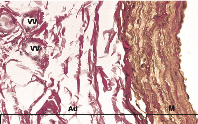
Te made of DICT w/ vasa vasorum
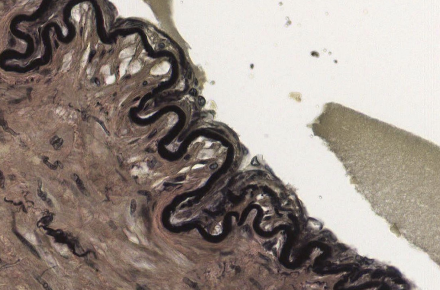
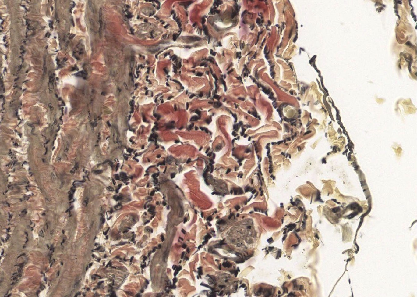
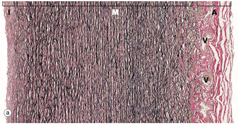
Identify the Vessel and the Layer pictured
Large/Elastic Artery Ti
Identify the Vessel and the Layer pictured
Large/Elastic Artery Tm!
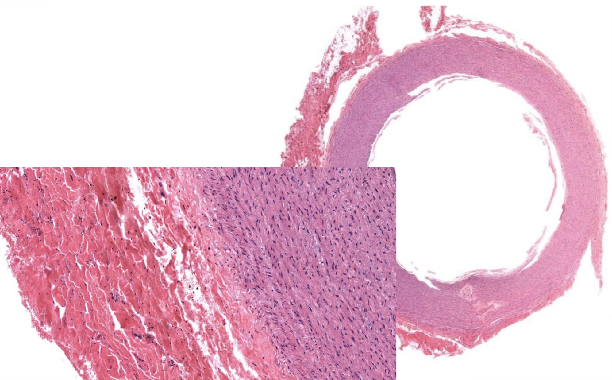
Identify the Vessel and the Layer pictured
Large/Elastic Artery Ta
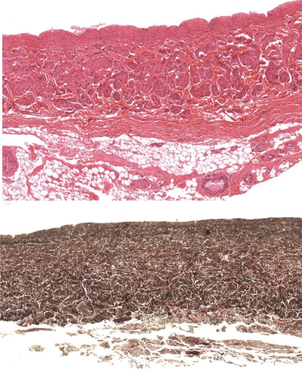
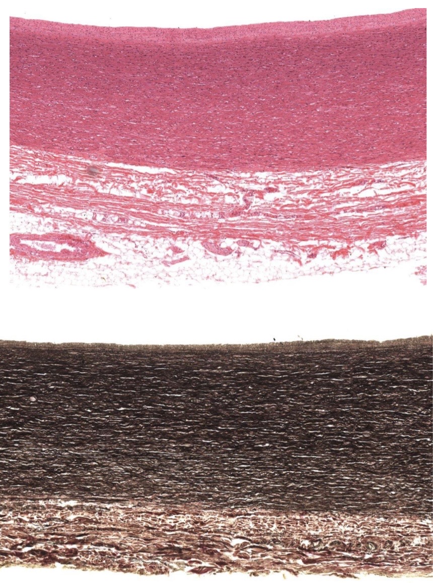
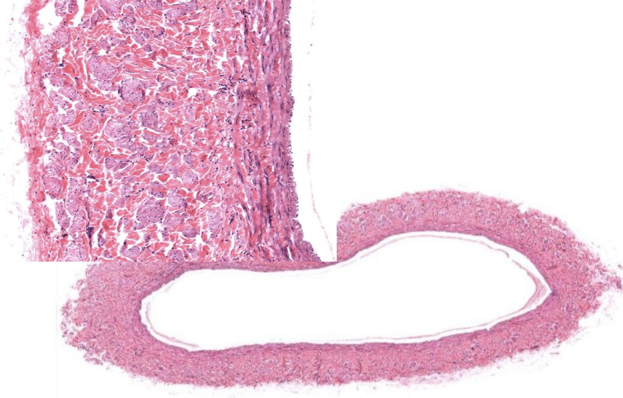
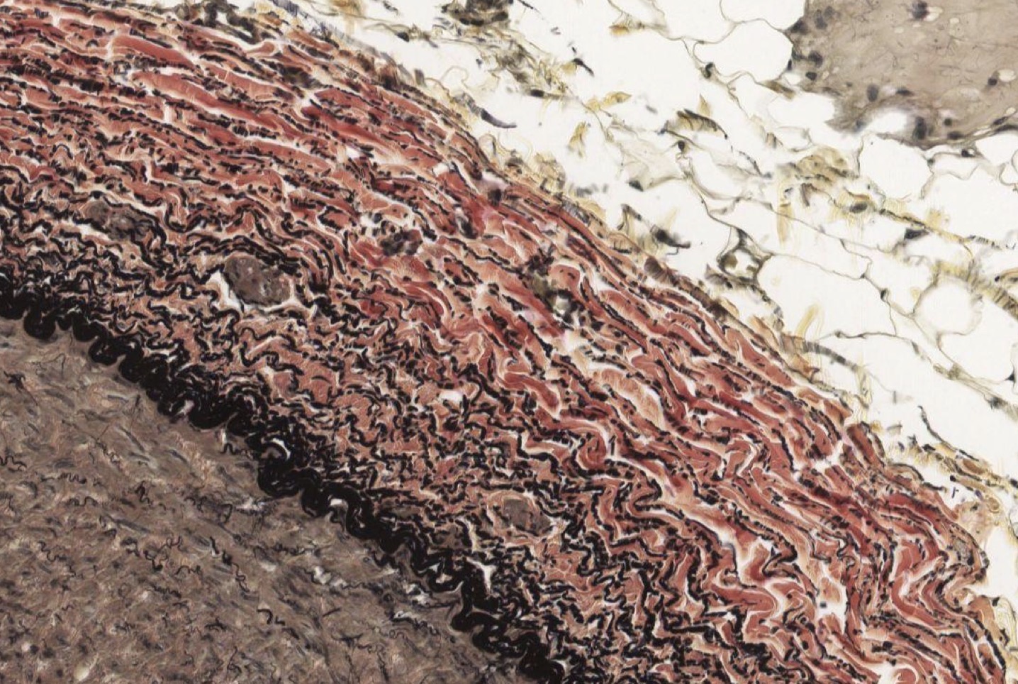
Idenitfy the Vessel and what are the Properties?
Large Vein
returns blood to heart
Ti made of endot
Tm made of some elastic F + 5 layers of Smooth M
Te made of DICT bundles/swirls! w/ vasa vasorum
Identify the Vessel and the Layer pictured
Large Vein Ti
Identify the Vessel and the Layer pictured
Large Vein Tm
Identify the Vessel and the Layer pictured
Large Vein Ta!
Identify the Vessel and the Layer pictured
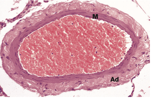
Medium Artery n/a
Identify the Vessel and the Layer pictured
Medium Artery Ti
Identify the Vessel and the Layer pictured
Medium Artery Tm!
Identify the Vessel and the Layer pictured
Medium Artery Ta
Identify the Vessel and the Layer pictured
Medium Vein n/a
Identify the Vessel and the Layer pictured
Medium Vein Ti
Identify the Vessel and the Layer pictured
Medium Vein Tm
Identify the Vessel and the Layer pictured
Medium Vein Ta!
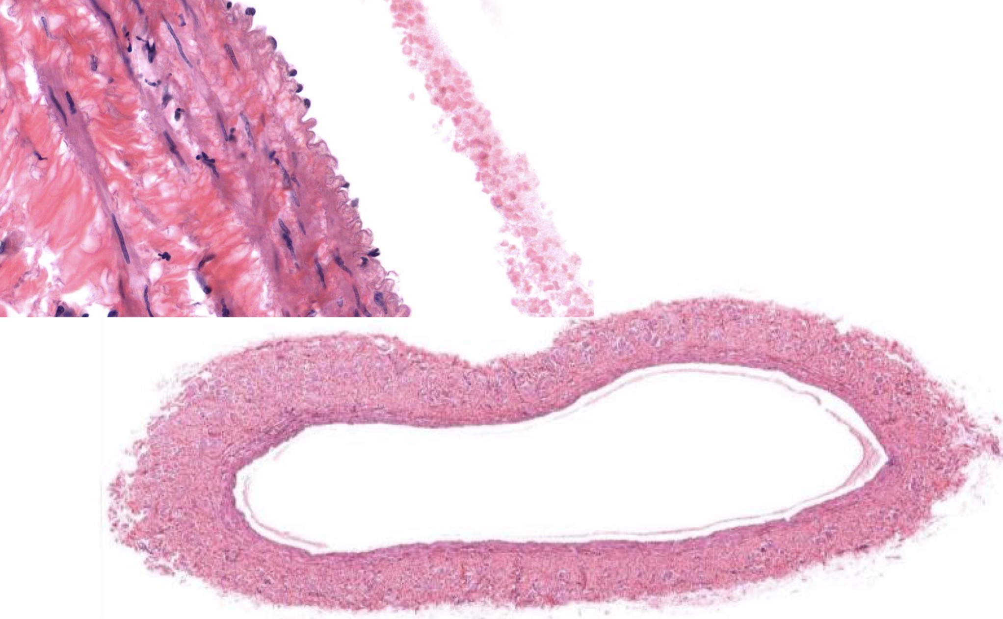
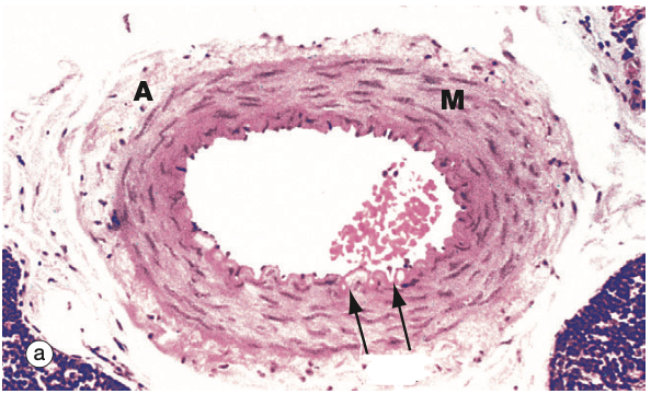
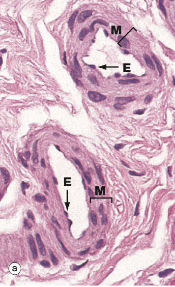
Identify the Vessel and the Layer pictured
Arteriole and Tm!
Identify the Vessel and the Layer pictured (Top and Bottom in both)
Top = Venule and Ta!
Bottom = Arteriole and Tm!
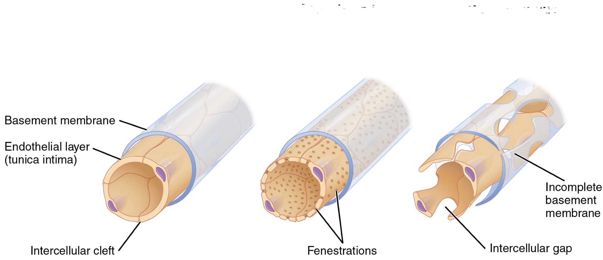
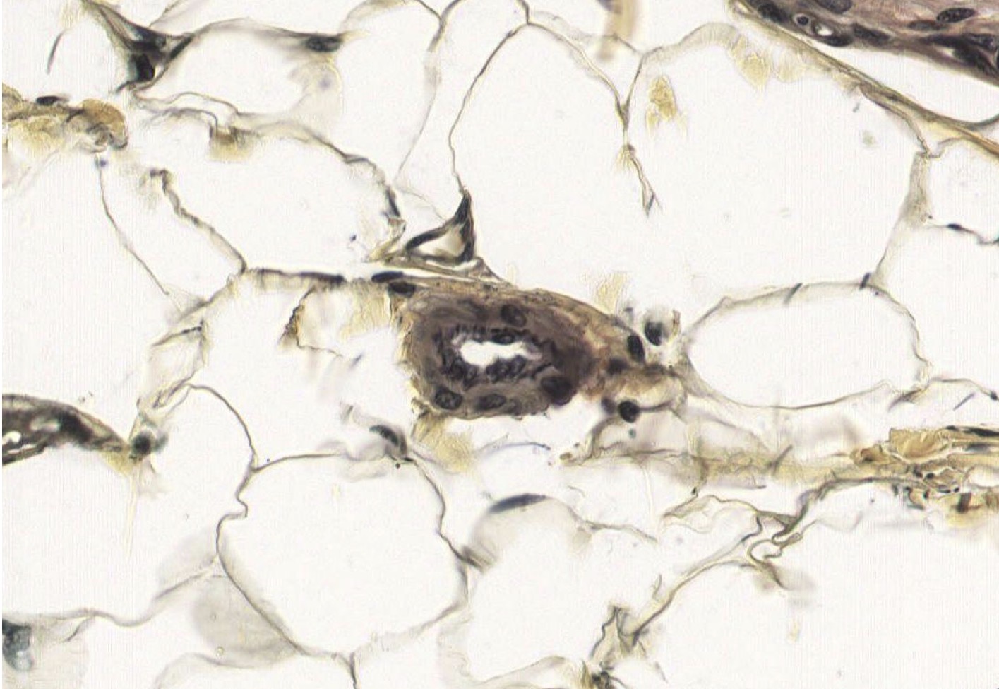
Identify the Structure(s)
Continuous, Fenestrated, Discontinuous
Identify the Structure(s)
Continuous Capillary
Identify the Structure(s)
Fenestrated Capillary
Identify the Structure(s)
Fenestrated Capillary
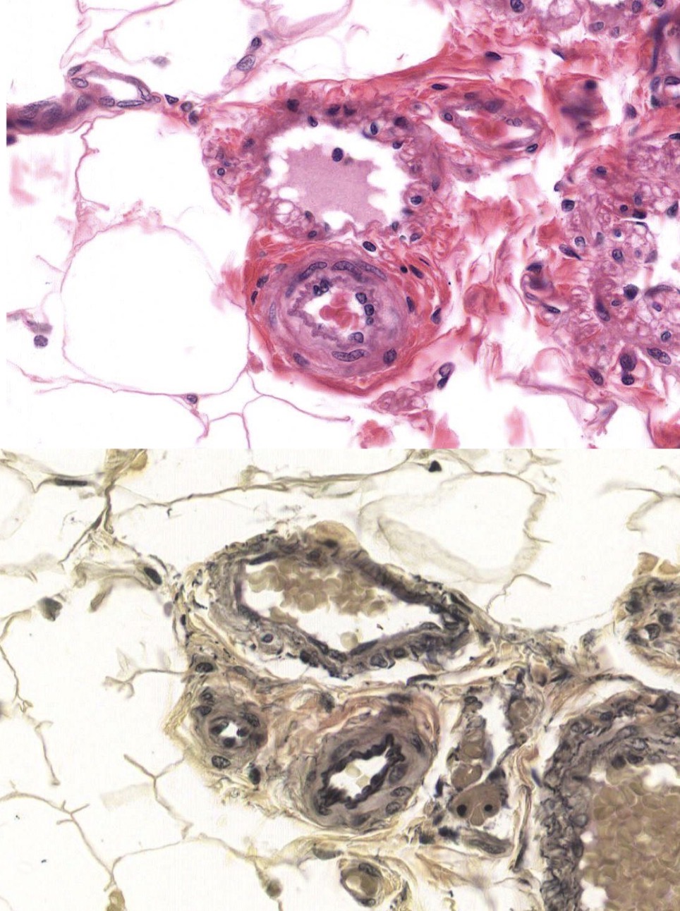
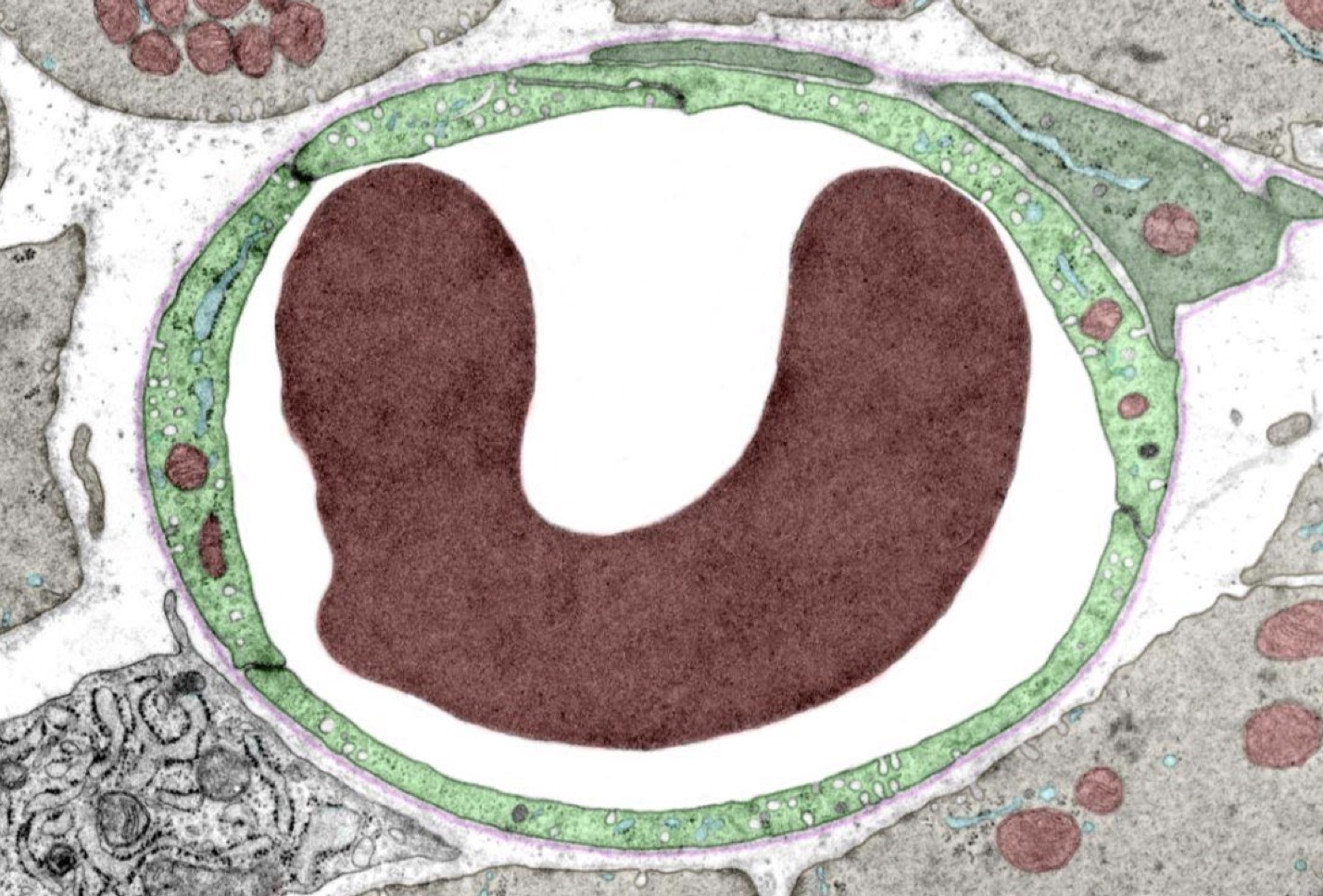
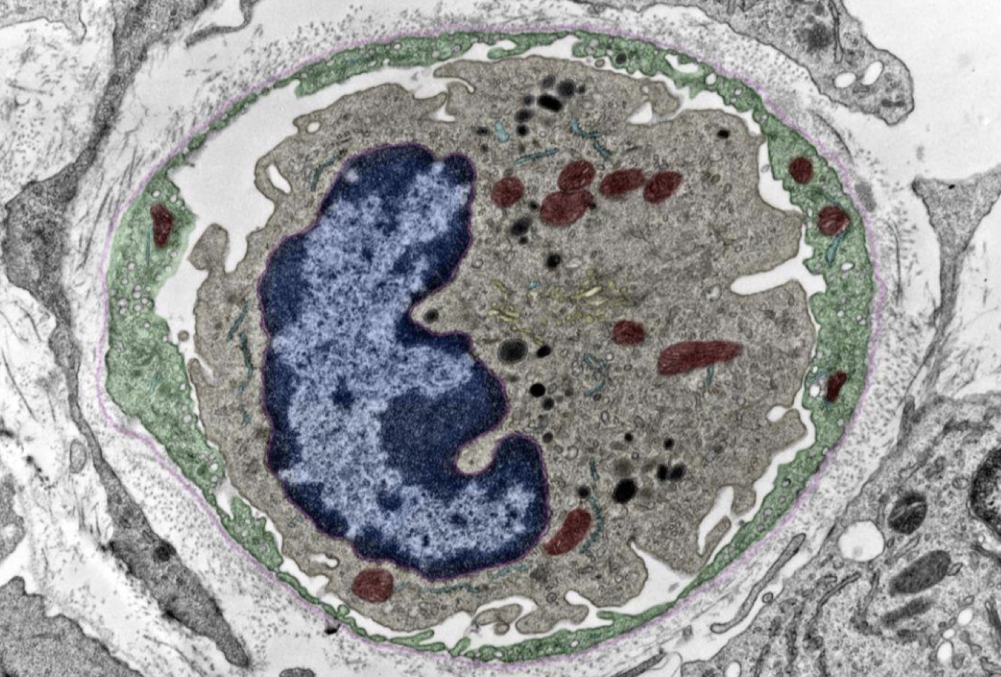
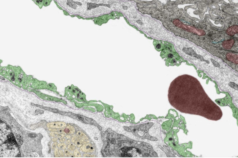
Identify the Structure(s)
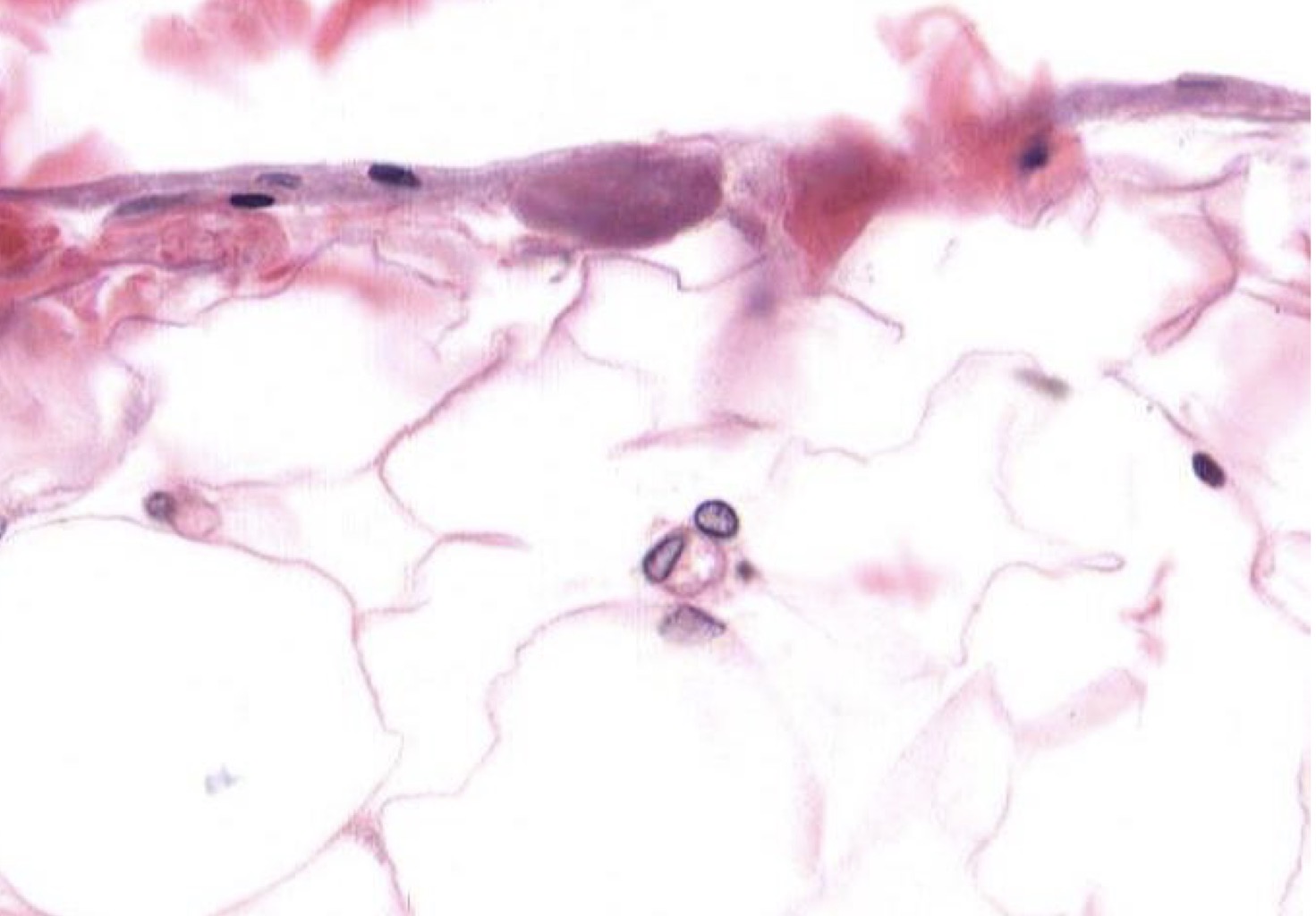
Capillary
Identify the Structure(s)
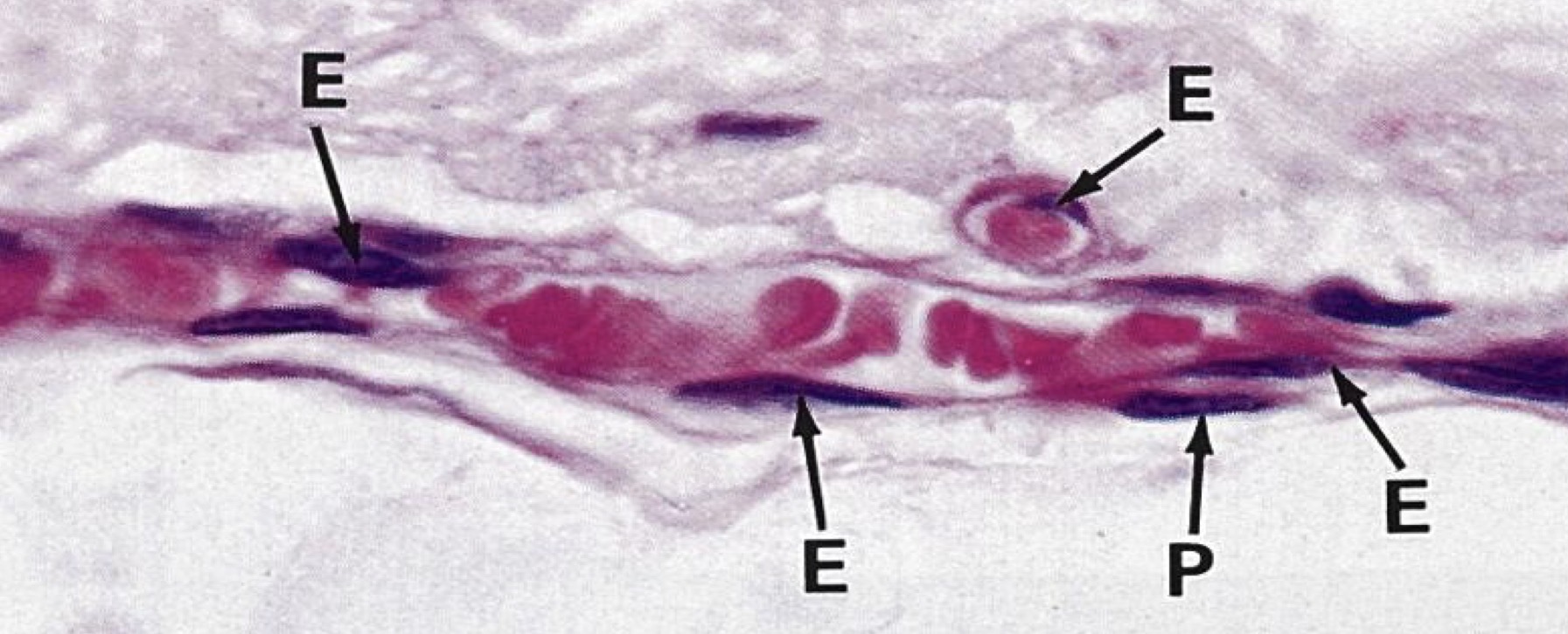
Capillary w/ Pericyte
Identify Which partition this is and Where?
Myocardium
Identify Which partition this is and Where?
Myocardium
Identify Which partition this is and Where?
Endocardium
Identify the Vessel and the Layer pictured
Large/Elastic Artery Ti, Tm!, and Ta
Identify the Vessel and the Layer pictured
Medium Artery Ti, Tm!, and Ta
Identify the Vessel and the Layer pictured
Medium/Small? Artery Ti, Tm!, and Ta
Identify the Vessel and the Layer pictured
Arteriole Ti, Tm!, and Ta
Identify the Vessel and the Layer pictured
Large Vein Ta!
Identify the Vessel and the Layer pictured
Medium Vein Ti, Tm, and Ta!