lecture 3 pelvic limb part 1
1/109
Earn XP
Description and Tags
equine pelvic limb sparks
Name | Mastery | Learn | Test | Matching | Spaced | Call with Kai |
|---|
No analytics yet
Send a link to your students to track their progress
110 Terms
how is the pelvis connected to the trunk
through a bony connection via the sacroiliac joint and with the pelvic limb via the coxal joint
what is the pelvis comprised of
left and right os coxae that are joined at the ventral midline by the pelvic symphysis
each os coxae is comprised of 3 bones what are they
ilium
ischium
pubis
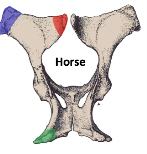
what is the red
tuber sacrale (sacral tuber)

what is the blue
tuber coxae (caxal tuber)

what is the green
tuber ischii (ischial tuber, ischiatic tuberosity)

what is 1
tuber sacrale

what is 2
tuber coxae
what makes up the wing of the ilium
tuber sacrale and tuber coxae

what is 3
tuber ischii
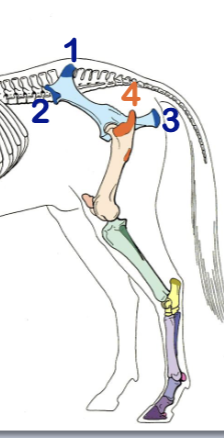
what is 4
greater trochanter
where is the greater trochanter caudal part located
2/3 of distance between tuber coxae and tuber ischii
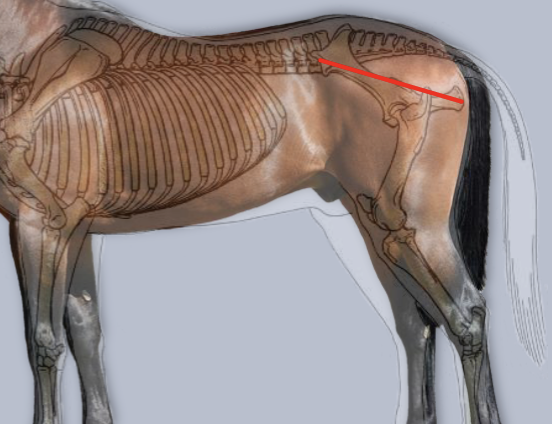
what is the red line
slope of pelvis

what is A
base of tail

what is B
anus
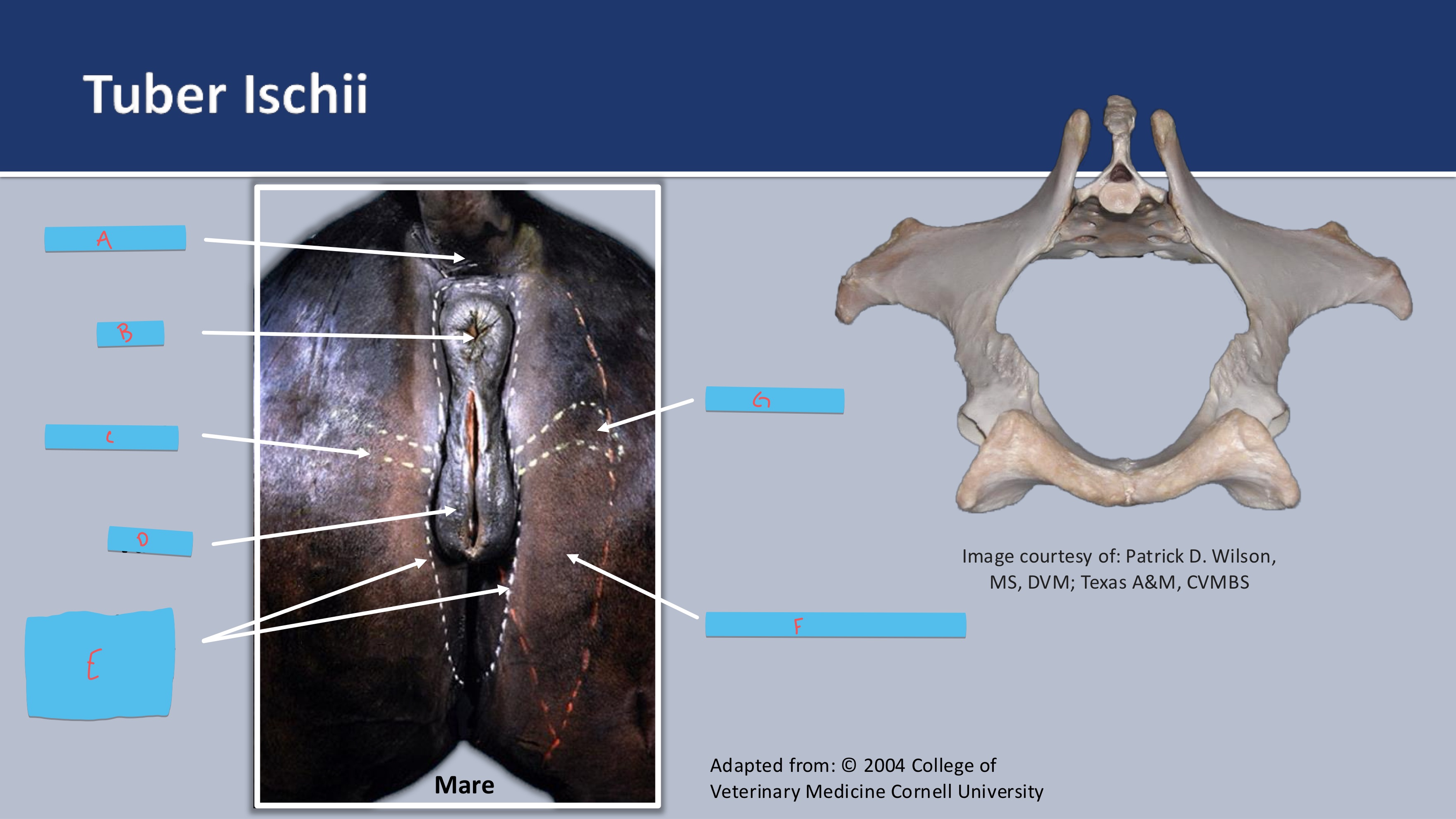
what bone prominence is found at C
ischial arch

what is D
vulva

what boundary is E
superficial boundary of perineum

what muscle is F
semimembranosus m.

what boney prominence is G
tuber ischii
how do horses sacrotuberous ligament compare to canine
horses are very broad and tehye attach to ischiatic spine
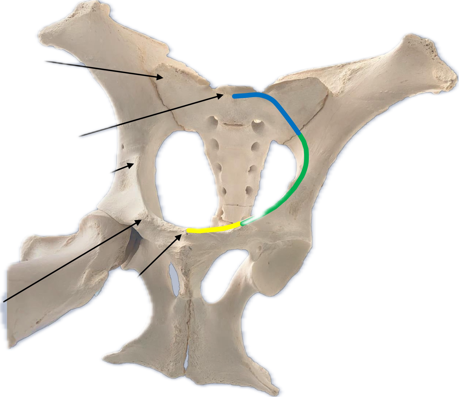
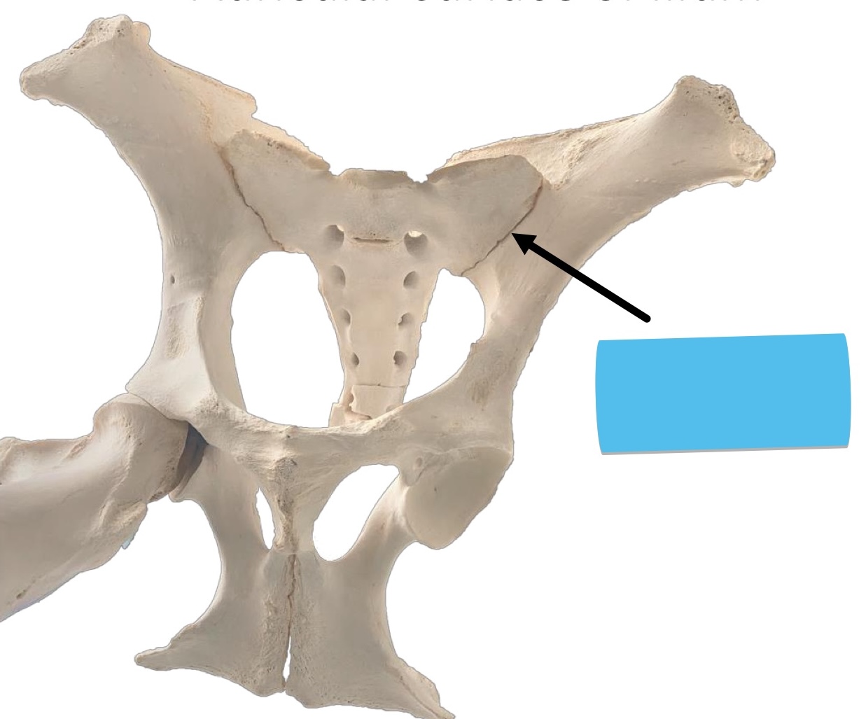
what is the yellow
ventral border known as the pecten pubis. this is located between the pubic tubercle and iliopubic eminence

what is the green
lateral border known as arcuate line this ridge extends between the iliopubic eminence and the auricular surface of the ilium

what is the blue
sacral promontory and wing of sacrum

what is A
wing of sacrum

what is B
sacral promontory

what is C
Psoas Tuberosity
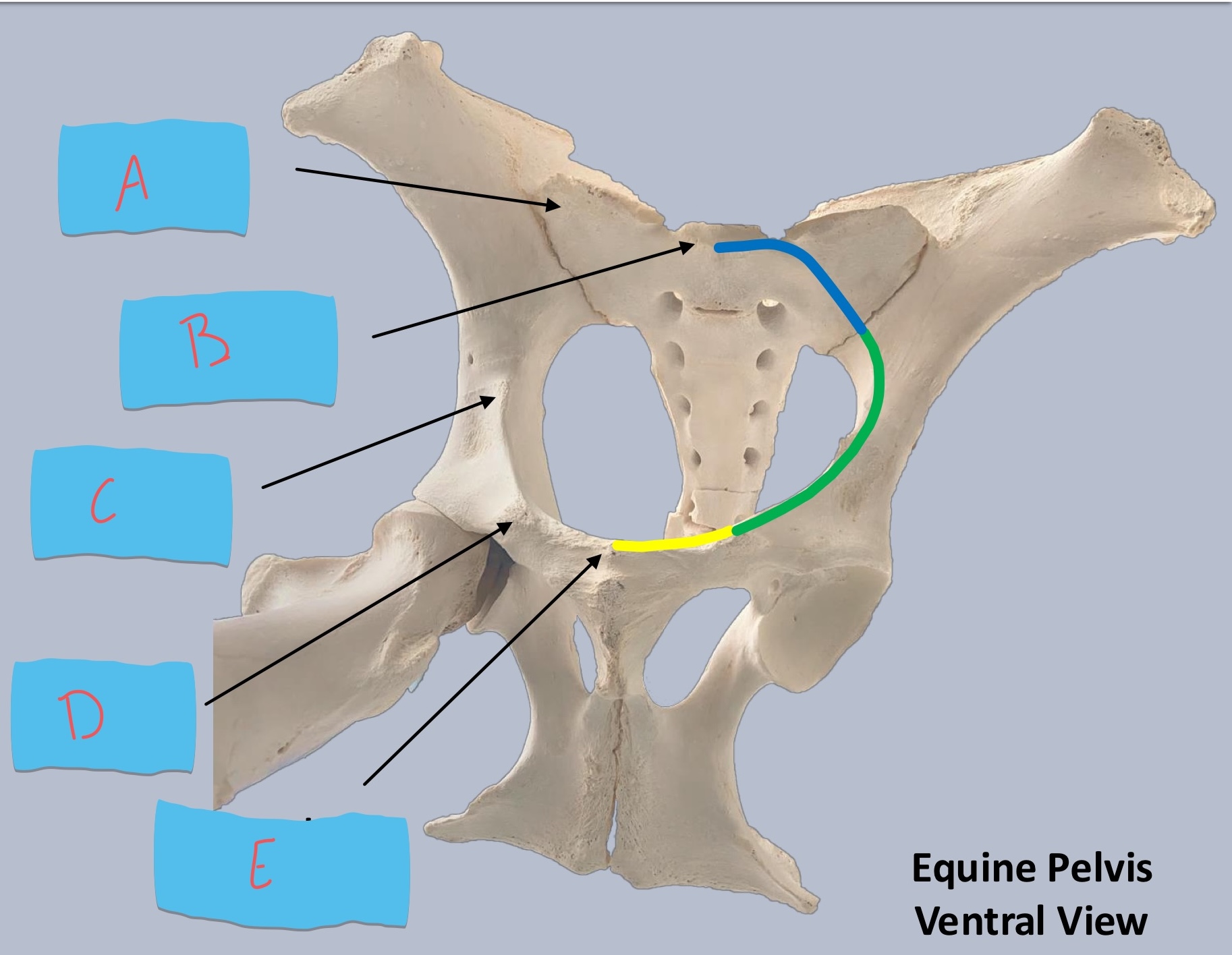
what is D
iliopubic eminence

what is E
pubic tubercle
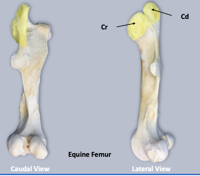
what is yellow on this
greater trochanter w
what is affiliated with the cranial part of the greater trochanter
trochanteric bursa
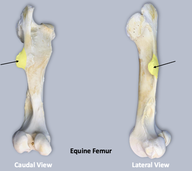
what is yellow
third trochanter
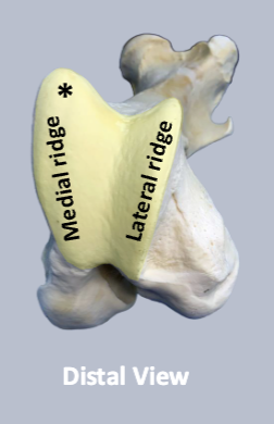
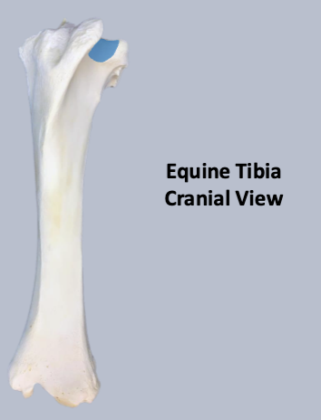
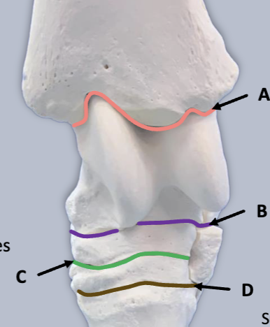
what is yellow
trochlea
what is the trochlea comprised of
medial and lateral ridges with a deep depression
which trochlear ridge is larger
medial

what is A
gliding surface

what is B
resting surface
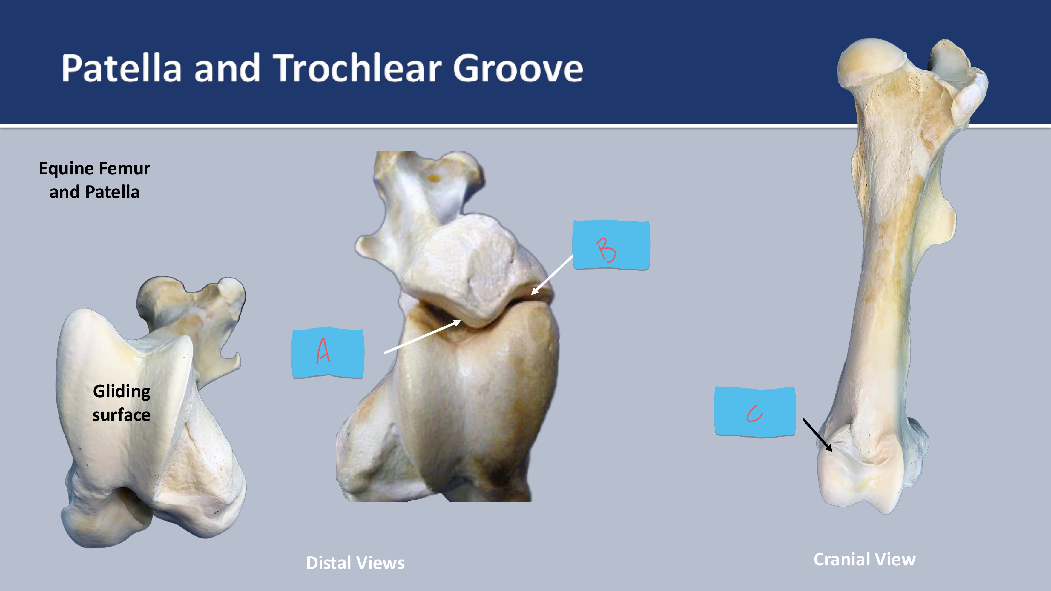
what is C
resting surface
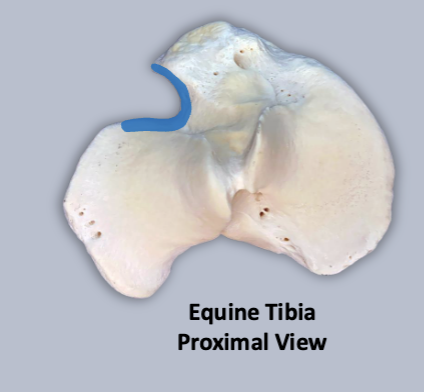
what is blue
extensor groove
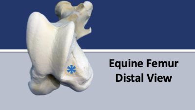
what is the blue star
extensor fossa
what is blue
extensor groove
where does the long digital extensor m. originate from
extensor fossa of the femur and passes throug hte extensor groove of the tibia
what other muscle outside of the long digital extensor m. also passes through the extensor groove
peroneus (fibularis) tertius
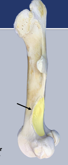
what is the yellow
supracondylar fossa

what is the arrow pointing to
lateral supracondylar tuberosity
what is special about the fibula in horses
it is reduced not elongated
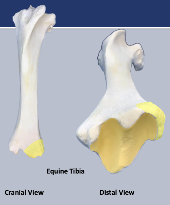
what is the yellow
lateral malleolus
the intermediate ridge and cochlear grooves of the tibia are oblique orientation what does this allow
outward rotation of pes upon flexion of tibiotalar joint
why is the oblique orientation of the tibia important
crucial for balancing the signifiant forces generated during fast running
what is the weight bearing digit for the pes
3
what is the tarsus made up of
▪ Fused tarsal bones I & II articulate with MT II & III
▪ Tarsal bone III articulates with MT III
▪ Tarsal bone IV articulates with MT III & IV
what is the metatarsus made up of
▪ MT III (cannon bone)
▪ MT II and IV (medial/lateral splint bones)
what is the digit made up of
▪ Proximal phalanx (P1, long pastern)
▪ Middle phalanx (P2, short pastern)
▪ Distal phalanx (P3, coffin)w
what are the sesamoid bones of the pes and where are they found
▪ Proximal, paired (at metatarsophalangeal jt.)
▪ Distal (at distal interphalangeal jt.)

what is 1
greater trochanter

what is 2
third trochanter


what is 3
patella

what is 4
tibial tuberosity

what is 5
tibial crest


what is 6
medial malleolus
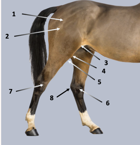

what is 7
lateral malleolus

what is 8
tuber calcaneii
what is this
sacroiliac joint
excessive strain of the sacroiliac joint can lead to
hunter’s bump due to ligament tears and dislocation
what is the coxal joint articulation of
femoral head with acetabulum of os coxae
what is a deep depression that receives the head of the femur in formation of the coxal joint
acetabulum
what is the acetabulum composed of
ilium, ischium, pubis, and acetabular bone
what is the labrum
the fibrocartilage that extends from the rim of the acetabulum
what is the acetabulum in life spanned by
transverse acetabular ligament
what is the articular surface of the acetabulum
semilunar
what is the attachment site of the ligament of the femoral head
acetabular fossa

what is the purple
semilunar
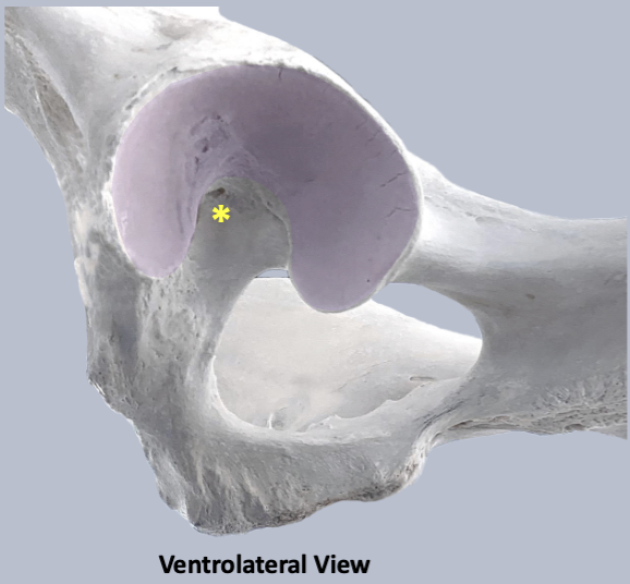

what is the star
acetabular fossa

where do the femoral ligaments pass
deep to the transverse acetabular ligament and insert on the fovea capitis of the heaad of the femur
the ligament of femoral head extends from what to what
fovea capitis of femur to acetabular fossa
what does the accessory ligament detach from
prepubic tendon
what is the main purposes of accessory ligament
restricts movement and stablizes the jointT
T/F coxal joint luxations are very common
false they are rare
what kind of joint is the stifle joint
compound jointw
what joints are within the stifle joint
▪ Femorotibial articulation
▪ Femoropatellar articulation
▪ Proximal tibiofibular articulation
bony articular surfaces of the stifle joint are
incongruent and unstable

what is A
medial collateral ligament


what is B
medial meniscus

what is C
caudal cruciate ligament

what is D
meniscofemoral ligament

what is E
popliteus m.
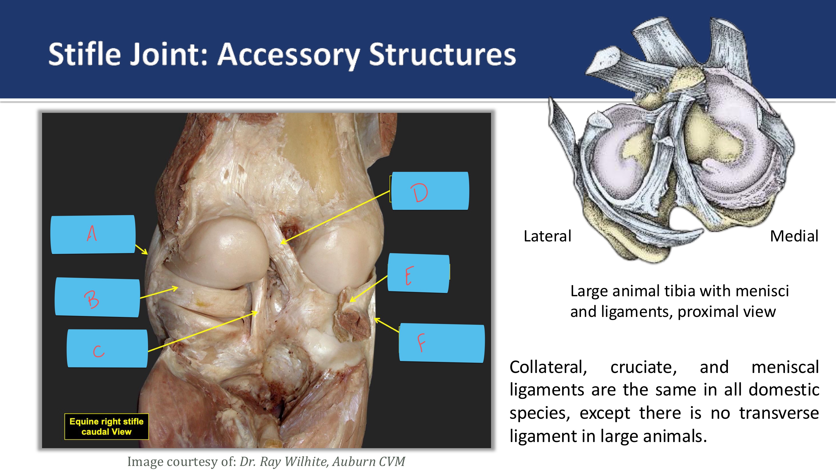
what is F
lateral collateral ligament
in the horse the patella is attached to the tibial tuberosity by what 3 patellar ligaments
medial, intermediate (middle), lateral
what is found between the medial patellar ligament and patella
parapatellar fibrocartilage
what is the parapatellar fibrocartilage important for
patellar locking mechanism
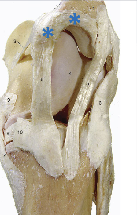
what is the blue star
parapatellar fibrocartilage
what forms a loop around the medial trochlear ridge of the femur
medial, intermediate patellar ligament
patella and parapatellar fibrocartilage
what does the loop around the medial trochlear ridge of the femur aid in
locking of the patella on the resting surface of the tubercle of the medial trochlear ridge of the femur
what are the two synovial compartments of the femorotibial joint
medial and lateral
what compartments of the synovial compartment of stifle joint usually communicate
femoropatellar and medial femortibial
what compartments of the synovial compartment of the stifle joint sometimes communicate (25%)
femoropatellar and lateral femorotibialwh
what synovial compartments of the stifle joint never communicate
medial femorotibial and lateral femorotibial
what is the located between the joint capsule of the femoropatellar joint and the patellar ligaments
fat pad
what can be used for locating patellar ligaments
tibial tuberosity
what joint is peach
tibotarsal joint (btwn tibia and talus)